. 2016 Aug 5;17(1):30.
doi: 10.1186/s12860-016-0108-6.

Aneuploidy screening of embryonic stem cell clones by metaphase karyotyping and droplet digital polymerase chain reaction

Affiliations

Aneuploidy screening of embryonic stem cell clones by metaphase karyotyping and droplet digital polymerase chain reaction

Gemma F Codner et al. BMC Cell Biol. .

Abstract

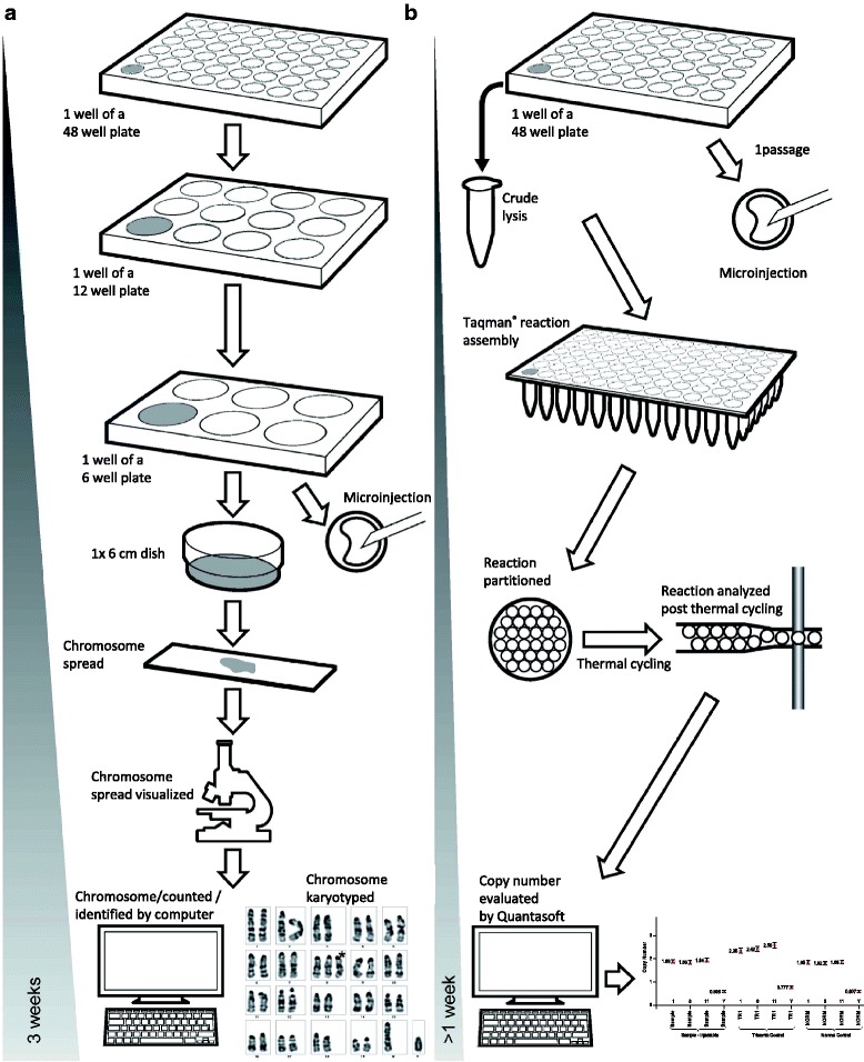
Background: Karyotypic integrity is essential for the successful germline transmission of alleles mutated in embryonic stem (ES) cells. Classical methods for the identification of aneuploidy involve cytological analyses that are both time consuming and require rare expertise to identify mouse chromosomes.

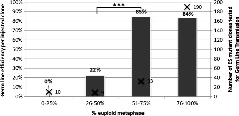
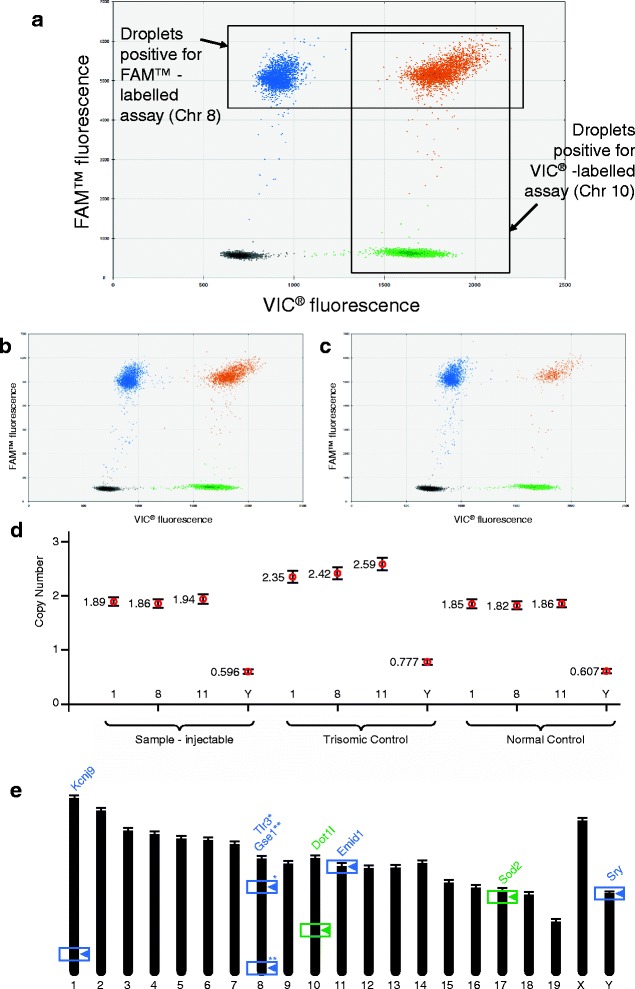
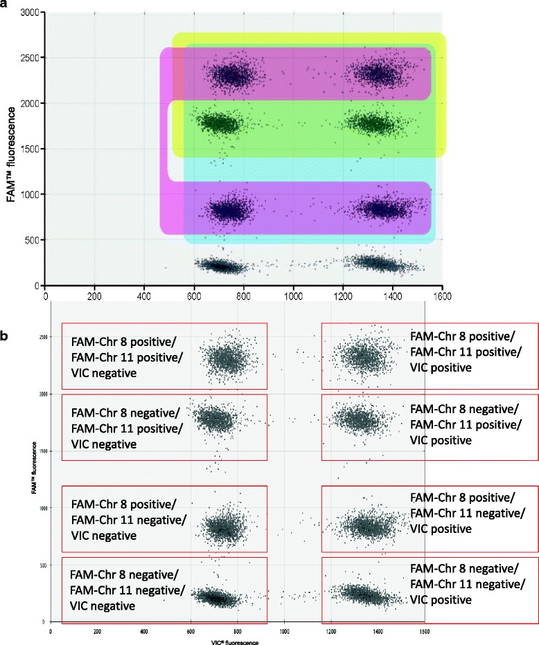
Results: As part of the International Mouse Phenotyping Consortium, we gathered data from over 1,500 ES cell clones and found that the germline transmission (GLT) efficiency of clones is compromised when over 50 % of cells harbour chromosome number abnormalities. In JM8 cells, chromosomes 1, 8, 11 or Y displayed copy number variation most frequently, whilst the remainder generally remain unchanged. We developed protocols employing droplet digital polymerase chain reaction (ddPCR) to accurately quantify the copy number of these four chromosomes, allowing efficient triage of ES clones prior to microinjection. We verified that assessments of aneuploidy, and thus decisions regarding the suitability of clones for microinjection, were concordant between classical cytological and ddPCR-based methods. Finally, we improved the method to include assay multiplexing so that two unstable chromosomes are counted simultaneously (and independently) in one reaction, to enhance throughput and further reduce the cost.

Conclusion: We validated a PCR-based method as an alternative to classical karyotype analysis. This technique enables laboratories that are non-specialist, or work with large numbers of clones, to precisely screen ES cells for the most common aneuploidies prior to microinjection to ensure the highest level of germline transmission potential. The application of this method allows early exclusion of aneuploid ES cell clones in the ES cell to mouse conversion process, thus improving the chances of obtaining germline transmission and reducing the number of animals used in failed microinjection attempts. This method can be applied to any other experiments that require accurate analysis of the genome for copy number variation (CNV).

Keywords: Aneuploidy; Cell culture; Chromosome number; Droplet digital PCR; Embryonic stem cells; Karyotype; Multiplex assay.


Figures

Fig. 1
Fig. 1
Contribution of euploid cells and GLT rate in all C57BL/6N-derived clones. Percentage of euploid metaphases observed by Giemsa staining metaphase spread-based karyotyping was compared to germ line efficiency obtained at ICS (left axis, grey). Number of ES mutant clones tested is indicated on the right axis (cross). Data was analysed using the Fisher Exact test and yielded a P value of 0.000806. False discovery rate (Q) calculated by the Benjamini-Hochberg procedure was 0.003224. This showed that clones with greater than 50 % euploid representation are preferable candidates for microinjection
Fig. 2
Fig. 2
Example of evaluation of copy number by ddPCR. Panel a shows an annotated example of FACS-like plot obtained with the QuantaSoft software, version 1.2.10.0 (Bio-Rad, CA, USA) taken from a CNV2 copy counting ddPCR experiment. Panel b and c show typical results obtained from quantifying Chr 8 in a euploid and a trisomic sample, respectively. Panel d shows copy numbers as calculated and presented in the CNV option obtained with known euploid (Normal) and trisomic for Chr 1, 8 and 11 (Trisomic) samples as external calibrators. A new sample of unknown quality is shown to be injectable. Vertical bars are Standard Errors. Panel e presents the distribution of the marker genes and their mouse chromosomal location and the assays that were employed in this study (* and ** show the position on Chr 8 of Tlr3 and Gse1, respectively)
Fig. 3
Fig. 3
Comparison of processes based on karyotyping of mitotic chromosome spreads and ddPCR chromosome counting. Panel a details the method including chromosome spreads that we used for karyotyping by chromosome counting of ES cell lines. Note that the ES cell amplification phase spans several culture passages, including intensive preparation and evaluation of samples. The overall length of the process covered a period of 3 weeks for each sample. Panel b details the alternative process based on the novel ddPCR method introduced in this article as implemented at MRC Harwell. Note the shortened cell culture period, less intensive wet laboratory time (PCR-based), a faster readout of copy numbers from raw data with an overall process time of less than 1 week for each sample. For operational reasons, the ddPCR screen is implemented at a later passage at ICS. A key aspect of the workflow is that the DNA extraction is performed from an ES cell passage number close to that at which the cells are injected
Fig. 4
Fig. 4
Evaluation of copy number by multiplexed ddPCR. The figure describes the structure of a FACS-like plot obtained with multiplexed ddPCR analysed with the QuantaSoft software, as in version 1.2.10.0 (Bio-Rad, CA, USA). a The area in blue shows the droplets positive for either or both unstable chromosomes analysed. The area highlighted in yellow shows droplets positive for the assay of Chr 11, whilst the area shaded in pink shows the droplets positive for the other unstable chromosome analysed (8); (b) a similar plot where each droplet populations are annotated

References

    1. Skarnes WC, Rosen B, West AP, Koutsourakis M, Bushell W, Iyer V, et al. A conditional knockout resource for the genome-wide study of mouse gene function. Nature. 2011;474:337–42. doi: 10.1038/nature10163. - DOI - PMC - PubMed
    1. White JK, Gerdin A-K, Karp NA, Ryder E, Buljan M, Bussell JN, et al. Genome-wide generation and systematic phenotyping of knockout mice reveals new roles for many genes. Cell. 2013;154:452–64. doi: 10.1016/j.cell.2013.06.022. - DOI - PMC - PubMed
    1. de Angelis MH, Nicholson G, Selloum M, White JK, Morgan H, Ramirez-Solis R, et al. Analysis of mammalian gene function through broad-based phenotypic screens across a consortium of mouse clinics. Nat Genet. 2015;47:969–78. doi: 10.1038/ng.3360. - DOI - PMC - PubMed
    1. Brown SDM, Moore MW. The International Mouse Phenotyping Consortium: past and future perspectives on mouse phenotyping. Mamm Genome Off J Int Mamm Genome Soc. 2012;23:632–40. doi: 10.1007/s00335-012-9427-x. - DOI - PMC - PubMed
    1. Liu X, Wu H, Loring J, Hormuzdi S, Disteche CM, Bornstein P, et al. Trisomy eight in ES cells is a common potential problem in gene targeting and interferes with germ line transmission. Dev Dyn Off Publ Am Assoc Anat. 1997;209:85–91. - PubMed

Publication types

LinkOut - more resources