

Cerebral blood flow in small vessel disease: A systematic review and meta-analysis
- PMID: 27496552
- PMCID: PMC5076792
- DOI: 10.1177/0271678X16662891
Cerebral blood flow in small vessel disease: A systematic review and meta-analysis
Abstract
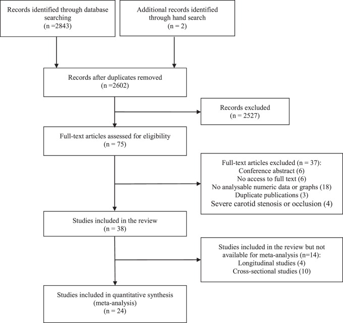
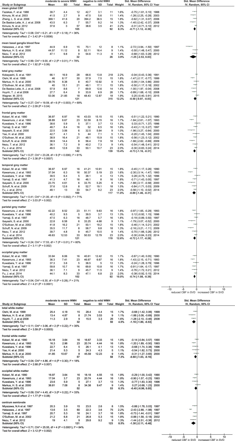
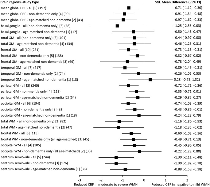
White matter hyperintensities are frequent on neuroimaging of older people and are a key feature of cerebral small vessel disease. They are commonly attributed to chronic hypoperfusion, although whether low cerebral blood flow is cause or effect is unclear. We systematically reviewed studies that assessed cerebral blood flow in small vessel disease patients, performed meta-analysis and sensitivity analysis of potential confounders. Thirty-eight studies (n = 4006) met the inclusion criteria, including four longitudinal and 34 cross-sectional studies. Most cerebral blood flow data were from grey matter. Twenty-four cross-sectional studies (n = 1161) were meta-analysed, showing that cerebral blood flow was lower in subjects with more white matter hyperintensity, globally and in most grey and white matter regions (e.g. mean global cerebral blood flow: standardised mean difference-0.71, 95% CI -1.12, -0.30). These cerebral blood flow differences were attenuated by excluding studies in dementia or that lacked age-matching. Four longitudinal studies (n = 1079) gave differing results, e.g., more baseline white matter hyperintensity predated falling cerebral blood flow (3.9 years, n = 575); cerebral blood flow was low in regions that developed white matter hyperintensity (1.5 years, n = 40). Cerebral blood flow is lower in subjects with more white matter hyperintensity cross-sectionally, but evidence for falling cerebral blood flow predating increasing white matter hyperintensity is conflicting. Future studies should be longitudinal, obtain more white matter data, use better age-correction and stratify by clinical diagnosis.
Keywords: Cerebral blood flow; cerebral small vessel disease; meta-analysis; systematic review; white matter hyperintensities.
© The Author(s) 2016.
Figures
References
-
- Jeerakathil T, Wolf PA, Beiser A, et al. Stroke risk profile predicts white matter hyperintensity volume: The Framingham Study. Stroke 2004; 35: 1857–1861. - PubMed
-
- Yata K, Tomimoto H. Chronic cerebral hypoperfusion and dementia. Neurol Clin Neurosci 2014; 2: 129–134.
-
- Ibayashi S, Nagao T, Kuwabara Y, et al. Mechanism for decreased cortical oxygen metabolism in patients with leukoaraiosis: Is disconnection the answer? J Stroke Cerebrovasc Dis 2000; 9: 22–26.
Publication types
MeSH terms
Grants and funding
LinkOut - more resources
Full Text Sources
Other Literature Sources
Medical
Miscellaneous
