

Growth inhibition in a brain metastasis model by antibody delivery using focused ultrasound-mediated blood-brain barrier disruption
- PMID: 27496633
- PMCID: PMC5014601
- DOI: 10.1016/j.jconrel.2016.08.001
Growth inhibition in a brain metastasis model by antibody delivery using focused ultrasound-mediated blood-brain barrier disruption
Abstract
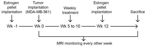
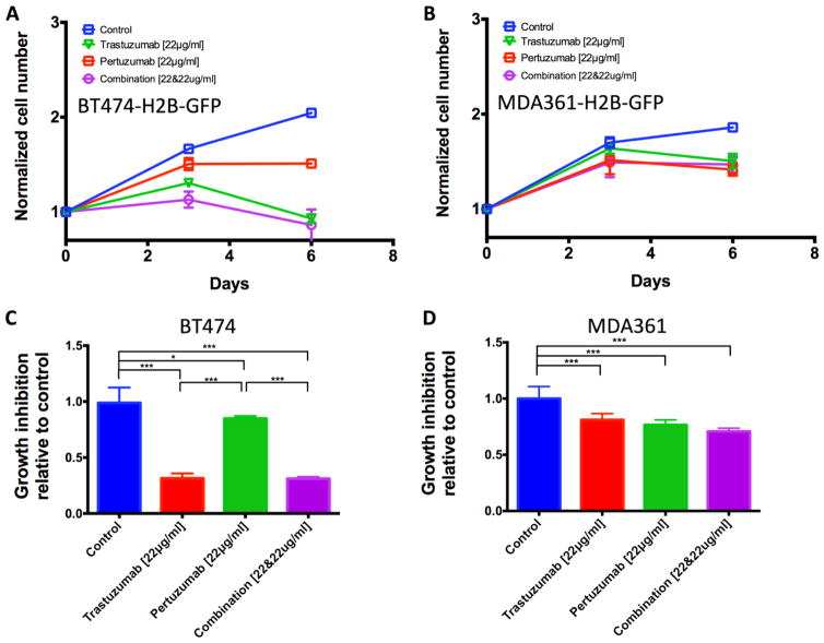
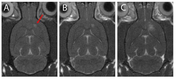
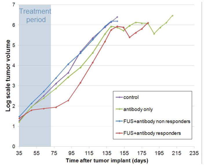
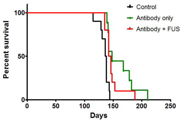
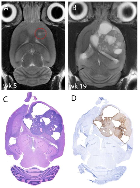
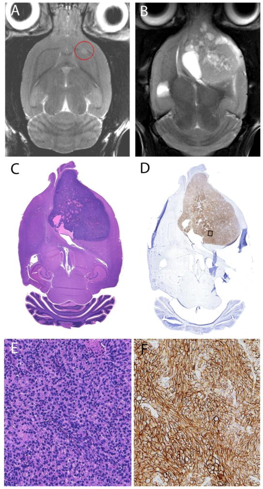
HER2-targeting antibodies (i.e. trastuzumab and pertuzumab) prolong survival in HER2-positive breast cancer patients with extracranial metastases. However, the response of brain metastases to these drugs is poor, and it is hypothesized that the blood-brain barrier (BBB) limits drug delivery to the brain. We investigated whether we could improve the response by temporary disruption of the BBB using focused ultrasound in combination with microbubbles. To study this, we inoculated 30 nude rats with HER2-positive cells derived from a brain metastasis of a breast cancer patient (MDA-MB-361). The animals were divided into three groups: a control-group that received no treatment; an antibody-only group that received six weekly treatments of trastuzumab and pertuzumab; and an ultrasound+antibody group that received trastuzumab and pertuzumab in combination with six weekly sessions of BBB disruption using focused ultrasound. In two animals, the leakiness of the tumors before disruption was evaluated using contrast-enhanced T1-weighted magnetic resonance imaging and found that the tumors were not leaky. The same technique was used to evaluate the effectiveness of BBB disruption, which was successful in all sessions. The tumor in the control animals grew exponentially with a growth constant of 0.042±0.011mm(3)/day. None of the antibody-only animals responded to the treatment and the growth constant was 0.033±0.009mm(3)/day during the treatment period. Four of the ten animals in the ultrasound+antibody-group showed a response to the treatment with an average growth constant of 0.010±0.007mm(3)/day, compared to a growth constant 0.043±0.013mm(3)/day for the six non-responders. After the treatment period, the tumors in all groups grew at similar rates. As the tumors were not leaky before BBB disruption and there were no responders in the antibody-only group, these results show that at least in some cases disruption of the BBB is necessary for a response to the antibodies in these brain metastases. Interestingly, only some of the rats responded to the treatment. We did not observe a difference in tumor volume at the start of the treatment, nor in HER2 expression or in contrast-enhancement on MRI between the responders and non-responders to explain this. Better understanding of why certain animals respond is needed and will help in translating this technique to the clinic. In conclusion, we demonstrate that BBB disruption using focused ultrasound in combination with antibody therapy can inhibit growth of breast cancer brain metastasis.
Keywords: Blood-brain barrier; Brain metastasis; Focused ultrasound; HER2-targeting antibodies; Targeted drug delivery.
Copyright © 2016 Elsevier B.V. All rights reserved.
Figures
Similar articles
-
Ultrasound-mediated blood-brain/blood-tumor barrier disruption improves outcomes with trastuzumab in a breast cancer brain metastasis model.J Control Release. 2012 Nov 10;163(3):277-84. doi: 10.1016/j.jconrel.2012.09.007. Epub 2012 Sep 18. J Control Release. 2012. PMID: 23000189 Free PMC article.
-
Trastuzumab uptake and its relation to efficacy in an animal model of HER2-positive breast cancer brain metastasis.Breast Cancer Res Treat. 2017 Aug;164(3):581-591. doi: 10.1007/s10549-017-4279-4. Epub 2017 May 10. Breast Cancer Res Treat. 2017. PMID: 28493046 Free PMC article.
-
Mechanisms of enhanced drug delivery in brain metastases with focused ultrasound-induced blood-tumor barrier disruption.Proc Natl Acad Sci U S A. 2018 Sep 11;115(37):E8717-E8726. doi: 10.1073/pnas.1807105115. Epub 2018 Aug 27. Proc Natl Acad Sci U S A. 2018. PMID: 30150398 Free PMC article.
-
Therapeutic approaches for HER2-positive brain metastases: circumventing the blood-brain barrier.Cancer Treat Rev. 2013 May;39(3):261-9. doi: 10.1016/j.ctrv.2012.05.006. Epub 2012 Jun 22. Cancer Treat Rev. 2013. PMID: 22727691 Free PMC article. Review.
-
Systemic Therapy for HER2-Positive Central Nervous System Disease: Where We Are and Where Do We Go From Here?Curr Oncol Rep. 2015 Oct;17(10):46. doi: 10.1007/s11912-015-0471-z. Curr Oncol Rep. 2015. PMID: 26314739 Review.
Cited by
-
Repeated blood-brain barrier opening with a nine-emitter implantable ultrasound device in combination with carboplatin in recurrent glioblastoma: a phase I/II clinical trial.Nat Commun. 2024 Feb 23;15(1):1650. doi: 10.1038/s41467-024-45818-7. Nat Commun. 2024. PMID: 38396134 Free PMC article. Clinical Trial.
-
Applications of focused ultrasound in the brain: from thermoablation to drug delivery.Nat Rev Neurol. 2021 Jan;17(1):7-22. doi: 10.1038/s41582-020-00418-z. Epub 2020 Oct 26. Nat Rev Neurol. 2021. PMID: 33106619 Review.
-
Breaching the Blood-Brain Tumor Barrier for Tumor Therapy.Cancers (Basel). 2021 May 15;13(10):2391. doi: 10.3390/cancers13102391. Cancers (Basel). 2021. PMID: 34063335 Free PMC article. Review.
-
Low-Intensity MR-Guided Focused Ultrasound Mediated Disruption of the Blood-Brain Barrier for Intracranial Metastatic Diseases.Front Oncol. 2018 Aug 28;8:338. doi: 10.3389/fonc.2018.00338. eCollection 2018. Front Oncol. 2018. PMID: 30211117 Free PMC article. Review.
-
Therapeutic Effect of Cabazitaxel and Blood-Brain Barrier opening in a Patient-Derived Glioblastoma Model.Nanotheranostics. 2019 Feb 7;3(1):103-112. doi: 10.7150/ntno.31479. eCollection 2019. Nanotheranostics. 2019. PMID: 30899638 Free PMC article.
References
-
- Aryal Muna, Vykhodtseva Natalia, Zhang Yong-Zhi, Park Juyoung, McDannold Nathan. Multiple Treatments with Liposomal Doxorubicin and Ultrasound-Induced Disruption of Blood–tumor and Blood–brain Barriers Improve Outcomes in a Rat Glioma Model. Journal of Controlled Release. 2013;169(1–2):103–11. doi: 10.1016/j.jconrel.2013.04.007. - DOI - PMC - PubMed
Publication types
MeSH terms
Substances
Grants and funding
LinkOut - more resources
Full Text Sources
Other Literature Sources
Medical
Research Materials
Miscellaneous
