. 2016 Aug 16;45(2):255-66.
doi: 10.1016/j.immuni.2016.06.015. Epub 2016 Aug 2.

The AIM2-like Receptors Are Dispensable for the Interferon Response to Intracellular DNA

Affiliations

The AIM2-like Receptors Are Dispensable for the Interferon Response to Intracellular DNA

Elizabeth E Gray et al. Immunity. .

Abstract

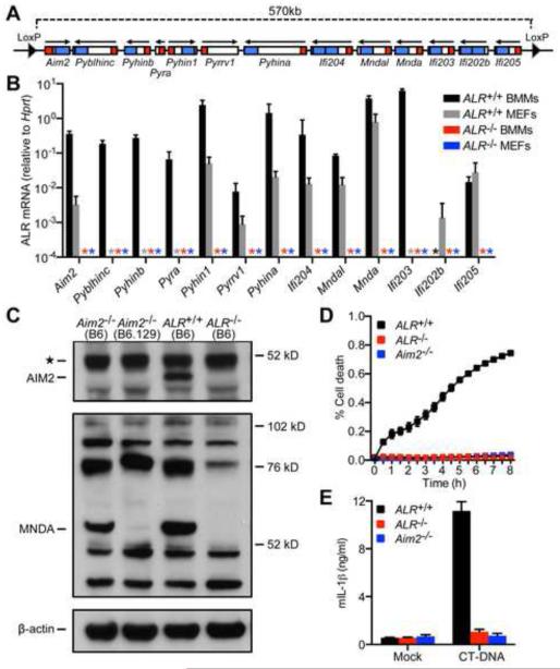
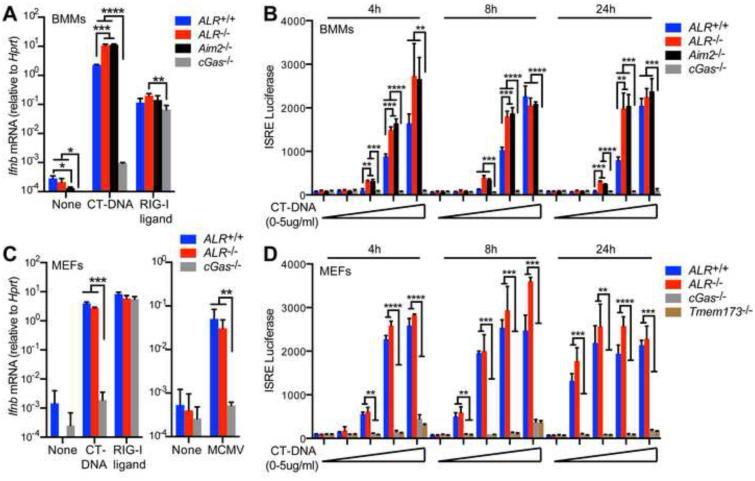
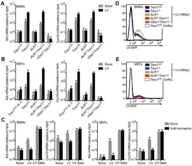
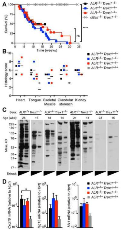
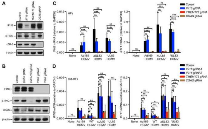
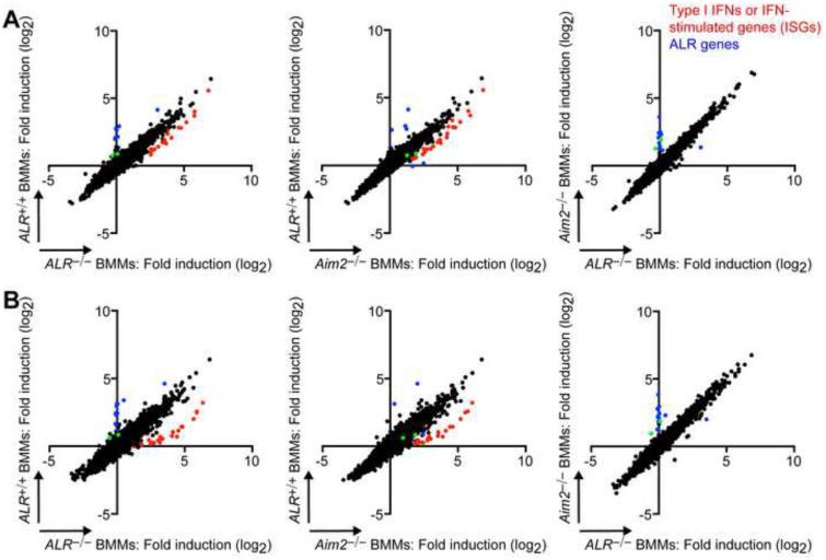
Detection of intracellular DNA triggers activation of the STING-dependent interferon-stimulatory DNA (ISD) pathway, which is essential for antiviral responses. Multiple DNA sensors have been proposed to activate this pathway, including AIM2-like receptors (ALRs). Whether the ALRs are essential for activation of this pathway remains unknown. To rigorously explore the function of ALRs, we generated mice lacking all 13 ALR genes. We found that ALRs are dispensable for the type I interferon (IFN) response to transfected DNA ligands, DNA virus infection, and lentivirus infection. We also found that ALRs do not contribute to autoimmune disease in the Trex1(-/-) mouse model of Aicardi-Goutières Syndrome. Finally, CRISPR-mediated disruption of the human AIM2-like receptor IFI16 in primary fibroblasts revealed that IFI16 is not essential for the IFN response to human cytomegalovirus infection. Our findings indicate that ALRs are dispensable for the ISD response and suggest that alternative functions for these receptors should be explored.


Figures

Figure 1
Figure 1. Generation of ALR-deficient mice
(A) Schematic of the LoxP-flanked mouse ALR locus for Cre-mediated deletion of all 13 mouse ALR genes. Pyrin and HIN domains are indicated in red and blue, respectively. (B) Quantification by RT-PCR of mouse ALR gene transcripts in bone marrow-derived macrophages (BMMs) or embryonic fibroblasts (MEFs) from ALR+/+ and ALR−/− mice (* = not detected). (C) AIM2 and MNDA protein expression evaluated in IFNβ-primed BMMs cultured from mice of the indicated genotype. Note that the band at 76kD (indicated by **) was not consistently observed in multiple experiments and does not match the expected size of IFI204. (D) Cell death of BMMs from ALR+/+, ALR−/−, or Aim2−/− mice transfected with calf-thymus DNA (CT-DNA) quantified using an IncuCyte imaging system. (E) Quantification of IL-1β secretion by BMMs from ALR+/+, ALR−/−, or Aim2−/− mice transfected with CT-DNA. Error bars represent mean ± SD. Data are representative of one experiment (B,C) or two experiments (D,E). See also Figure S1 and S2.
Figure 2
Figure 2. ALRs are dispensable for the IFN response to transfected DNA
(A) Quantification of Ifnb mRNA induction in BMMs from ALR+/+,ALR−/−, Aim2−/−, or cGas−/− mice transfected with CT-DNA or RIG-I ligand for 4h. (B) Quantification of type I IFN production using an IFN bioassay with supernatants from BMMs (A) of the indicated genotype transfected with 0-5μg CT-DNA (0μg, 0.01μg, 0.1μg, 1μg, or 5μg) for 4, 8, or 24h. (C) Quantification of Ifnb mRNA induction in mouse embryonic fibroblasts (MEFs) from ALR+/+,ALR−/−, or cGas−/− mice transfected with nucleic acid ligands as in (A) for 4h (left panel) or infected with MCMV (MOI=3) for 5h (right panel). (D) Quantification of type I IFN production using an IFN bioassay with supernatants from MEFs of the indicated genotype transfected with CT-DNA as in (B). Error bars represent mean ± SD. Data are representative of three experiments (A), two experiments (B,C), or one experiment (D). Statistical analysis was performed using an unpaired t-test corrected for multiple comparisons using the Holm-Sidak method. *p<0.05. **p < 0.01, ***p<0.001, ****p < 0.0001.
Figure 3
Figure 3. ALRs are dispensable for the IFN response to lentivirus infection
(A,B) Quantification of Ifnb and Cxcl10 mRNA induction by RT-PCR in BMMs (A) and MEFs (B) from Trex1+/+, Trex1/−, ALR−/−, ALR−/−Trex1−/−, and cGas−/−Trex1−/− mice uninfected (“None”) or infected with a VSVg-pseudotyped, self-inactivating lentivirus (“LV”, MOI=1) for 20h. (C) Quantification of Ifnb and Cxcl10 mRNA induction by RT-PCR in BMMs (left panels) and MEFs (right panels) from Trex1/− cells treated with the reverse-transcriptase inhibitor nevirapine and infected with lentivirus (MOI=1) or transfected with 5μg CT-DNA for 20h. (D,E) Flow cytometric quantification of the frequency of infected (GFP+) BMMs (D) and MEFs (E) from mice of the indicated genotype 48h after infection with lentivirus (MOI=1). Error bars represent mean ± SD. All data are representative of two independent experiments. See also Figure S3.
Figure 4
Figure 4. AIM2-like receptors are not required to drive autoimmune disease in the Trex1-deficient mouse model of Aicardi Goutières Syndrome
(A) Survival curves of ALR+/+Trex1−/− (n=29), ALR+/−Trex1−/− (n=25), ALR−/−Trex1−/− (n=19), and cGas−/−Trex1−/− (n=29, monitored during a similar time period in our colony and published elsewhere (Gray et al., 2015)). All mice were on a pure C57BL/6 background. Statistical analysis was performed with a Log-rank (Mantel-Cox) test. (B) Histological scores of inflammation in the indicated tissues from ALR+/+Trex1−/−, ALR+/−Trex1−/−, ALR−/−Trex1−/−, and ALR−/−Trex1+/+ mice. All histological analysis was performed in a blinded manner. (C) Autoantibodies against heart antigens evaluated by blotting Rag2−/−Trex1−/− mouse heart extracts (neat and 1:5 diluted) with sera from mice of the indicated genotype. (D) Quantification by RT-PCR of IFN-stimulated gene transcripts in total peripheral blood cells from mice of the indicated genotype. Error bars represent mean ± SD. Data are representative of one experiment with 2-5 mice of each genotype.
Figure 5
Figure 5. IFI16 is not required for the IFN response to HCMV infection
(A,B) Primary human fibroblasts (HFs, A) or telomerase-immortalized HFs (tert-HFs, B) were transduced with LentiCRISPR lentivirus encoding Cas9 and the indicated guide RNAs (gRNA) and selected for three days in puromycin. Cells were harvested for evaluation of IFI16, STING, and cGAS protein expression by Western blot at least 12 days after transduction. (C,D) Quantification of IFNB and IFIT1 mRNA induction by RT-PCR in targeted HFs (C) and tert-HFs (D) described in panels (A) and (B), infected with the indicated HCMV viral strains (MOI = 3) for 6h. Error bars represent mean ± SD. Data are representative of five independent experiments (with at least two experiments with each gRNA). Statistical analysis was performed comparing control cells to cGAS, STING, and IFI16-targeted cells using an unpaired t-test corrected for multiple comparisons using the Holm-Sidak method. *p < 0.05, **p < 0.01, ***p < 0.001, ****p < 0.0001. See also Figure S4.
Figure 6
Figure 6. ALR-deficient and AIM2-deficient macrophages have an enhanced IFN response to transfected DNA
Microarray analysis of gene expression changes in ALR+/+,ALR−/−, and Aim2−/− BMMs transfected with 5μg calf-thymus DNA (CT-DNA) for 4h (A) or 8h (B) plotted as fold induction relative to mock-treated cells (treated with lipofectamine alone). Selected type I IFN, cytokines, and IFN-stimulated genes are highlighted in red, and ALR genes are highlighted in blue. Nrap and Rab3d are indicated in green. Data represent one experiment. See also Figures S5 and S6.
Figure 7
Figure 7. Unbiased exploration of ALR ligands and functions
(A) Schematic of human ALR genes with Pyrin domains indicated in red and HIN domains indicated in blue. Chimeric ALRs were constructed in which the AIM2 Pyrin domain was fused to the HIN domains of IFI16, PYHIN1, or MNDA. (B,C) Cell death of AIM2-deficient THP1 cells reconstituted with lentiviral constructs expressing chimeric ALRs (B) or wild type ALRs (C) transfected CT-DNA or ISD 100-mer oligonucleotides (Stetson and Medzhitov, 2006) quantified using an IncuCyte imaging system. Error bars represent mean ± SD. Data are representative of three independent experiments. (D) Schematic of dimerizable ALR constructs in which two FV domains were fused to the C-terminus of each human ALR gene. (E) Cell death of THP1 cells (left panel) or tert-HFs (right panel) reconstituted with lentiviral constructs expressing ALR-2xFV dimerizable proteins treated with 3nM or 30nM of the AP1 dimerizer drug. Error bars represent mean ± SD. Data are representative of two (THP1 cells) or one (tert-HFs) independent experiment. (F) Microarray analysis of gene expression changes in ALR-2xFV-expressing THP1 cells 4h or 12h after treatment with 30nM AP1 (plotted as raw expression in vehicle-treated (Mock, X-axis) relative to AP1-treated cells (Y-axis). Data are representative of one experiment. See also Figure S7.

Comment in

Similar articles

Cited by

References

    1. Abate DA, Watanabe S, Mocarski ES. Major human cytomegalovirus structural protein pp65 (ppUL83) prevents interferon response factor 3 activation in the interferon response. J Virol. 2004;78:10995–11006. - PMC - PubMed
    1. Browne EP, Shenk T. Human cytomegalovirus UL83-coded pp65 virion protein inhibits antiviral gene expression in infected cells. Proc Natl Acad Sci U S A. 2003;100:11439–11444. - PMC - PubMed
    1. Brunette RL, Young JM, Whitley DG, Brodsky IE, Malik HS, Stetson DB. Extensive evolutionary and functional diversity among mammalian AIM2-like receptors. J Exp Med. 2012;209:1969–1983. - PMC - PubMed
    1. Burckstummer T, Baumann C, Bluml S, Dixit E, Durnberger G, Jahn H, Planyavsky M, Bilban M, Colinge J, Bennett KL, Superti-Furga G. An orthogonal proteomic-genomic screen identifies AIM2 as a cytoplasmic DNA sensor for the inflammasome. Nat Immunol. 2009;10:266–272. - PubMed
    1. Cagliani R, Forni D, Biasin M, Comabella M, Guerini FR, Riva S, Pozzoli U, Agliardi C, Caputo D, Malhotra S, et al. Ancient and recent selective pressures shaped genetic diversity at AIM2-like nucleic acid sensors. Genome biology and evolution. 2014;6:830–845. - PMC - PubMed

MeSH terms

Supplementary concepts