. 2016 Dec;26(12):1284-1296.
doi: 10.1093/glycob/cww075. Epub 2016 Jul 22.

LARGE2-dependent glycosylation confers laminin-binding ability on proteoglycans

Affiliations

LARGE2-dependent glycosylation confers laminin-binding ability on proteoglycans

Kei-Ichiro Inamori et al. Glycobiology. 2016 Dec.

Abstract

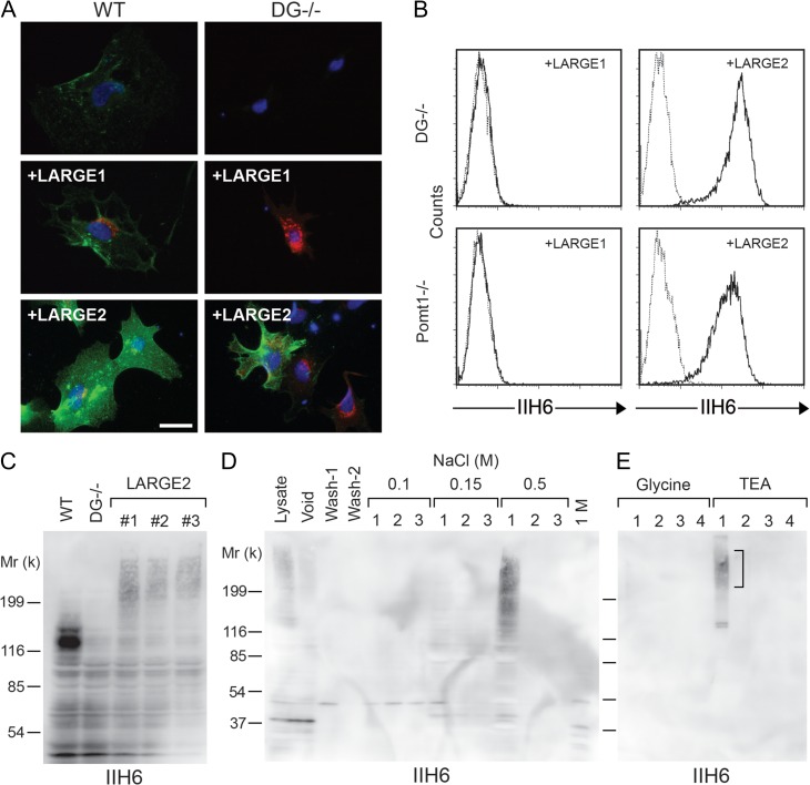
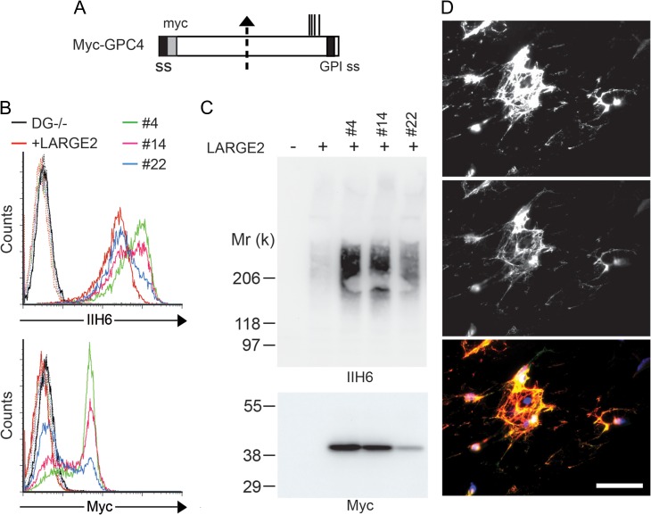
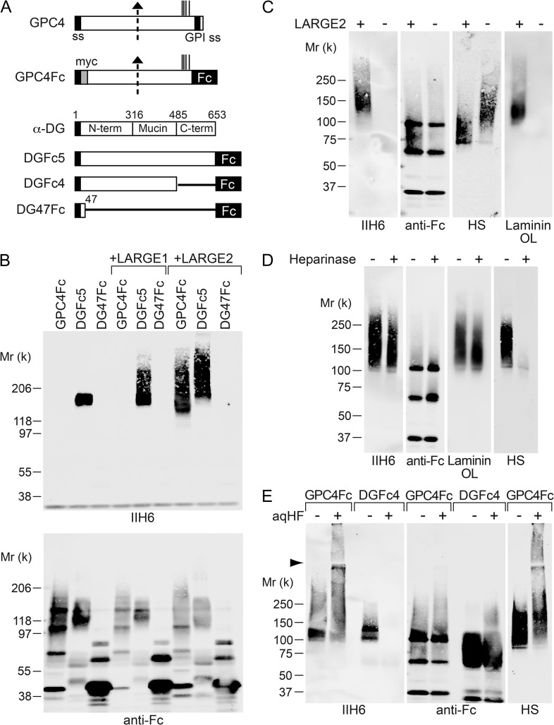
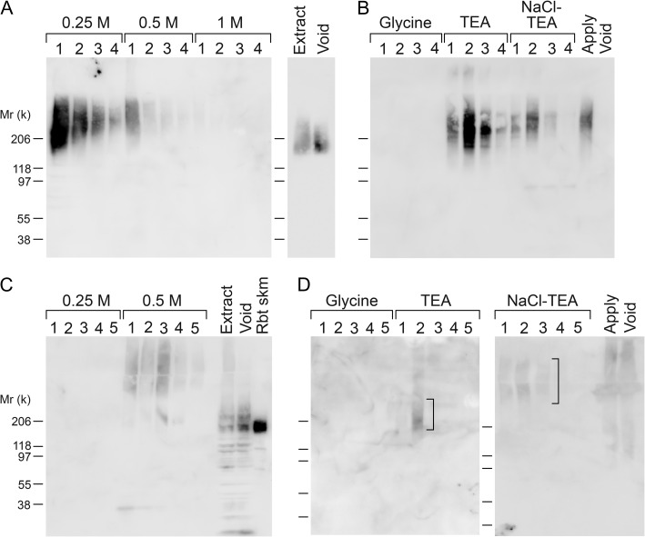
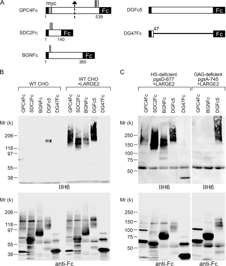
Both LARGE1 (formerly LARGE) and its paralog LARGE2 are bifunctional glycosyltransferases with xylosy- and glucuronyltransferase activities, and are capable of synthesizing polymers composed of a repeating disaccharide [-3Xylα1,3GlcAβ1-]. Post-translational modification of the O-mannosyl glycan of α-dystroglycan (α-DG) with the polysaccharide is essential for it to act as a receptor for ligands in the extracellular matrix (ECM), and both LARGE paralogs contribute to the modification in vivo. LARGE1 and LARGE2 have different tissue distribution profiles and enzymatic properties; however, the functional difference of the homologs remains to be determined, and α-DG is the only known substrate for the modification by LARGE1 or LARGE2. Here we show that LARGE2 can modify proteoglycans (PGs) with the laminin-binding glycan. We found that overexpression of LARGE2, but not LARGE1, mediates the functional modification on the surface of DG-/-, Pomt1-/- and Fktn-/- embryonic stem cells. We identified a heparan sulfate-PG glypican-4 as a substrate for the LARGE2-dependent modification by affinity purification and subsequent mass spectrometric analysis. Furthermore, we showed that LARGE2 could modify several additional PGs with the laminin-binding glycan, most likely within the glycosaminoglycan (GAG)-protein linkage region. Our results indicate that LARGE2 can modify PGs with the GAG-like polysaccharide composed of xylose and glucuronic acid to confer laminin binding. Thus, LARGE2 may play a differential role in stabilizing the basement membrane and modifying its functions by augmenting the interactions between laminin globular domain-containing ECM proteins and PGs.

Keywords: dystroglycan, glycosaminoglycan, laminin binding, LARGE2, proteoglycan.


Figures

Fig. 1.
Fig. 1.
LARGE2 but not LARGE1 confers IIH6 immunoreactivity to DG−/− ES cells. (A) WT and DG−/− ES cells were infected with adenovirus to express LARGE1 or LARGE2. Cells were incubated with IIH6 (antibody that recognizes the functional glycan epitope of α-DG) and either anti-LARGE1 or anti-LARGE2 antibody, and then probed with Alexa-conjugated secondary antibodies (green pseudo-color: IIH6; red: LARGE1 or LARGE2; blue: DAPI). Scale bar, 50 μm. (B) Flow cytometry detecting surface IIH6 labeling of DG−/− cells or Pomt1−/− cells stably expressing either LARGE1 or LARGE2. Dashed line, secondary antibody alone. (C) Immunoblotting of whole cell lysate with IIH6. The WT sample shows a robust 120-kD band that corresponds to α-DG and is not present in DG−/− samples. Independent DG−/− cell clones stably expressing LARGE2 (#1–3) show a high-molecular weight smear that is absent in WT and the DG−/− cells. (D) IIH6 immunoblotting of eluted fractions from a diethylaminoethyl (DEAE)-cellulose column. Lysate of the DG−/− cells stably expressing LARGE2 was applied to the column. After two washes (Wash-1 and Wash-2), bound proteins were released by three sequential elutions with increasing concentrations of NaCl. (E) IIH6 immunoblotting of eluted fractions from a IIH6-Sepharose column. Bound proteins were eluted by four sequential glycine buffer washes, and then with triethanolamine (TEA) buffer. The high-molecular weight smear (square bracket) and two bands ~130-kD were excised from a SYPRO Ruby-stained gel, and were then subjected to in-gel trypsin digestion and mass spectrometric (MS) analysis (Table I). This figure is available in black and white in print and in color at Glycobiology online.
Fig. 2.
Fig. 2.
Overexpression of GPC4 enhances the IIH6 immunoreactivity of the LARGE2-expressing DG−/− cells. (A) Schematic representation of the myc-tagged GPC4 (Myc-GPC4) construct. The suggested proteolytic cleavage site and potential GAG attachment sites are indicated by an arrow and vertical lines, respectively. ss, signal sequence. GPI ss, GPI-anchoring signal sequence. The DG−/− cells stably expressing LARGE2 were transfected with Myc-GPC4. (B) Clones stably expressing myc (#4, #14 and #22) were analyzed by flow cytometry for cell-surface staining with IIH6 or anti-myc antibody. (C) Lysates of these cells enriched for Myc-GPC4 by DEAE-enrichment were immunoblotted with IIH6 or anti-myc antibody. Note that the enhanced intensity of the IIH6 staining is correlated with that of anti-myc staining. (D) Representative immunofluorescence micrograph of the DG−/− cells stably co-expressing LARGE2 and Myc-GPC4 (clone #4). The cell surface was stained with IIH6 and anti-myc antibody in the absence of permeabilization. Scale bar, 50 μm. The top image shows IIH6 immunofluorescence, the middle image shows myc immunofluorescence, and the bottom image shows IIH6 (red) merged with Myc (green) and DAPI (blue). All myc staining colocalizes with IIH6 as no ‘green only’ fluorescence is seen. This figure is available in black and white in print and in color at Glycobiology online.
Fig. 3.
Fig. 3.
LARGE2 can modify GPC4 with the laminin-binding glycan. (A) Schematic representation of Fc-fusion constructs. Dotted arrow, suggested proteolytic cleavage site. Vertical lines, potential GAG attachment sites. ss, signal sequence. GPI ss, GPI-anchoring signal sequence. (B) LARGE2 can modify GPC4Fc in CHO cells. Immunoblotting of the Fc fusion proteins transiently expressed in, and purified from, the media of CHO cells with or without stable expression of LARGE1 or LARGE2. (C) Immunoblotting or laminin overlay (OL) of GPC4Fc purified from serum-free CHO culture with or without stable expression of LARGE2. Treatment with neither heparinase (D) nor aqHF (E) removed the functional modification from GPC4Fc.
Fig. 4.
Fig. 4.
Purification of IIH6+ proteins from rabbit kidney. IIH6 immunoreactivity of rabbit kidney extracts (A and B, Triton extract; C and D, urea extract) applied to columns (A and C, DEAE-cellulose; B and D, IIH6-Sepharose), and samples separated by the columns (the void and eluted fractions). The fractions TEA1-3 and NaCl-TEA1-3 derived from the Triton extract were subjected to trypsin digestion and analyzed by MS (Table II). In the cases of the TEA-2 and NaCl-TEA1-3 fractions derived from urea extract, the areas in the SDS-PAGE gels indicated by squared brackets were excised and analyzed by in-gel trypsin digestion and subsequent MS (Table III).
Fig. 5.
Fig. 5.
LARGE2 can modify not only GPC4 but also other PGs. (A) Schematic representation of Fc-fusion constructs. Dotted arrow, suggested proteolytic cleavage site. Vertical lines, potential GAG attachment sites. (B) LARGE2 can modify PGs. Immunoblotting of Fc fusion proteins transiently expressed in the WT CHO cells with or without stable expression of LARGE2. (C) Immunoblotting of Fc-fusion proteins transiently expressed in HS- or GAG-deficient CHO mutant cells that stably express LARGE2. In the GAG-deficient cells, LARGE2 could not modify GPC4Fc or BGNFc.
Fig. 6.
Fig. 6.
Sulfation is dispensable for the LARGE2-dependent modification. Flow cytometry of chlorate-treated DG−/− ES cells stably expressing LARGE2. HS staining was reduced by the chlorate treatment, and this effect was negated by the addition of magnesium sulfate. In contrast, IIH6 staining was not reduced by the treatment.

Similar articles

Cited by

References

    1. Aguilan JT, Sundaram S, Nieves E, Stanley P. 2009. Mutational and functional analysis of large in a novel CHO glycosylation mutant. Glycobiology. 19:971–986. - PMC - PubMed
    1. Ashikov A, Buettner FF, Tiemann B, Gerardy-Schahn R, Bakker H. 2013. LARGE2 generates the same xylose- and glucuronic acid-containing glycan structures as LARGE. Glycobiology. 23:303–309. - PubMed
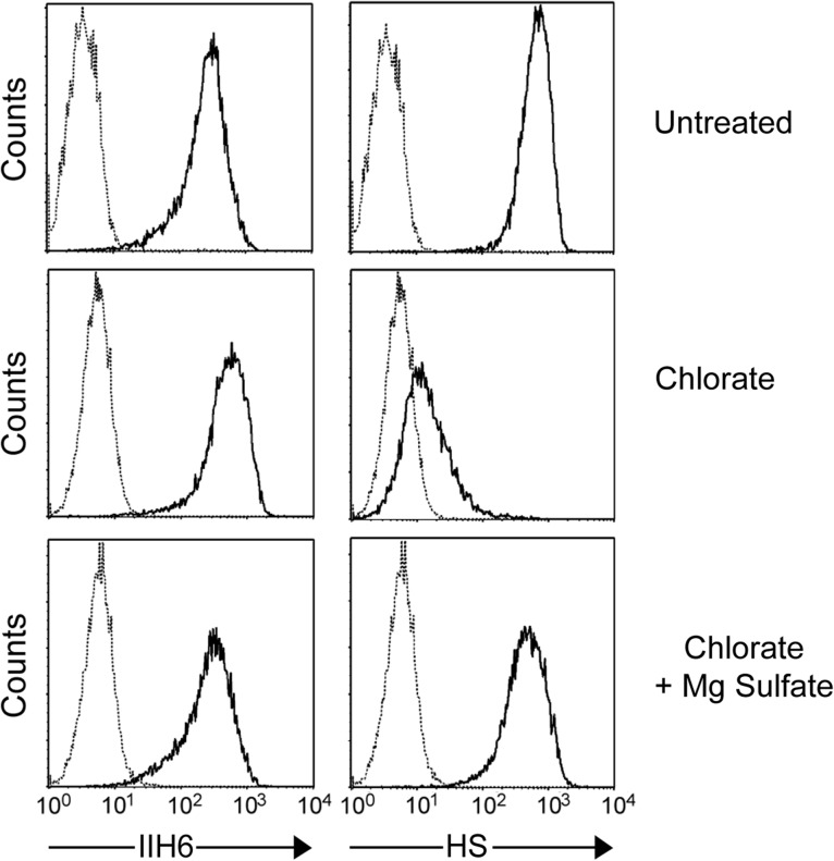
    1. Baeuerle PA, Huttner WB. 1986. Chlorate—A potent inhibitor of protein sulfation in intact cells. Biochem Biophys Res Commun. 141:870–877. - PubMed
    1. Barresi R, Campbell KP. 2006. Dystroglycan: From biosynthesis to pathogenesis of human disease. J. Cell Sci.. 119:199–207. - PubMed
    1. Beedle AM, Turner AJ, Saito Y, Lueck JD, Foltz SJ, Fortunato MJ, Nienaber PM, Campbell KP. 2012. Mouse fukutin deletion impairs dystroglycan processing and recapitulates muscular dystrophy. J Clin Invest. 122:3330–3342. - PMC - PubMed