. 2016 Sep 1;311(3):H815-21.
doi: 10.1152/ajpheart.00948.2015. Epub 2016 Aug 5.

CXCL16 regulates renal injury and fibrosis in experimental renal artery stenosis

Affiliations

CXCL16 regulates renal injury and fibrosis in experimental renal artery stenosis

Zhiheng Ma et al. Am J Physiol Heart Circ Physiol. .

Abstract

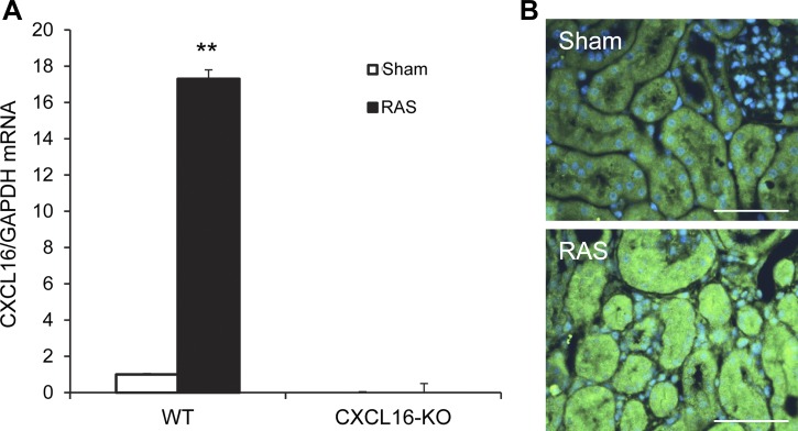
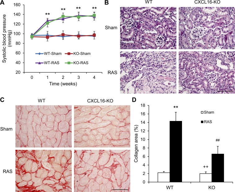
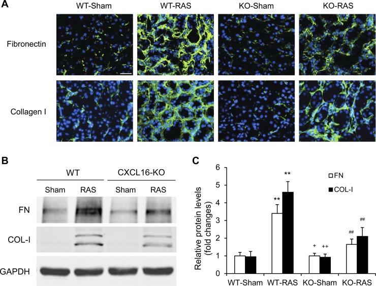
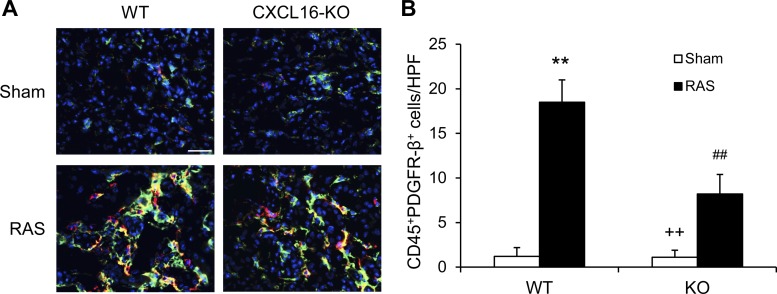
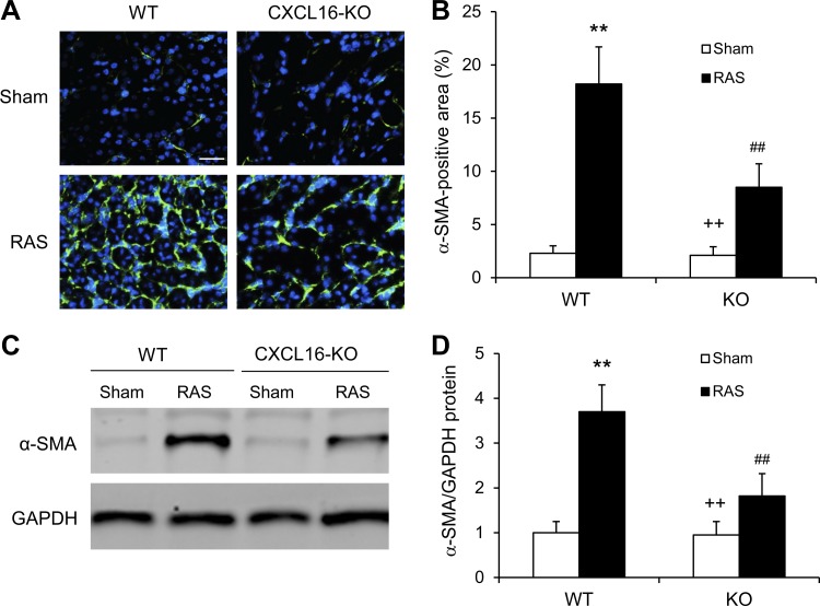
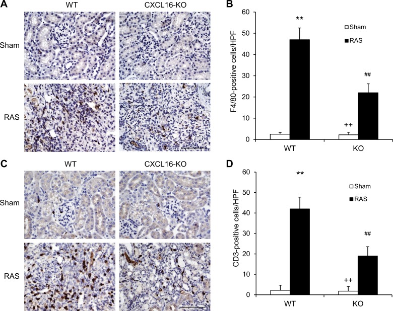
Recent studies have shown that inflammation plays a critical role in the initiation and progression of hypertensive kidney disease, including renal artery stenosis. However, the signaling mechanisms underlying the induction of inflammation are poorly understood. We found that CXCL16 was induced in the kidney in a murine model of renal artery stenosis. To determine whether CXCL16 is involved in renal injury and fibrosis, wild-type and CXCL16 knockout mice were subjected to renal artery stenosis induced by placing a cuff on the left renal artery. Wild-type and CXCL16 knockout mice had comparable blood pressure at baseline. Renal artery stenosis caused an increase in blood pressure that was similar between wild-type and CXCL16 knockout mice. CXCL16 knockout mice were protected from RAS-induced renal injury and fibrosis. CXCL16 deficiency suppressed bone marrow-derived fibroblast accumulation and myofibroblast formation in the stenotic kidneys, which was associated with less expression of extracellular matrix proteins. Furthermore, CXCL16 deficiency inhibited infiltration of F4/80(+) macrophages and CD3(+) T cells in the stenotic kidneys compared with those of wild-type mice. Taken together, our results indicate that CXCL16 plays a pivotal role in the pathogenesis of renal artery stenosis-induced renal injury and fibrosis through regulation of bone marrow-derived fibroblast accumulation and macrophage and T-cell infiltration.

Keywords: chemokine; inflammation; renal artery stenosis; renal fibrosis.


Figures

Fig. 1.
Fig. 1.
CXCL16 is induced in the kidney with RAS. A: RAS induces CXCL16 mRNA in the kidneys. Values are means ± SE; n = 6 per group. **P < 0.01 vs. sham controls. B: representative photomicrographs of kidney sections stained for CXCL16 (green) and counterstained with 4,6-diamidino-2-phenylindole (DAPI; blue). Scale bar, 50 μm.
Fig. 2.
Fig. 2.
CXCL16 deficiency does not affect blood pressure, but suppresses renal injury and fibrosis. A: CXCL16 deficiency has no effect on RAS-induced elevation of blood pressure. Values are means ± SE; n = 6 per group. **P < 0.01 between RAS groups and sham control groups. B: representative photomicrographs of hematoxylin- and eosin-stained sections showing RAS-induced kidney damage in WT and CXCL16 KO mice. Scale bar, 50 μm. C: representative photomicrographs of kidney sections from WT and CXCL16 KO mice 4 wk after sham or RAS stained with Sirius red for assessment of total collagen deposition. Scale bar, 50 μm. D: quantitative analysis of interstitial collagen content in the kidneys of WT and CXCL16 KO mice. Values are means ± SE; n = 6 per group. **P < 0.01 vs. WT controls. ++P < 0.01 vs. KO RAS. ##P < 0.01 vs. WT RAS.
Fig. 3.
Fig. 3.
CXCL16 deficiency reduces fibronectin and collagen I expression. A: representative photomicrographs of immunofluorescence staining for fibronectin and collagen in the kidneys of WT and CXCL16 KO mice 4 wk after sham or RAS. Scale bar, 25 μm. B: representative Western blots show the protein levels of fibronectin and collagen I in the kidneys of WT and CXCL16 KO mice 4 wk after sham or RAS. C: quantitative analysis of fibronectin and collagen I protein expression in the kidneys of WT and CXCL16 KO mice. Values are means ± SE; n = 6 per group. **P < 0.01 vs. WT controls. ++P < 0.01 vs. KO RAS. +P < 0.05 vs. KO RAS. ##P < 0.01 vs. WT RAS.
Fig. 4.
Fig. 4.
CXCL16 deficiency suppresses bone marrow-derived fibroblast accumulation. A: representative photomicrographs of kidney sections from WT and CXCL16 KO mice 2 wk after sham or RAS stained for CD45 (red), procollagen I (green), and DAPI (blue). Scale bar, 25 μm. B: quantitative analysis of CD45+ and procollagen I+ fibroblasts in kidneys of WT and CXCL16 KO mice 2 wk after sham or RAS. Values are means ± SE; n = 6 per group. **P < 0.01 vs. WT controls. ++P < 0.01 vs. KO RAS. ##P < 0.01 vs. WT RAS.
Fig. 5.
Fig. 5.
CXCL16 deficiency inhibits myofibroblast formation in the kidney. A: representative photomicrographs of kidney sections stained for α-SMA. Scale bar, 25 μm. B: quantitative analysis of α-SMA positive area in kidneys of WT and CXCL16 KO mice. Values are means ± SE; n = 6 per group. **P < 0.01 vs. WT controls. ++P < 0.01 vs. KO RAS. ##P < 0.01 vs. WT RAS. C: representative Western blots show the levels of α-SMA protein expression in the kidneys of WT and CXCL16 KO mice 4 wk after sham or RAS. D: quantitative analysis of α-SMA protein expression in the kidneys of WT and CXCL16 KO mice. Values are means ± SE; n = 6 per group. **P < 0.01 vs. WT controls. ++P < 0.01 vs. KO RAS. ##P < 0.01 vs. WT RAS.
Fig. 6.
Fig. 6.
CXCL16 deficiency reduces macrophage and T-cell infiltration. A: representative photomicrographs of kidney sections stained for F4/80 (a macrophage marker; brown) and counterstained with hematoxylin (blue) in WT and CXCL16 KO mice 4 wk after sham or RAS. Scale bar, 50 μm. B: quantitative analysis of F4/80+ macrophages in the kidneys of WT and CXCL16 KO mice. Values are means ± SE; n = 6 in each group. **P < 0.01 vs. WT controls. ##P < 0.01 vs. WT RAS. ++P < 0.01 vs. KO RAS. C: representative photomicrographs of kidney sections stained for CD3 (a T-lymphocyte marker; brown) and counterstained with hematoxylin (blue) in WT and CXCL16 KO mice 4 wk after sham or RAS. Scale bar, 50 μm. D: quantitative analysis of CD3+ T cells in the kidneys of WT and CXCL16 KO mice. Values are means ± SE; n = 6 in each group. **P < 0.01 vs. WT controls. ##P < 0.01 vs. WT RAS. ++P < 0.01 vs. KO RAS.

References

    1. ASTRAL Investigators, Wheatley K, Ives N, Gray R, Kalra PA, Moss JG, Baigent C, Carr S, Chalmers N, Eadington D, Hamilton G, Lipkin G, Nicholson A, Scoble J. Revascularization vs. medical therapy for renal-artery stenosis. N Engl J Med 361: 1953–1962, 2009. - PubMed
    1. Bidani AK, Griffin KA. Pathophysiology of hypertensive renal damage: implications for therapy. Hypertension 44: 595–601, 2004. - PubMed
    1. Broekema M, Harmsen MC, van Luyn MJ, Koerts JA, Petersen AH, van Kooten TG, van Goor H, Navis G, Popa ER. Bone marrow-derived myofibroblasts contribute to the renal interstitial myofibroblast population and produce procollagen I after ischemia/reperfusion in rats. J Am Soc Nephrol 18: 165–175, 2007. - PubMed
    1. Bucala R, Spiegel LA, Chesney J, Hogan M, Cerami A. Circulating fibrocytes define a new leukocyte subpopulation that mediates tissue repair. Mol Med 1: 71–81, 1994. - PMC - PubMed
    1. Chen G, Lin SC, Chen J, He L, Dong F, Xu J, Han S, Du J, Entman ML, Wang Y. CXCL16 recruits bone marrow-derived fibroblast precursors in renal fibrosis. J Am Soc Nephrol 22: 1876–1886, 2011. - PMC - PubMed

Publication types

MeSH terms

LinkOut - more resources