. 2016 Sep;139(Pt 9):2441-55.
doi: 10.1093/brain/aww187. Epub 2016 Aug 7.

Hyperphosphorylated tau in patients with refractory epilepsy correlates with cognitive decline: a study of temporal lobe resections

Affiliations

Hyperphosphorylated tau in patients with refractory epilepsy correlates with cognitive decline: a study of temporal lobe resections

Xin You Tai et al. Brain. 2016 Sep.

Abstract

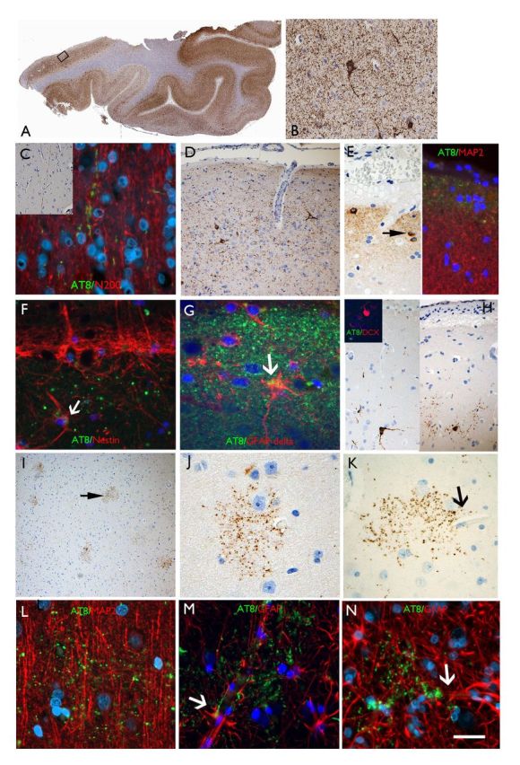
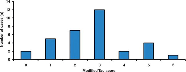
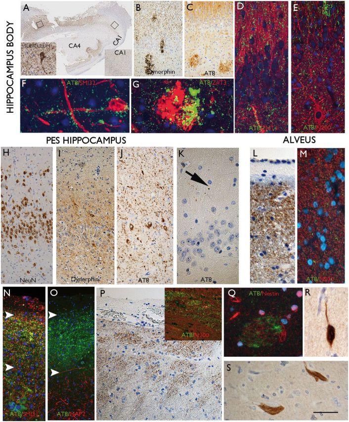
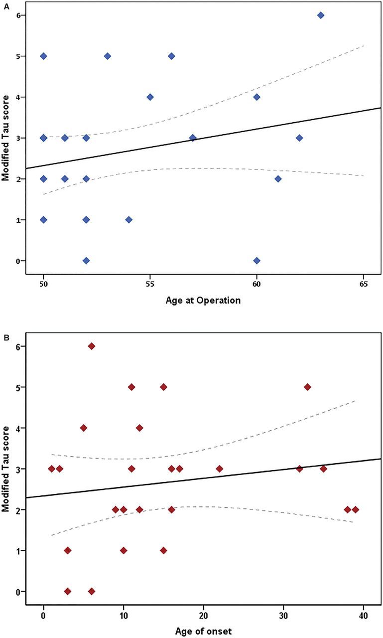
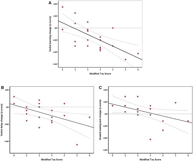
SEE BERNASCONI DOI101093/AWW202 FOR A SCIENTIFIC COMMENTARY ON THIS ARTICLE: Temporal lobe epilepsy, the most prevalent form of chronic focal epilepsy, is associated with a high prevalence of cognitive impairment but the responsible underlying pathological mechanisms are unknown. Tau, the microtubule-associated protein, is a hallmark of several neurodegenerative diseases including Alzheimer's disease and chronic traumatic encephalopathy. We hypothesized that hyperphosphorylated tau pathology is associated with cognitive decline in temporal lobe epilepsy and explored this through clinico-pathological study. We first performed pathological examination on tissue from 33 patients who had undergone temporal lobe resection between ages 50 and 65 years to treat drug-refractory temporal lobe epilepsy. We identified hyperphosphorylated tau protein using AT8 immunohistochemistry and compared this distribution to Braak patterns of Alzheimer's disease and patterns of chronic traumatic encephalopathy. We quantified tau pathology using a modified tau score created specifically for analysis of temporal lobectomy tissue and the Braak staging, which was limited without extra-temporal brain areas available. Next, we correlated tau pathology with pre- and postoperative cognitive test scores and clinical risk factors including age at time of surgery, duration of epilepsy, history of secondary generalized seizures, history of head injury, handedness and side of surgery. Thirty-one of 33 cases (94%) showed hyperphosphorylated tau pathology in the form of neuropil threads and neurofibrillary tangles and pre-tangles. Braak stage analysis showed 12% of our epilepsy cohort had a Braak staging III-IV compared to an age-matched non-epilepsy control group from the literature (8%). We identified a mixture of tau pathology patterns characteristic of Alzheimer's disease and chronic traumatic encephalopathy. We also found unusual patterns of subpial tau deposition, sparing of the hippocampus and co-localization with mossy fibre sprouting, a feature of temporal lobe epilepsy. We demonstrated that the more extensive the tau pathology, the greater the decline in verbal learning (Spearman correlation, r = -0.63), recall (r = -0.44) and graded naming test scores (r = -0.50) over 1-year post-temporal lobe resection (P < 0.05). This relationship with tau burden was also present when examining decline in verbal learning from 3 months to 1 year post-resection (r = -0.54). We found an association between modified tau score and history of secondary generalized seizures (likelihood-ratio χ(2), P < 0.05) however there was no clear relationship between tau pathology and other clinical risk factors assessed. Our findings suggest an epilepsy-related tauopathy in temporal lobe epilepsy, which contributes to accelerated cognitive decline and has diagnostic and treatment implications.

Keywords: Alzheimer’s disease; dementia; neurofibrillary tangles; tau; temporal lobe epilepsy.


Figures

None
See Bernasconi (doi:10.1093/aww202) for a scientific commentary on this article. Temporal lobe epilepsy (TLE) is associated with a high prevalence of cognitive decline. Tai et al. reveal the presence of hyperphosphorylated tau in resected temporal lobe tissue from patients with TLE. The quantity of pathological tau correlates with cognitive decline and its distribution suggests an epilepsy-related tauopathy.
Figure 1
Figure 1
AT8 patterns in TLE/HS cases in the temporal neocortex. (A) Section of temporal lobe from case with maximal modified tau score of 6 showing dense accumulation of tau with AT8 immunohistochemistry in all gyri. The rectangle is shown at higher magnification in B, highlighting neuropil threads and neuronal labelling. (C) Axonal labelling with AT8 in the cortex (Case 24) in inset and double labelling with neurofilament (N200) showing beaded-like AT8 staining along the trajectory of radial cortical axons. (D) CTE-like pattern with focal increased AT8 in threads and small neurons in the superficial cortical layers. (E) Subpial granular band of labelling and positive neurons, reminiscent of Cajal-Retzius cells (arrow); double labelling with MAP2 (dendritic marker) on the right side of the panel, shows AT8 labelling in the compartment above dendritic MAP2 labelling supporting this more likely represents superficial subpial axonal projections. (F) Double labelling between AT8 and nestin in layer I showed no overlap with the granular, axonal-like AT8 labelling and the nestin in the subpial glia with occasional possible co-localization (arrow). (G) Layer I in a further case with AT8 showing a mainly beaded axonal staining pattern and only rare possible expression in GFAP-delta positive subpial astroglial cell. (H) Small neurons at interface of layer I and II were AT8 positive but double-labelling with doublecortin (DCX) in selected cases (inset) did not shown any AT8 positivity in these immature cell types that are known to reside in this cortical layer in the temporal lobe. (I) ‘Granular aggregates’ of tau were noted in the temporal cortex in five cases scattered in the cortex pattern but not typical of neuritic plaques of Alzheimer’s disease or glial inclusions. (J) Occasionally these aggregates surrounding cortical neurons but without definite labelling of the neuronal cell body. (K) Granular aggregates were observed in the vicinity of small capillaries (arrow). (L) The granular aggregates did not appear to co-localize with dendrites on double-labelling with MAP2 (a neuronal dendritic marker). (M and N) Labelling with GFAP confirmed that the AT8-granular aggregates were not in astroglia but through highlighting the glial foot processes along the vessels, indicated their proximity to vascular channels. Supplementary Fig. 3 contains a panel of similar immunofluorescence images captured with a non-red/green colour spectrum. Scale bars = 500 µm in A, 70 µm in I, 50 µm in B and D, 20 µm in C, E, F, G, J, K L, M and N.
Figure 2
Figure 2
Quantifying tau pathology in temporal lobe tissue of epilepsy patients. Histogram showing modified tau score (described in Table 1) distribution across study cohort. Thirty-one of 33 cases showed AT8 labelling with modified tau score 3 being most common.
Figure 3
Figure 3
AT8 patterns in TLE/HS in the hippocampus, pes and amygdala. (A) Hippocampus body from case with highest tau load in the cortex. The hippocampus, which showed typical features of longstanding hippocampal sclerosis and gliosis with neuronal loss in CA1 and CA4. More AT8 labelling was seen in the subiculum compared to the CA1 subfield (inserts). (B) In another case of HS, mossy fibre sprouting was show with dynorphin staining and granular aggregates noted within the CA4 region and in C. A similar pattern was observed with AT8. (D) Double labelling of AT8 with ZnT3 and (E) neurofilament (N200), confirmed some overlap of labelling in the molecular layer of the dentate gyrus. (F) AT8-positive granules were observed in close proximity, surrounding a CA4 neuron and its processes and in (G) overlap of ZnT3 and AT8 was noted in the CA4 region corresponding to mossy fibre end terminals. (H) Pes hippocampus from one case showed marked dispersion of the granule cells on NeuN compared to the body also with a greater degree of mossy fibre sprouting on dynorphin stain as shown in I. (J) Abundant AT8 labelling in the granule cells and axons in the molecular layer was noted compared to very little staining in the adjacent CA1 subfield (not shown) and (K) the hippocampus body of the same cases, where only rare AT8-positive threads were noted. (L) An axonal-like pattern of labelling was noted in the alveus beneath the ependymal of the lateral ventricle and confirmed with double-labelling with N200 neurofilament marker (M) showing association of AT8-positive grains with axons. (N) In the subpial surface of the pes, a band of AT8 was noted in some case (between arrows), comparable to the subpial band in the cortex; this again had an axonal appearance with localization with non-phosphorylate neurofilament (SMI3 shown in N), but not dendritic marker (MAP2 shown in O), which labelled the processes in underlying neurons only. (P) Bundles of axons were also noted in the peri-ventricular region of the pes hippocampus white matter. (Q) Occasional fibre bundles were noted in the amygdala with AT8, which were weakly nestin-positive. (R) Labelling of neurons with 3R and (S) 4R tau isoforms was confirmed. Supplementary Fig. 3 contains a panel of similar immunofluorescence images captured with a non-red/green colour spectrum. Scale bars: A = 500 µm; B and C = 200 µm; H, I and J = 75 µm; D, E, K, N, 0, P = 50 µm; F, G, Q, R, S = 25 µm.
Figure 4
Figure 4
The effect of (A) age at time of surgery and (B) age of onset of epilepsy on the modified tau score in the epilepsy cohort. Scatter plot graphs with linear correlation curve fitted. Spearman correlation shows a weak relationship between both clinical factors (r = 0.39 and r = 0.21, respectively) and are not statistically significant (P > 0.05). Individual points may overlap. Interrupted line indicates 95% confidence interval for the mean modified tau score for the given age.
Figure 5
Figure 5
Correlation between modified tau score and change in verbal memory domains and graded naming test scores. Decline in cognitive scores from preoperative assessment to 1 year post-temporal lobe resection shows a significant negative relationship with increasing tau burden for verbal memory domains and graded naming (Spearman coefficient r = −0.66, *P < 0.01, for verbal learning; r = −0.44, *P < 0.05, for verbal recall and r = −0.50, *P < 0.05, for graded naming test). Interrupted line indicates 95% confidence interval for the mean cognitive score for the given modified tau score.

Comment in

References

    1. Amador-Ortiz C, Lin W-L, Ahmed Z, Personett D, Davies P, Duara R, et al. . TDP-43 immunoreactivity in hippocampal sclerosis and Alzheimer’s disease [Internet]. Ann Neurol 2007; 61: 435–45. - PMC - PubMed
    1. Amatniek JC, Hauser WA, DelCastillo-Castaneda C, Jacobs DM, Marder K, Bell K, et al. . Incidence and predictors of seizures in patients with Alzheimer’s disease [Internet]. Epilepsia 2006; 47: 867–72. - PubMed
    1. Bernhardt BC, Worsley KJ, Kim H, Evans AC, Bernasconi A, Bernasconi N. Longitudinal and cross-sectional analysis of atrophy in pharmacoresistant temporal lobe epilepsy [Internet]. Neurology 2009; 72: 1747–54. - PMC - PubMed
    1. Black LC, Schefft BK, Howe SR, Szaflarski JP, Yeh H, Privitera MD. The effect of seizures on working memory and executive functioning performance [Internet]. Epilepsy Behav 2010; 17: 412–9. - PubMed
    1. Braak H, Alafuzoff I, Arzberger T, Kretzschmar H, Del Tredici K. Staging of Alzheimer disease-associated neurofibrillary pathology using paraffin sections and immunocytochemistry. [Internet]. Acta Neuropathol 2006; 112: 389–404. - PMC - PubMed

Publication types