Review
. 2017 Jan;1862(1):8-24.
doi: 10.1016/j.bbalip.2016.08.001. Epub 2016 Aug 4.

Known unknowns of cardiolipin signaling: The best is yet to come

Affiliations
Review

Known unknowns of cardiolipin signaling: The best is yet to come

John J Maguire et al. Biochim Biophys Acta Mol Cell Biol Lipids. 2017 Jan.

Abstract

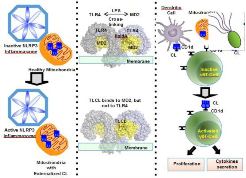
Since its discovery 75years ago, a wealth of knowledge has accumulated on the role of cardiolipin, the hallmark phospholipid of mitochondria, in bioenergetics and particularly on the structural organization of the inner mitochondrial membrane. A surge of interest in this anionic doubly-charged tetra-acylated lipid found in both prokaryotes and mitochondria has emerged based on its newly discovered signaling functions. Cardiolipin displays organ, tissue, cellular and transmembrane distribution asymmetries. A collapse of the membrane asymmetry represents a pro-mitophageal mechanism whereby externalized cardiolipin acts as an "eat-me" signal. Oxidation of cardiolipin's polyunsaturated acyl chains - catalyzed by cardiolipin complexes with cytochrome c. - is a pro-apoptotic signal. The messaging functions of myriads of cardiolipin species and their oxidation products are now being recognized as important intracellular and extracellular signals for innate and adaptive immune systems. This newly developing field of research exploring cardiolipin signaling is the main subject of this review. This article is part of a Special Issue entitled: Lipids of Mitochondria edited by Guenther Daum.

Keywords: Apoptosis; Cardiolipin oxidation; Cardiolipin signaling; Innate and adaptive immunity; Mitophagy; Peroxidase.


Figures

Fig. 1
Fig. 1. Highly diversified molecular speciation of cardiolipin are typical of fish tissues compared to the high uniformity of CLs in bovine heart
A. LC-MS spectra of CLs from brain, skeletal muscle and heart from a Northern Red Snapper (Lutjanus campechanus) and bovine heart CL. The m/z shown are for the most abundant species in each cluster of CL. The CL species with m/z of 1447.9653 (72:8) is the major species in bovine heart but not in fish heart. B. Spider graph plots of the number (left panel) and amounts (right panel) CL molecular species in fish and bovine hearts. Species are presented as a ratio of carbon to double bonds number (C:DB). For clarity, the scales for the number of CL species in bovine heart (pale pink, left panel) and for the amount of CL species in fish heart (pale blue, right panel) were magnified in 4 and 5 times respectively. C. Cardiolipin structure. A pro-chiral carbon (1) is located on the central glycerol. The 4 acyl chains (R1, R2, R3 and R4) are linked to 2 glycerols each with a chiral carbon with the same (R) chirality on the left and right side of the molecule (2,3), highlighted by the shading. The phosphates are charged at physiological pH.
Fig. 2
Fig. 2. Expansion of the diversity of CLs
The initially synthesized 4 CL acyl chains can be saturated, mono- unsaturated or poly-unsaturated. Remodeling is initiated by deacylation of one or more acyl chains and with enzymatic precision a wide variety of fatty acids can re-acylate the lyso-CLs forming organ and tissue specific variations of CL content.
Fig. 3
Fig. 3. Detailed structural information can be obtained by contemporary high resolution MS equipment: The Orbitrap™ high energy collision dissociation (HCD) MS2-spectra of cardiolipins with the [M-H]- ion at m/z 1371.9359
Two isomers CL(18:1/18:1/18:1/18:2) and CL(16:0/18:1/18:0/20:4) were identified. CL(18:1/18:1/18:1/18:2) (in blue): The characteristic structural fragments includes: [PG(18:1/18:1)+Phosphate-H] and [PG(18:1/18:2)+Phosphate-H] ions of m/z 835 and m/z 833, respectively; neutral loss (NL) of phosphate group from above moieties generating [PG-H] ions with m/z 755 and m/z 753, respectively. [PA(18:1/18:1)-H] and [PA(18:1/18:2)-H] ions of m/z 699 and m/z 697, respectively; NL of C18:1 from above PA moieties generating m/z 417 and m/z 415 (box), respectively. The individual fatty acid carboxylate anion was also observed with m/z 281 and m/z 279, corresponding to C18:1 and C18:2, respectively. CL(16:0/18:1/18:0/20:4) (in red): The characteristics structural fragments includes: [PG(16:0/18:1)+Phosphate-H] and [PG(18:0/20:4)+Phosphate-H] ions of m/z 809 and m/z 859, respectively; [PA(16:0/18:1)-H] and [PA(18:0/20:4)-H] ions of m/z 673 and m/z 723, respectively; neutral loss (NL) of fatty acids from above PA moieties generating m/z 417, 391, 439 and 419 (box). The individual fatty acid carboxylate anion was also observed with m/z 255, 281, 283 and 303, corresponding to C16:0, C18:1, C18:0 and C20:4, respectively. m/z 153 was ion of glycerol-3-phosphoate with NL of H2O, the characteristic ion of phospholipids. Shared fragments were marked in black in MS/MS spectra. Abbreviation: NL, neutral loss; PA, phosphatidic acid; PG, phosphatidylglycerol
Fig. 4
Fig. 4. MS2/MS3 fragmentation analysis leads to detailed structural characterization of oxidized CLs
An example showing the increased resolution of the structural characterization of CL-(C18:2)4 hydroperoxy molecular species formed in an AAPH (2,2'-azobis-2-methyl-propanimidamide, dihydrochloride) driven reaction using a Tribrid™ MS Fusion Lumos (Thermo Fisher Scientific, San Jose, CA). A. Full, MS2 and MS3 spectra of (hydroperoxy-C18:2)1(C18:2)3. Daughter molecular ions formed during MS2 analysis corresponding to the oxygenated C18:2 are shown in red. Characteristic fragments of 9-hydroperoxy-C18:2 and 13-hydroperoxy-C18:2 formed during MS3 analysis are shown in blue. B. The structures of the detected nascent CL (C18:2)4 and two of its oxidized products, (9-hydroperoxy-C18:2)1(C18:2)3-CL and (13-hydroperoxy-C18:2)1(C18:2)3-CL as identified and characterized using MS2/MS3.
Fig. 5
Fig. 5. Biochemical microscopy by MALDI-MS imaging reveals the differential distribution of CL species in various anatomical areas of the brain
Serial rat brain coronal sections were prepared. The left panel shows an optical image from a hematoxylin and eosin stained section using a Nikon-90i™ microscope. MALDI-MS imaging detects 23 species of CL at a lateral resolution (pixel size) of 50 microns, two of these species are shown in the middle and right panels. Predominantly saturated species of CL such as CL(70:4) (middle panel) are strongest in the hippocampal region (H), especially the dentate gyrus (DG), and moderately less intense in the cortex (C) and ventricles ( V). Highly unsaturated species of CL such as CL(76:12) (right panel) are strongest in the cortex and ventricles (C, V) and less intense in the hippocampus (H). The images were acquired on a Bruker Daltonics-RapiFlex™ MALDI, using the protocol from [101]. Ions detected as [M-H]1- and intensities are relative with respect to the given ion. The white bar = 1 mm.
Fig. 6
Fig. 6. Computer modeling illustrates the major modes of CL interactions with proteins
Structural details of the interaction between CL and: (A) the ATP/ADP carrier (PDB 1okc); (B) the cytochrome C oxidase (PDB 3w7g) and cytochrome C1 heme protein (PDB 1P84). The CL molecule is shown in sticks, oxygen atoms in red, phosphorus atoms in orange and carbon atoms in pale gray. When super-secondary structural motifs (loops) were identified near the CL molecule, these are highlighted in cyan (helix) and light-pink (coiled coil) colors (A and B) the closest protein region is shown in light blue (C). Over the cartoon representation of secondary structures, a mesh network displays physico-chemical properties of amino-acids neighboring the CL molecule: in orange, hydrophobic; in blue: positively charged; in red, negatively charged. The key CL-binding motif [KR]-(X)-G is highlighted using sticks representations and lime-green color for Gly and blue-purple color for Arg/Lys.
Fig. 7
Fig. 7. Molecular dynamic simulations illustrate different modes of cyt c interactions with CL-containing lipid bilayers
Two separate binding orientations are shown. A) Productive binding involves tighter interactions and deeper penetration into the membrane facilitated by CL sequestration leading to peroxidase activation. B) Unproductive binding engages smaller amounts of sequestered CL and does not lead to peroxidase activation.
Fig. 8
Fig. 8. Partial x-ray structure of cyt c illustrates the proximity of a candidate Tyr residue involved in peroxidase activation by CL
The upper panel shows the positions of the cyt c tyrosines (Y48, Y67, Y74, and Y94). Y67 is located in the closest proximity to the heme-iron (lower panel). Y67 acts as a donor of an electron for a porphyrin cation-radical generated as a reactive peroxidase intermediate. In the oxygenase half-reaction of the cyt c /CL peroxidase complex, Y67 radical acts as an electron acceptor from oxidizable CL.
Fig. 9
Fig. 9. Externalization of CLs to the mitochondrial surface occurs during mitophagy whereas oxidation of CLs is necessary for the execution of apoptosis
In mitophagy CL is translocated by NDPK-D which binds to both the IMM and OMM, facilitating CL movement to the OM where it can bind and activate LC3-II which initiates autophagosome formation leading to mitophagy. In the figure the CL is colored blue, other lipids are brown and oxidized CL is colored red. In apoptosis CL forms a complex with cyt c converting it to a peroxidase, H2O2 is used as the oxidant of CL and the net result is the release of cyt c through the OMM and into the cytoplasm, as well as externalization of CL to the OMM resulting in apoptotic death.
Fig. 10
Fig. 10. Intra- and Extracellular CLs are involved in the regulation of the immune responses
The left panel shows that mitochondrial externalized CLs are involved in the activation of NLRP3 inflammasomes, which in turn activates caspase-1; The middle panel shows that LPS (which contains lipid A) cross-links two TLR4-MD2 complexes to activate an inflammatory response. However, CL only binds to MD2, but cannot cross-links TLR4's; In the right panel, mitochondria with externalized CL have characteristics of bacteria. CD1d protein is able to bind and present mammalian or bacterial CL to CL-responsive γδ T cells that exist in the spleen and liver of healthy mice. In response to CL these cells proliferate in a dose-dependent manner, and secrete the cytokines IFN-γ and RANTES.

References

    1. Lane N. Bioenergetic constraints on the evolution of complex life. Cold Spring Harbor perspectives in biology. 2014;6:a015982. - PMC - PubMed
    1. Lane N, Martin W. The energetics of genome complexity. Nature. 2010;467:929–934. - PubMed
    1. Martin WF, Garg S, Zimorski V. Endosymbiotic theories for eukaryote origin, Philosophical transactions of the Royal Society of London. Series B. Biological sciences. 2015;370:20140330. - PMC - PubMed
    1. Lyons TW, Reinhard CT, Planavsky NJ. The rise of oxygen in Earth's early ocean and atmosphere. Nature. 2014;506:307–315. - PubMed
    1. Westbroek P. Life as a geological force — dynamics of the earth. W. W. Norton; New York, London: 1991.

Publication types

LinkOut - more resources