. 2016 Aug 8:6:30976.
doi: 10.1038/srep30976.

Regulation of proinflammatory genes by the circulating microRNA hsa-miR-939

Affiliations

Regulation of proinflammatory genes by the circulating microRNA hsa-miR-939

Marguerite K McDonald et al. Sci Rep. .

Abstract

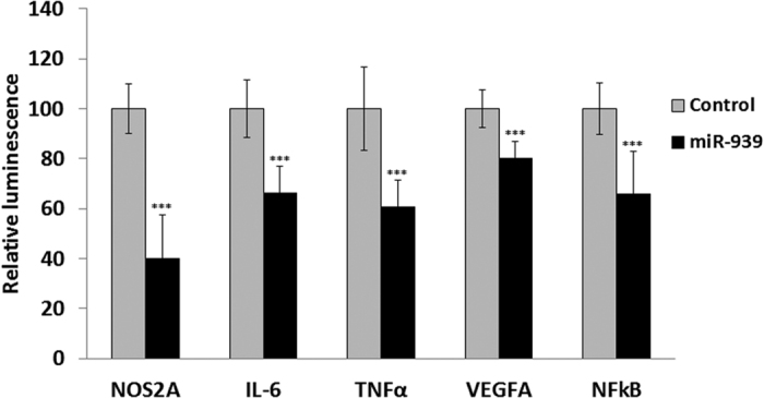
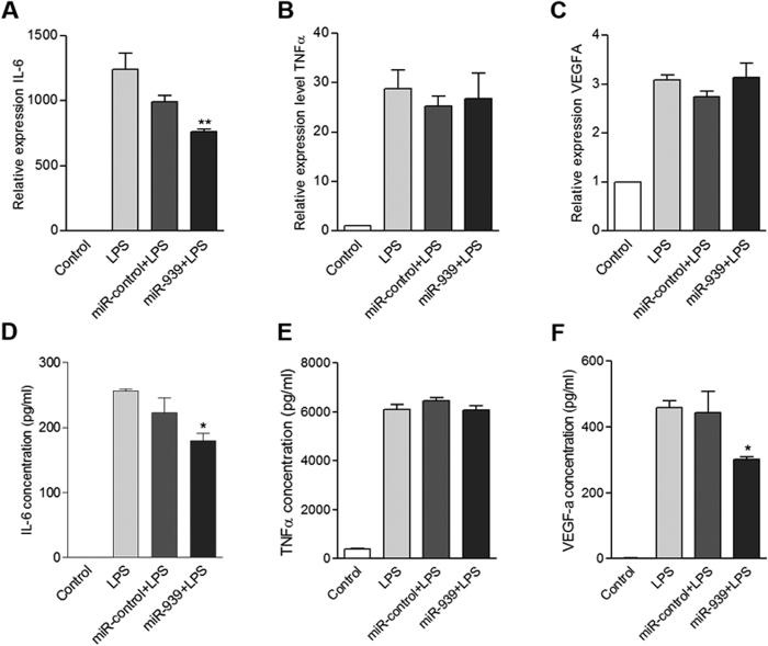
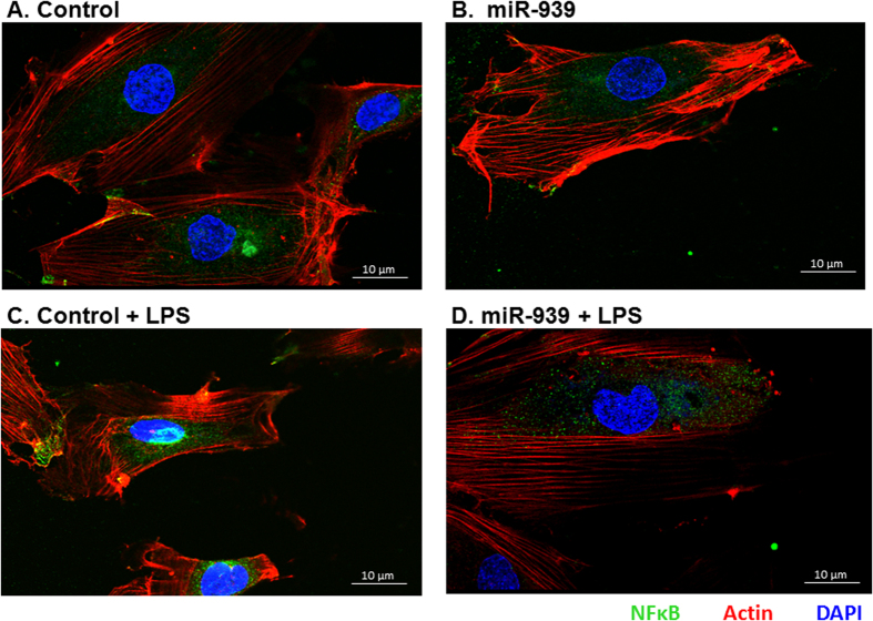
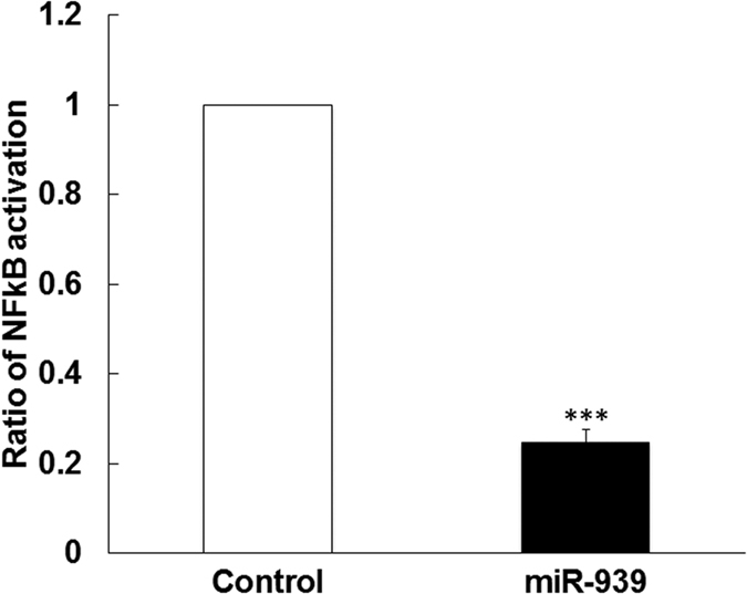
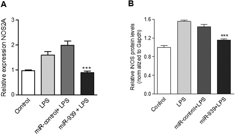
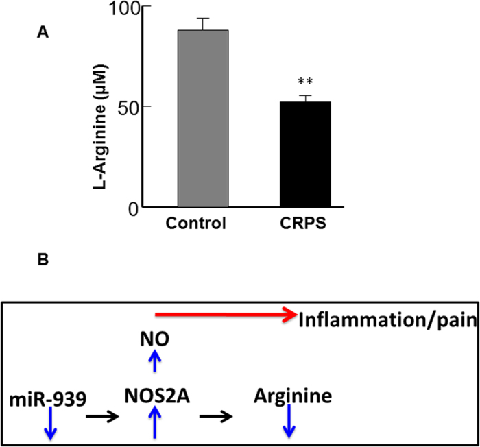
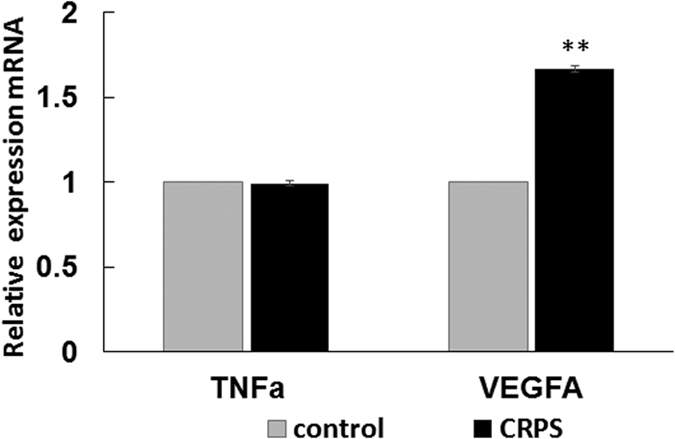
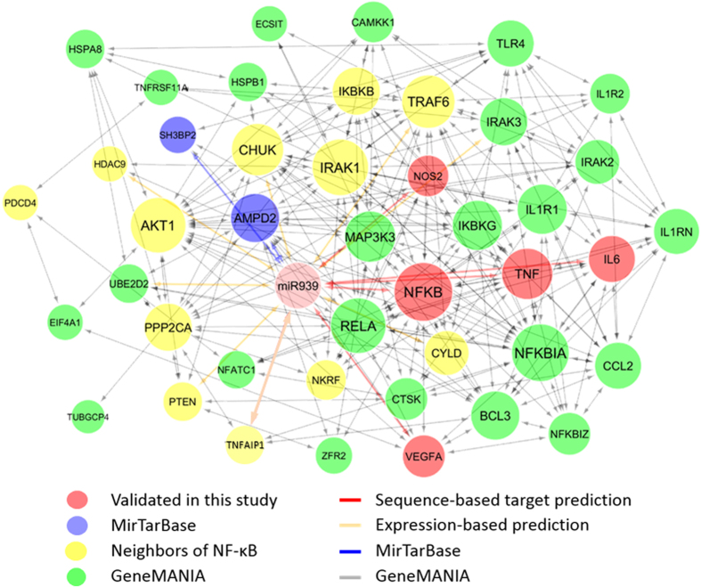
Circulating microRNAs are beneficial biomarkers because of their stability and dysregulation in diseases. Here we sought to determine the role of miR-939, a miRNA downregulated in patients with complex regional pain syndrome (CRPS). Hsa-miR-939 is predicted to target several proinflammatory genes, including IL-6, VEGFA, TNFα, NFκB2, and nitric oxide synthase 2 (NOS2A). Binding of miR-939 to the 3' untranslated region of these genes was confirmed by reporter assay. Overexpression of miR-939 in vitro resulted in reduction of IL-6, NOS2A and NFκB2 mRNAs, IL-6, VEGFA, and NOS2 proteins and NFκB activation. We observed a significant decrease in the NOS substrate l-arginine in plasma from CRPS patients, suggesting reduced miR-939 levels may contribute to an increase in endogenous NOS2A levels and NO, and thereby to pain and inflammation. Pathway analysis showed that miR-939 represents a critical regulatory node in a network of inflammatory mediators. Collectively, our data suggest that miR-939 may regulate multiple proinflammatory genes and that downregulation of miR-939 in CRPS patients may increase expression of these genes, resulting in amplification of the inflammatory pain signal transduction cascade. Circulating miRNAs may function as crucial signaling nodes, and small changes in miRNA levels may influence target gene expression and thus disease.


Figures

Figure 1
Figure 1. Luciferase assay to determine miR-939 binding to the 3′UTR of predicted mRNA targets.
Plasmids with a target 3′UTR cloned downstream of the luciferase open-reading frame were cotransfected with plasmids encoding precursor miR-939 or control miRNA in HEK293 cells. Luciferase activity was measured 48 hours after transfection, and the data expressed as percentage of control. Firefly luciferase measurements normalized to Renilla was used as a transfection control. The average of 3 independent experiments is shown. Statistically significant difference from control was calculated using Student t-test ***p < 0.001.
Figure 2
Figure 2. Expression levels of miR-939 target genes IL-6, TNFα and VEGFA in THP-1 cells transfected with miR-939 followed by 6 hours of stimulation with LPS.
(A–C) Taqman analysis of endogenous levels of IL-6, TNFα and VEGFA, after LPS induction, showed that transfection with miR-939 reduced IL-6 transcripts compared to miR- control. (D–F) Overexpression of miR-939 decreased secreted IL-6, and VEGFA. ELISA using cell culture supernatants of THP-1 cells stimulated with LPS showed lower levels of these two proinflammatory mediators secreted by miR-939 transfected cells compared to control miRNA transfection. Significance was determined by one-way ANOVA with Dunnet’s post hoc test *p < 0.05, **p < 0.01.
Figure 3
Figure 3. miR-939 reduced the translocation of functional NFκB to nucleus in response to LPS stimulation.
HUVEC cells were transfected with miR-939 for 24 h, followed by LPS stimulation for 6 h and fixed. Immunostaining was performed with anti-NFκB antibody (green), Actin (red) and DAPI (blue). While LPS stimulation of HUVEC cells results in the translocation of NFκB to the nucleus (C) in comparison to untreated control (A), this is impaired in cells transfected with miR-939 (D), suggesting that a potential regulation of NFκB2 subunit by miR-939 can influence the dimerization and function of NFκB.
Figure 4
Figure 4. miR-939 reduced the activation of NFκB in response to LPS.
THP-1XBlue cells, which have an inducible, chromosomally integrated secreted alkaline phosphatase (SEAP) gene downstream of the NFκB promoter, were transfected with miR-939 and then stimulated with LPS for 1 hour. The presence of high levels of miR-939 reduced the amount of SEAP detected in the media indicating less NFκB activity. Statistical significance was calculated using Student t-test ***p < 0.001.
Figure 5
Figure 5. Overexpression of miR-939 decreases NOS2A mRNA and protein levels in HUVEC cells.
(A) Taqman analysis of endogenous levels of NOS2A mRNA in HUVEC cells determined 24 hours after transfection with miR-939, and LPS treatment for 1 h. GAPDH was used as a normalizer. (B) ELISA for NOS2 in HUVEC cells transfected with miR-939 followed by LPS treatment for 6 h, show that NOS2 levels were lower after miR-939 transfection compared to control. Values are the mean of three experiments. Significance was determined by one-way ANOVA with Dunnet’s post hoc test ***p < 0.001.
Figure 6
Figure 6. Arginine levels in plasma from CRPS patients and control individuals.
The NOS2 substrate, l-arginine, was downregulated in plasma from CRPS patients (n = 41) compared to control individuals (n = 20). Statistical significance was calculated using Student t-test **p < 0.01. Downregulation of miR-939 may be linked to inflammation in CRPS patients (diagram). Increase in NOS2 protein, with the consequent decrease in its substrate, l-arginine, may result in enhanced NO levels, leading to pain and inflammation.
Figure 7
Figure 7. Relative expressions of VEGFA and TNFα mRNA in whole blood from CRPS patients and control individuals.
Taqman analysis of target mRNAs of miR-939 that were consistently detectable in whole blood, showed a significant increase of VEGFA mRNA in patients with CRPS compared to control samples. TNFα mRNA levels were not significant between CRPS and control samples. GAPDH was used as the normalizer. Data represent mean ± SEM CRPS (n = 37) and control (n = 18). Statistical significance was calculated using Student t-test **p < 0.01.
Figure 8
Figure 8. The inflammatory gene network representing miR-939 involvement.
A seed list of genes constructed from validated targets of miR-939 and interaction neighbors of NFκB, was enriched with functionally associated genes, using GeneMANIA. The nodes are colored according to the source of information. The connections between genes indicate a functional association; those between miR-939 and genes indicate validated or computationally predicted targets (Arrowheads suggest association, but do not indicate directionality in the interaction).

References

    1. De Guire V. et al. Circulating miRNAs as sensitive and specific biomarkers for the diagnosis and monitoring of human diseases: promises and challenges. Clin Biochem 46, 846–860, doi: 10.1016/j.clinbiochem.2013.03.015 (2013). - DOI - PubMed
    1. Bartel D. P. MicroRNAs: target recognition and regulatory functions. Cell 136, 215–233, doi: S0092-8674(09)00008-7 (2009). - PMC - PubMed
    1. Wilczynska A. & Bushell M. The complexity of miRNA-mediated repression. Cell death and differentiation 22, 22–33, doi: 10.1038/cdd.2014.112 (2015). - DOI - PMC - PubMed
    1. Schwartzman R. J., Erwin K. L. & Alexander G. M. The natural history of complex regional pain syndrome. The Clinical journal of pain 25, 273–280, doi: 10.1097/AJP.0b013e31818ecea5 (2009). - DOI - PubMed
    1. Bruehl S. An update on the pathophysiology of complex regional pain syndrome. Anesthesiology 113, 713–725, doi: 10.1097/ALN.0b013e3181e3db38 (2010). - DOI - PubMed

Publication types

MeSH terms