

Review
doi: 10.1038/nri.2016.88.
Epub 2016 Aug 8.
Immunological aspects of intestinal mucus and mucins
Affiliations
- PMID: 27498766
- PMCID: PMC6435297
- DOI: 10.1038/nri.2016.88
Item in Clipboard
Review
Immunological aspects of intestinal mucus and mucins
Nat Rev Immunol.
2016 Oct.
Abstract
A number of mechanisms ensure that the intestine is protected from pathogens and also against our own intestinal microbiota. The outermost of these is the secreted mucus, which entraps bacteria and prevents their translocation into the tissue. Mucus contains many immunomodulatory molecules and is largely produced by the goblet cells. These cells are highly responsive to the signals they receive from the immune system and are also able to deliver antigens from the lumen to dendritic cells in the lamina propria. In this Review, we will give a basic overview of mucus, mucins and goblet cells, and explain how each of these contributes to immune regulation in the intestine.
Figures
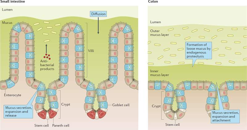
In the small intestine the mucus is not attached and forms a diffusion barrier with antibacterial products that limit penetration by bacteria. In colon bacteria are compartmentalized to the outer loose mucus layer while the inner attached layer is almost free of bacteria and protect the epithelium.
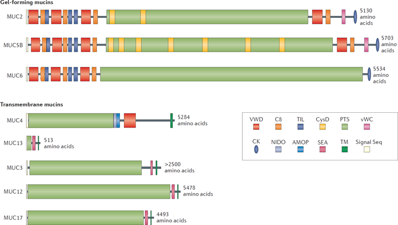
The gel-forming mucins and the transmembrane mucins are presented group vise to scale. The PTS domains become heavily O-glycosylated to form the mucin domains. These are rod-like and extended, looking like a bottle brush. The non-PTS parts of the gel-forming mucins are rich in Cys amino acids and form compact structures.
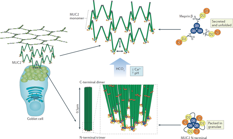
MUC2 is packed in goblet cell granulae and secreted into the lumen. Mucus is secreted into a bicarbonate-rich environment as generated by the CFTR channel in the small intestine, and by raising pH and lowering Ca2+ concentration it allows the packed molecules to expand into net-like sheets. The expanded conformation allows the protease Meprinβ to digest the N-terminal part of MUC2 releasing the attached mucus from the epithelium. This process is important for the clearance of mucus that has trapped bacteria.
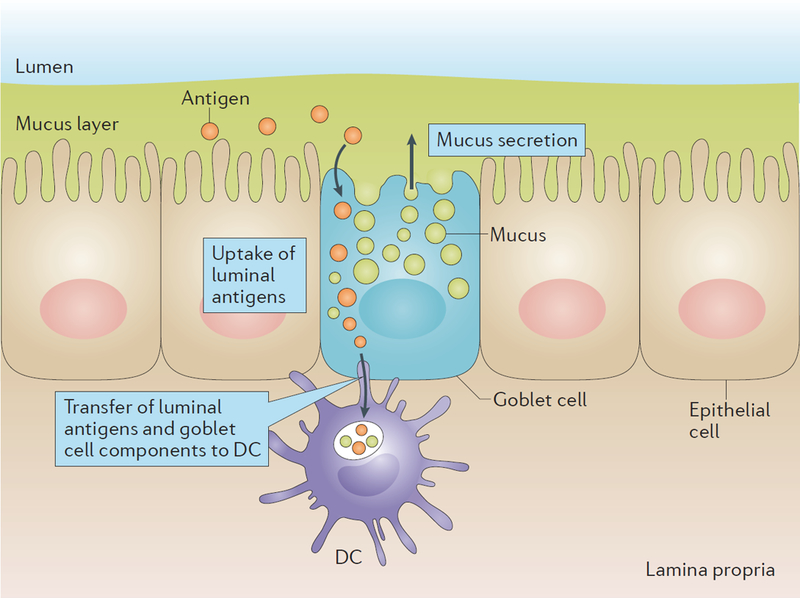
During mucus secretion small intestinal goblet cells sample the luminal content. How this uptake occurs is not known, but it could be proposed to occur through endocytosis and vesicle transport or by free diffusion in the cytoplasm. The luminal material is transferred together with goblet cell components to CD103+ DCs in the lamina propria that docks up against the secreting goblet cell.
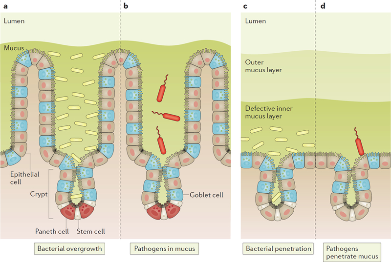
a, Defective release of the mucus from the epithelium in the small intestine results in stagnant mucus that allows bacteria to grow in the mucus (overgrowth). b, Pathogens that infect the small intestine must be motile and swim against the mucus flow and have an advantage if able to degrade the mucus. c, Defects in the inner mucus layer properties in colon allow bacteria to penetrate and reach the epithelium and penetrate into the crypts, something that triggers inflammation. d, Pathogens that infect colon must produce proteases able to degrade the inner mucus layer to be able to penetrate and reach the epithelial cells.
References
Publication types
MeSH terms
Substances
Grants and funding
LinkOut - more resources
Full Text Sources
Other Literature Sources
