. 2016 Sep 6;24(9):1573-81.
doi: 10.1016/j.str.2016.06.022. Epub 2016 Aug 4.

Tankyrase Sterile α Motif Domain Polymerization Is Required for Its Role in Wnt Signaling

Affiliations

Tankyrase Sterile α Motif Domain Polymerization Is Required for Its Role in Wnt Signaling

Amanda A Riccio et al. Structure. .

Abstract

Tankyrase-1 (TNKS1/PARP-5a) is a poly(ADP-ribose) polymerase (PARP) enzyme that regulates multiple cellular processes creating a poly(ADP-ribose) posttranslational modification that can lead to target protein turnover. TNKS1 thereby controls protein levels of key components of signaling pathways, including Axin1, the limiting component of the destruction complex in canonical Wnt signaling that degrades β-catenin to prevent its coactivator function in gene expression. There are limited molecular level insights into TNKS1 regulation in cell signaling pathways. TNKS1 has a sterile α motif (SAM) domain that is known to mediate polymerization, but the functional requirement for SAM polymerization has not been assessed. We have determined the crystal structure of wild-type human TNKS1 SAM domain and used structure-based mutagenesis to disrupt polymer formation and assess the consequences on TNKS1 regulation of β-catenin-dependent transcription. Our data indicate the SAM polymer is critical for TNKS1 catalytic activity and allows TNKS1 to efficiently access cytoplasmic signaling complexes.

Keywords: Wnt/β-catenin signaling; X-ray crystallography; poly(ADP-ribose) polymerase; tankyrase.


Figures

Figure 1
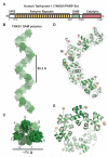
Figure 1. Crystal Structure of the TNKS1 SAM domain polymer
(A) Human TNKS1 domain organization. (B, C) Two views of the TNKS1 SAM polymer highlighting the polymer pitch of 95.5 Å and width of ~71 Å. (D, E) Two views of the seven SAM domains within the crystal asymmetric unit. The N-terminus of each SAM monomer is marked by a tan sphere, and the C-terminus of each SAM monomer is marked by a red sphere. All structure images were made using PYMOL (www.pymol.org).
Figure 2
Figure 2. Structure-based mutagenesis of TNKS1 SAM domain polymer
(A) Residues contributing to the head-to-tail interface of two SAM domain monomers. EH – end-helix. ML – mid-loop. (B) Electrostatic surface potential for the EH and ML contact surfaces calculated using APBS (Baker et al., 2001). Each monomer from panel A was rotated 90 degrees away from the interface. (C) left, Gel filtration elution profiles for SUMO-SAM constructs: WT (residues 1026-1092, G1074D, V1056R, and V1056H/G1074D. right, SDS-PAGE analysis of the designated fractions. M – molecular weight markers; 25, 20, and 15 kDa. (D) HeLa cells transfected with the indicated GFP-TNKS1 constructs. (E) HeLa cells transfected with the indicated GFP-SAM constructs.
Figure 3
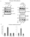
Figure 3. SAM polymerization is required for TNKS1 function in Wnt signaling
(A) HEK293 cells were transfected with the indicated versions of GFP-TNKS1: WT/G1074D (residues 1-1327), SAM (deletion of residues 1026-1092). Molecular weight markers (in kDa) are indicated on the left of each panel. left, Whole cell lysates were loaded at equal volumes (2.5% of total) and then immunoblotted. top right, Soluble lysates were loaded at equal volumes (2.5% of total) and probed with anti-GFP and anti-alpha tubulin. bottom right, After GFP immunopurification, GFP-TNKS1 or SAM polymer mutants were loaded in equal volumes (10% of total) and immunoblotted. (B) TOP luciferase reporter assay measuring β-catenin dependent transcription in cells transfected with control (GFP only) or the designated GFP-TNKS1 variants. The reported data represent the average of three independent experiments and the error bars represent the associated standard deviations.
Figure 4
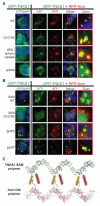
Figure 4. The SAM domain promotes efficient TNKS1 assembly into signaling complexes
(A and B) HEK293 cells transfected with the designated GFP-TNKS1 variants alone or co-transfected with RFP-Axin. WT TNKS1 (residues 1-1327), HPS (residues 174-1327), CAT (residues 1-1092), HPS/ankyrin repeats (residues 1-961), SAM (deletion of residues 1026-1092), SAM (residues 1026-1092. (C) The TNKS1 SAM polymer determined in this study (PDB code 5kni) is shown next to the Axin1 DIX polymer (Schwarz-Romond et al., 2007; PDB code 1wsp) to illustrate the relative positioning of TNKS1 N- and C-terminal domains and the key contact point between the TNKS1 ankyrin repeats and the N-terminus of Axin1 (Morrone et al., 2012).

References

    1. Baker NA, Sept D, Joseph S, Holst MJ, McCammon JA. Electrostatics of nanosystems: application to microtubules and the ribosome. Proc. Natl. Acad. Sci. U. S. A. 2001;98:10037–10041. - PMC - PubMed
    1. Bienz M. Signalosome assembly by domains undergoing dynamic head-to-tail polymerization. Trends Biochem. Sci. 2014;39:487–495. - PubMed
    1. Chang P, Coughlin M, Mitchison TJ. Tankyrase-1 polymerization of poly(ADP-ribose) is required for spindle structure and function. Nat. Cell Biol. 2005;7:1133–1139. - PubMed
    1. Chang P, Coughlin M, Mitchison TJ. Interaction between Poly(ADP-ribose) and NuMA contributes to mitotic spindle pole assembly. Mol. Biol. Cell. 2009;20:4575–4585. - PMC - PubMed
    1. Chi NW, Lodish HF. Tankyrase is a golgi-associated mitogen-activated protein kinase substrate that interacts with IRAP in GLUT4 vesicles. J. Biol. Chem. 2000;275:38437–38444. - PubMed

MeSH terms

LinkOut - more resources