. 2016 Oct 10;63(18):1840-1885.
doi: 10.1080/09500340.2016.1178820. Epub 2016 May 16.

Fifteen years of cold matter on the atom chip: promise, realizations, and prospects

Affiliations

Fifteen years of cold matter on the atom chip: promise, realizations, and prospects

Mark Keil et al. J Mod Opt. .

Abstract

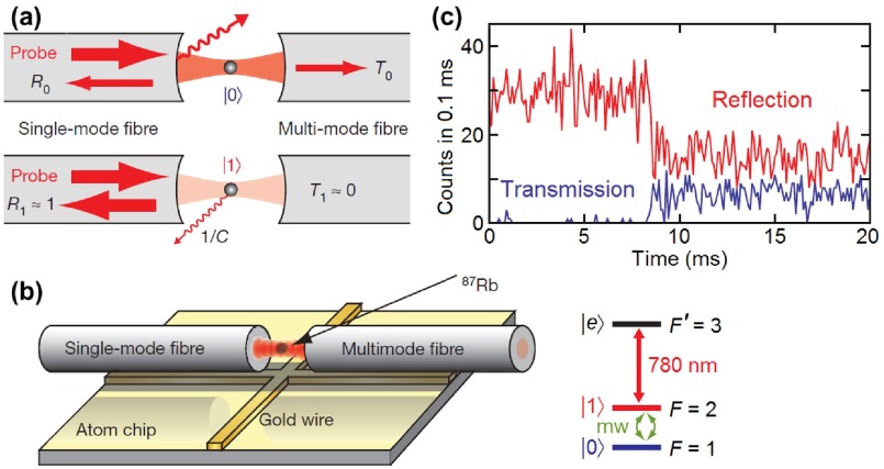
Here we review the field of atom chips in the context of Bose-Einstein Condensates (BEC) as well as cold matter in general. Twenty years after the first realization of the BEC and 15 years after the realization of the atom chip, the latter has been found to enable extraordinary feats: from producing BECs at a rate of several per second, through the realization of matter-wave interferometry, and all the way to novel probing of surfaces and new forces. In addition, technological applications are also being intensively pursued. This review will describe these developments and more, including new ideas which have not yet been realized.

Keywords: Atom chip; Bose-Einstein condensate; ultracold atomic physics; matter-wave quantum technology; quantum optics; atomtronics.


Conflict of interest statement

The authors declare that they have no competing financial interests.

Figures

Figure 1.
Figure 1.
Futuristic visions of highly integrated atom chips. (a) A single substrate (gray shading) holds all the light sources (yellow), atom sources (purple), photonics, and micromagnetic traps. Successive stages of cooling and experimentation proceed in a series of miniature vacuum chambers (unshaded) from left to right. Conceptual sketch courtesy of Tim Freegarde (Southampton) and adapted from [1], with permission from Springer Science+Business Media. (b) An integrated design currently being realized incorporates the vacuum system, atom source and optical components in a permanently sealed microlitre system [2]. The device is designed for magneto-optical trapping and cooling and is capable of passively maintaining ultrahigh vacuum for >1000days. The composite chip size will be 20×24×5mm3. Schematic courtesy of Matt Himsworth (Southampton). Compact modular systems developed by Dana Anderson’s group (Boulder) are now capable of producing chip-based BECs [3,4].
Figure 2.
Figure 2.
Vehicle and payload of the DLR sounding rocket MAIUS-1 in launch configuration. The scientific payload developed by the consortium led by Ernst Rasel consists of an autonomous atom chip device for interferometry employing BECs, including the vacuum system housing a three-layer atom chip, solid-state laser system, electronics, control system and batteries. Courtesy of Stephan Seidel (Hannover), Jens Große (DLR-Bremen) and DLR-MORABA [18].
Figure 3.
Figure 3.
Ion chip produced at Ben-Gurion University before installation in the vacuum chamber at Mainz. Inset: Two ions trapped on the chip. Courtesy of Ferdinand Schmidt-Kaler (Mainz) and adapted from [1], with permission from Springer Science+Business Media.
Figure 4.
Figure 4.
Microwave spectroscopy of Rydberg atoms on a superconducting atom chip. (a) Scheme of the experimental setup. (b) Scheme of the superconducting atom chip. (c) Scheme of the field-ionization detection system. Note the different co-ordinate axis definitions in the three panels. The origin O is taken at the centre of the horizontal segment of the Z-wire connecting pads G and L. Adapted from [96], with permission ©  2014 by the American Physical Society.
Figure 5.
Figure 5.
Appearance of a degenerate Fermi gas of 40K on an atom chip. Owing to Pauli pressure, Fermi degenerate 40K clouds seem to stop getting colder, even when the reservoir temperature approaches zero. Data is compared with its classical expectation (dashed black line) and with a Gaussian fit of a theoretically generated ideal Fermi distribution (solid red line). Absorption images are shown for (a) kBT/EF=0.35 and (b) 0.95, where the white circle indicates the Fermi energy EF. Fit residuals in (c) of a radially averaged cloud profile (taken after 9ms time-of-flight) show a strong systematic deviation when assuming Boltzmann (blue circles) instead of Fermi (red diamonds) statistics. Courtesy of Marcius Extavour (Toronto) [102] and adapted from [101], with permission by Macmillan Publishers Ltd.
Figure 6.
Figure 6.
Trapping molecules on a chip. (a) A pulsed beam of CO molecules is prepared in the upper Λ-doublet level of the a3Π1 (v=0,J=1) state by direct laser excitation from the electronic ground state. (b) The molecules are collimated and then travel closely above the ‘molecule chip’ over its full 50-mm length. Upon arrival above the chip, the molecules are confined in tubular electric field microtraps centred 25μm above the chip that move with the molecular beam at a velocity of several hundred m/s. (c) An array of these miniaturized moving traps (blue tubes) is brought to a standstill over a distance of only a few centimetres by applying phase-shifted MHz-range potentials to the microstructured electrode array. After a certain holding time, the molecules are accelerated off the chip again for detection. Adapted from [115], with permission from AAAS.
Figure 7.
Figure 7.
Integrated ion chip. (a) Optical micrograph of the ion chip with integrated waveguides and couplers underneath at multiple trap zones; optical waveguides and couplers are visible via topography transfer to the metal. Ions are trapped at one of the positions marked by the red dots, 50μm above the electrodes, with appropriate potentials applied to the DC and RF electrodes. (b) Simulated electric field mode profile of the single quasi-TE mode (field oriented predominantly horizontally) waveguide used for routing. Quantum coherent operations are performed on the optical qubit transition in individual 88Sr + ions by visible light routed in and emitted from the SiN waveguides and couplers. (c–h) Sequence of images of 422nm fluorescence from a chain of five 88Sr + ions, with the middle ion aligned to the grating coupler’s focus and occasionally entering a dark state; the sequence spans 2 s with frames spaced evenly. Courtesy of Karan Mehta (MIT) and MIT Lincoln Laboratory [149].
Figure 8.
Figure 8.
Chip trap for electrons. (a) Genesis of the coplanar-waveguide (CPW) Penning trap. The figure shows the projection of a standard cylindrical five-pole Penning trap onto a plane. The projected segments are shielded with two outer ground planes. (b) Sketch of the trap, with the resulting cyclotron and axial motions of an electron (red dot). Adapted from [157], with permission [161] ©  IOP Publishing & Deutsche Physikalische Gesellschaft.
Figure 9.
Figure 9.
Permanent magnet chips. (a) Schematic of the magnetic microstructure used to create a periodic 1D lattice of magnetic microtraps. Contour lines are calculated equipotentials with contour intervals of 0.5G. (b) Part of the absorption image for an array of clouds of 87Rb |F=1,mF=-1 atoms trapped in the 10μm-period magnetic lattice, after evaporative cooling to below the critical temperature. Adapted from [168], with permission ©  2014 by the American Physical Society.
Figure 10.
Figure 10.
Superconducting atom chip. (a) Magnetic trap geometries formed above an YBCO superconducting square and an external bias magnetic field. (b) For low values of Bbias there exists one field zero above the square centre. (c) Higher values of Bbias bring the atoms closer to the chip surface and split the initial, central trap into four separate traps (two of the atom clouds are hidden due to the imaging angle). Adapted from [191], with permission ©  2012 by the American Physical Society.
Figure 11.
Figure 11.
Concept of the grating chip MOT. Linearly polarized light diverging from the output of an optical fibre is collimated and circularly polarized by the combination of a lens (grey arrow) and λ/4 waveplate. This single input beam diffracts from microfabricated gratings on the chip to produce the additional beams (small red arrows) needed to form a MOT. Three linear gratings (the inset shows one pattern) diffract the light into nx=±1 orders to form a four-beam MOT (only trapping beams are shown). A square array of cylindrical indentations (not shown in this adaptation) could instead be used to diffract the input into the nx=±1 and ny=±1 orders to form a five-beam MOT. Magnetic quadrupole coils are omitted for clarity. Adapted from [205], with permission from Macmillan Publishers Ltd.
Figure 12.
Figure 12.
On-chip photonics. Schematic of an integrated-waveguide atom chip. An expanded view of the trench at the centre of (b) is shown in (a). A silicon substrate supports a layer of silica cladding, within which 4μm-square-doped silica waveguide cores are embedded. There are 12 parallel waveguides (for clarity, only six are shown) spaced at the centre of the chip by 10μm. These flare out at the edges of the chip so that optical fibres can be connected. The top layer of the chip is coated with gold to reflect the laser light used for cooling the atoms. Current-carrying wires below the chip provide magnetic fields to trap and move the atoms. Adapted from [207], with permission by Macmillan Publishers Ltd.
Figure 13.
Figure 13.
On-chip integrated photonic crystal waveguides. The scanning electron microscope image shows a photonic crystal waveguide made from 200nm-thick SiN. Arrows indicate radiative processes of an atom (green circle) coupled to an incident electric field Ein. Adapted from [219], with permission by Macmillan Publishers Ltd.
Figure 14.
Figure 14.
On-chip lattice proposals. (a–c) Plasmonic lattice. (a–b) Illustration of how to engineer an optical dipole trap by driving on the blue side of the plasmon resonance. (c) y-z contours (z:y axis expanded by 6:1) of the calculated potential for Rb near a line of nine spheres in the centre of a 45×45 square lattice with a 60nm lattice spacing. Black regions are where the potential is negative due to van der Waals attraction, and spheres are shown in white. Adapted from [225], with permission ©  2012 by the American Physical Society. (d–f) Capacitive lattice. (d) A 2D array of point capacitors can be created when each layer of wires (purple) is connected to an opposite voltage. A prism delivering a blue-detuned evanescent repulsive potential is located below these two layers and the atoms are above. Wire spacings are 1μm horizontally and vertically. (e–f) Trap potentials calculated assuming that each capacitor is charged with one electron in the top layer and one positive hole in the bottom layer. The CP force is included. (e) Trap potentials as a function of surface light intensity (blue to yellow): 20,,60W. (f) Simulated potential wells (blue) at a distance of 644nm from the chip. Adapted from [1], with permission by Springer Science+Business Media.
Figure 15.
Figure 15.
On-chip MEMS. (a) Microcantilever with wires for magnetic trapping of atoms. Cantilever vibrations can be independently probed with a readout laser. (b) Photograph of the atom chip showing the MOT loading region and the cantilever sub-assembly with a piezo for cantilever excitation. Rectangle: region shown in (a). (c) Potential U=Um+Us for trap frequency ωz/2π=10.5kHz at d=1.5μm from the driven cantilever. Dashed red line: magnetic potential Um. The surface potential Us reduces the trap depth to U0. Gray lines: U during the extremum positions of the cantilever for an oscillation amplitude a=120nm. Blue line: BEC chemical potential μc for 600 atoms. Adapted from [229], with permission ©  2010 by the American Physical Society.
Figure 16.
Figure 16.
On-chip plasmonics. (a) Photograph of the dielectric prism with sapphire substrate and fabricated gold structures.(b) Schematic side view of the prism. A laser beam is internally reflected with an adjustable angle Θin. In this Kretschmann configuration, plasmons are excited at the angle Θin=Θpl. (c) Diffraction images of cold atoms, which are reflected from the nominally 500nm gold stripes for laser powers of the evanescent wave 107<P<217mW. Diffraction orders l are indicated by the vertical yellow lines. Adapted from [234], with permission by Macmillan Publishers Ltd.
Figure 17.
Figure 17.
Probing Johnson noise and the CP potential on a chip. (a) Paths chosen for trap lifetime measurements above a dielectric surface (A) and above a copper film (B). Line C is the measured contour line of 22ms lifetime near the metal. (b) Remaining atom fraction χ in a trap at distance d from the dielectric surface for a condensate (solid squares) and for thermal clouds at 2.1μK (open squares) and 4.6μK (triangles). The solid (dashed) lines are calculated with (without) CP potential for the condensate, 2.1μK, and 4.6μK clouds (left to right). The inset shows the trapping potentials for C4=8.2×10-56Jm4 (solid line) and C4=0 (dotted line). Adapted from [20], with permission ©  2004 by the American Physical Society.
Figure 18.
Figure 18.
Probing the temperature dependence of the CP force. The fractional change in the trap frequency due to the CP force is shown as a function of the distance to the chip. Three sets of data are presented, with accompanying theoretical curves having no adjustable parameters. Error bars represent the total uncertainty (statistical and systematic) of the measurement. Adapted from [24], with permission ©  2007 by the American Physical Society.
Figure 19.
Figure 19.
Probing dispersion forces using a chip-mounted CNT. (a) A multi-walled CNT (length, 10.25μm), immersed in an ultracold quantum gas, stands on a silicon substrate (nanochip). (b) Scanning electron microscope image of the CNT used for the experiments. (c) Exponential decay constant of atom number at the CNT (red points) and at the plane surface (blue points) plotted against distance between the surface and the centre of the condensate, d. The vertical dashed line indicates the position of the CNT tip. The red shaded area denotes the regime where the condensate is in partial overlap with the CNT. Inset: detailed view of the sharp onset of scattering losses once the BEC touches the CNT. Adapted from [268], with permission by Macmillan Publishers Ltd.
Figure 20.
Figure 20.
Probing electron transport with cold-atom magnetometry on a chip. (a) Readout of the information by measuring the atomic density with reflected absorption imaging. (b–d) Magnetic field angle fluctuations (color scale bars, mrad) above (b) 2.08μm-thick and (c–d) 0.28μm-thick polycrystalline gold films (grain sizes are (b) 60–80 nm, (c) 30–50 nm, and (d) 150–170 nm). These fluctuations are due to variations in the direction of the current flow, nominally along the x axis. The appearance of ±45 patterns is clearly observable and reflects a correlated scattering of the electrons. (a) Adapted from [289], with permission by Macmillan Publishers Ltd. and (b–d) adapted from [22], with permission by AAAS.
Figure 21.
Figure 21.
Probing the surface for non-Newtonian gravitational effects. An optical lattice is used to position cold 88Sr atoms near two adjacent test masses. (a) The atoms are first placed close to the transparent part of the test surface; a vertical translation of Δz=(λ/4π)ϕ is accomplished by adjusting the relative optical phase ϕ accumulated between the two laser beams (green arrows). (b) The counter-propagating (lower) lattice beam is then switched off adiabatically and the atoms remain trapped in the standing wave produced by the co-propagating beam and the weak reflected beam. (c) The lattice beam is translated laterally, placing the atoms just above the Casimir shield and close to the Al and Au masses. The width of the arrows represents the relative intensity of laser beams. Adapted from [41], with permission ©  2009 by the American Physical Society.
Figure 22.
Figure 22.
Bragg interferometery in microgravity on an atom chip at the Bremen drop tower. (a) The experimental sequence includes capturing cold atoms in a MOT, loading a Ioffe-Pritchard trap, creating a BEC, and applying delta-kick cooling (DKC) followed by adiabatic rapid passage (ARP). The remaining time before the capture of the capsule at the bottom of the tower is used for atom interferometry (AI) and imaging of the atoms. (b) The evolution of the BEC and the asymmetric Mach–Zehnder interferometer is visualized by a series of absorption images of the atomic densities separated by 1 ms. A π/2 pulse from two counter-propagating+ light beams of frequency ω and ω+δ creates (time T0) a superposition of two wave packets that drift apart with a two-photon recoil velocity vrec=11.8mm/s. After T they are redirected by a π pulse and partially recombined after T-δT by a second π/2 pulse. A nonzero value of δT leads to a spatial interference pattern after τ=53ms in free-fall. The fringe spacing is inversely proportional to the separation d=vrecδT of the wave packets. Reprinted from [310], with permission ©  2013 by the American Physical Society.
Figure 23.
Figure 23.
An RF potential Mach–Zehnder interferometer on an atom chip. (a) Schematic of the atom chip: DC currents in the trap wire and in two perpendicular wires (not shown), together with a uniform external field, create an elongated Ioffe-Pritchard trap 60μm below the chip. RF currents with a relative phase of π are applied on the dressing wires to perform the splitting along x. (b) A relative phase between the two arms is imprinted by tilting the double well during a time tϕ; the spacing between the two wells is then abruptly reduced and the potential barrier acts as a beam splitter for both wave packets, transforming the relative phase into a population imbalance read out after the two clouds are separated again. (c) The normalized population difference z=n/Nt as a function of tϕ exhibits damping of interference fringes due to phase diffusion. Grey dots: imbalance of individual experimental realizations; black dots: ensemble average z; red curve: theoretical prediction taking into account phase diffusion; dashed black line: expected signal for a classical coherent state. Adapted from [314], with permission by Macmillan Publishers Ltd.
Figure 24.
Figure 24.
A static field double-well potential on an atom chip.(a) The magnetic field is generated by DC currents superposed with a static homogeneous field B0. The minimum is located at a distance z0=μ0I0/2πBy from the I0 wire and the field at the minimum is Bx. This minimum is modified by I1=2.0mA and I2, whose fields are predominantly along x on the trap axis. For z0d, the two I1 wires together provide harmonic confinement along x. I2, of opposite polarity, creates the barrier of adjustable height and also determines the spacing between the two resulting wells. (b) Profile of the trapping potential along the splitting axis x, for I2=0mA (cooling trap, dashed line) and I2=2.4mA (solid line). (c) Barrier height Vb (top), trap frequencies (fx,fy,fz) (center), and position x0 of the right minimum (bottom) as functions of the current I2. Adapted from [35], with permission ©  2010 by the American Physical Society.
Figure 25.
Figure 25.
Atom chip for a tight double-well potential.(a–b) Central region of the multi-layer chip; wire layers are separated by a 15μm-thick layer of polyimide insulation, and the entire chip is covered by an additional gold mirror layer. (a) Arrows show the faint outline of the trapping structure in the lower layer. (b) Up to 9 central wires (1μm wide, 1μm gaps) in the upper layer can be used for barrier control. (c) Magnetic potentials produced 5.5μm from the chip surface, shown along the longitudinal axis (x^). The harmonic potential (dashed) is modified by using (blue) a single central wire and (green, red) adding opposing currents in successive pairs of adjacent wires. The barrier becomes progressively narrower as wire pairs are added, while the barrier height is maintained at 100nK and the transverse frequency (along y^) is maintained at 2.4kHz. Potential calculations and design courtesy of Shimon Machluf (Be’er Sheva) [320].
Figure 26.
Figure 26.
Double well on an atom chip using potentials created by microwave co-planar waveguides (CPWs). (a) Photograph of the chip assembly. The Si experimental chip has two layers of gold wires separated by a thin polyimide layer, with CPWs on the upper layer. It is glued and wire-bonded to an AlN carrier chip with a single gold layer. (b) Schematic close-up of the experimental region. The three central wires (red) form a CPW. All wires (including the CPW) can carry stationary currents for the generation of static magnetic traps. The position of the minimum of the static trap is indicated by the black cross. (c) State-selective splitting of the BEC. Absorption images of the adiabatically split BEC. By imaging both hyperfine states simultaneously (top), only F=1 (middle) or only F=2 (bottom), the state selectivity of the splitting is established. Adapted from [321], with permission by Macmillan Publishers Ltd.
Figure 27.
Figure 27.
Clock interferometry on a chip. A magnetic gradient pulse of duration TG is applied to induce a relative angle of rotation between two clock wave packets (inducing a clock ‘tick’ rate difference Δω simulating a gravitational red shift). (a–b) For TG=0, the clock rate is approximately the same in the two wave packets, and interference is visible. (c–d) Visibility of the clock spatial interference may be destroyed (ΔωTG=π) or restored (ΔωTG=2π) merely by adjusting TG. Optical density curves are data (blue) and fits (red) to a simple combination of a sine with a Gaussian envelope. The vertical axis z is relative to the chip surface. Adapted from [37], with permission by AAAS.
Figure 28.
Figure 28.
Preparation of the internal states of a BEC realized on an atom chip. Optimal control theory is used to design frequency-modulated RF pulses to prepare an arbitrary coherent superposition of states starting from a given initial state (fixed by the BEC preparation). Shown in the bottom right corner are the experimentally observed clouds of atoms that are initially in sub-level +2 and then split coherently into the sub-levels -1 and -2 via optimal control. Adapted from [338], with permission ©  2016 by the American Physical Society.
Figure 29.
Figure 29.
Vibrational state interferometry: schematic of the Ramsey interferometric sequence. (a) Representation of the BEC subjected to a fast displacement λ(t) in the y-direction. (b) Trapping potential and effective two-mode system. The anharmonicity in the y-direction leads to a unique transition frequency between (blue) the ground state |0 and (red) the lowest-lying excited state |1y, effectively almost isolating the two-level system |0-|1y. The other states (dashed line) have higher energies. (c) Example of an interferometric trajectory (blue dots) on the Bloch sphere representation of the two-level system. (1) is the first pulse that prepares a balanced coherent superposition; (2) is the phase accumulation time corresponding to a rotation around the vertical axis; (3) is the second pulse, which is equivalent to a π/2 pulse for the states on the equator and corresponds to a 90 counter-clockwise rotation around Jy. The red squares show the 15 points on which the second pulse was optimized. Adapted from [339], with permission [340].
Figure 30.
Figure 30.
High-flux atom chip-based BEC source. Schematic drawings of (a) the two-chamber vacuum setup and (b) the atom chip setup. A beam of pre-cooled Rb atoms is formed in a 2D+ MOT and injected into the 3D chamber. Atom interferometry, as well as detection of the atoms, is carried out in the 3D chamber. The three layers of the atom chip setup are shown in an exploded view. Adapted from [348], with permission [340] ©  IOP Publishing & Deutsche Physikalische Gesellschaft.
Figure 31.
Figure 31.
Imaging of microwave magnetic field components near the atom chip microwave source (a CPW structure, inset). The images show the measured probability p2(r) to find an atom in F=2 after applying the microwave pulse. Columns correspond to measurements on the three different transitions ωγ (γ=-,π,+); rows to three different orientations of B0. The imaging beam is reflected from the chip surface at a small angle. As a result, on each picture, the direct image and its reflection on the chip surface are visible. The dashed line separates the two. Adapted from [360], with permission ©  2010 by AIP Publishing LLC.
Figure 32.
Figure 32.
On-chip lattices. (a) Photo of an atom chip showing the optical lattice system. (b) Model of the on-chip optical lattice system. The two mirrors at the bottom of the image retro-reflect two of the incoming lattice beams, while the window is used to retro-reflect the third beam. (c) Schematic illustration of the vertically-oriented 1D lattice used to demonstrate Landau-Zener tunnelling. Adapted from [223], with permission from the Optical Society of America.
Figure 33.
Figure 33.
High-density electrode chip. (a) Die bond region showing the ball-grid array (BGA) trap and the interposer footprint; (b) side view; (c) fully packaged BGA trap. The long bond wire supplies the trap RF signal. (d) Schematic cross section through a BGA trap die and interposer (not to scale). Adapted from [371], with permission ©  2015 by AIP Publishing LLC.
Figure 34.
Figure 34.
2D array and trapped lattice atoms on a permanent-magnet chip. The upper images show alternating strips of etched and non-etched regions in a 200nm-thick layer of FePt that generates a magnetic lattice with (a) square translational symmetry and (b) hexagonal (equilateral triangular) symmetry. An absorption image of 87Rb is shown in (c) for atoms loaded simultaneously into the hexagonal (upper left) and square (lower right) lattices, with (d) showing the entire extent of the lattice. Atom chip fabricated at Ben-Gurion University. Adapted from [95], with permission ©  2014 by AIP Publishing LLC.
Figure 35.
Figure 35.
Optical fibre-based high-finesse cavity mounted on an atom chip, enabling cavity-assisted detection of an atomic qubit.(a) For an atom in the dark state |0 (top), probe light is either transmitted, reflected or lost by mirror imperfections. For the bright state |1 (bottom), most incident photons are reflected. In both cases, only a small fraction is scattered by the atom. (b) The cavity is formed by the coated end facets of two optical fibres. The qubit states (F=1,mF=0 and F=2,mF=0) can be coupled by a resonant microwave pulse. The cavity and the atomic transition |1|e are resonant with the π-polarized probe laser at 780nm wavelength. (c) Typical detection trace, showing cavity transmission (blue) and reflection (red) for an atom initially in |1 performing a quantum jump to |0 owing to spontaneous emission. Adapted from [385], with permission by Macmillan Publishers Ltd.
Figure 36.
Figure 36.
On-chip integrated resonator. (a) Schematic depiction of the single-photon Raman interaction. The two transitions in a three-level Λ-system (a single atom of 87Rb in this case) are coupled (b) via a microresonator to different directions of a nanofibre waveguide. A photon coming from the left is deterministically reflected (red arrows) due to destructive interference in the transmission (blue arrows), resulting in the Raman transfer of the atom from ground state α to β. The atom then becomes transparent to subsequent photons, which are therefore transmitted. Adapted from [216], with permission by Macmillan Publishers Ltd.

References

    1. Folman R. Material Science for Quantum Computing with Atom Chips. In: Folman R., editor. Special Issue on Neutral Particles. Quantum Inf. Process. 2011. pp. 995–1036.
    1. Rushton J.A., Aldous M., Himsworth M.D. The Feasibility of a Fully Miniaturized Magneto-optical Trap for Portable Ultracold Quantum Technology. Rev. Sci. Instrum. 2014:121501. - PubMed
    1. Salim E.A., DeNatale J., Farkas D.M., Hudek K.M., McBride S.E., Michalchuk J., Mihailovich R., Anderson D.Z. Compact, Microchip-based Systems for Practical Applications of Ultracold Atoms. In: Folman R., editor. Special Issue on Neutral Particles. Quantum Inf. Process. 2011. pp. 975–994.
    1. Farkas D.M., Salim E.A., Ramirez-Serrano J. Production of Rubidium Bose--Einstein Condensates at a 1 Hz Rate. 2014. arXiv:1403.4641v2.
    1. Reichel J., Hänsel W., Hänsch T.W. Atomic Micromanipulation with Magnetic Surface Traps. Phys. Rev. Lett. 1999:3398–3401.