. 2016 Mar 9;3(1):39-51.
doi: 10.1002/reg2.51. eCollection 2016 Feb.

Analogous cellular contribution and healing mechanisms following digit amputation and phalangeal fracture in mice

Affiliations

Analogous cellular contribution and healing mechanisms following digit amputation and phalangeal fracture in mice

Lindsay A Dawson et al. Regeneration (Oxf). .

Abstract

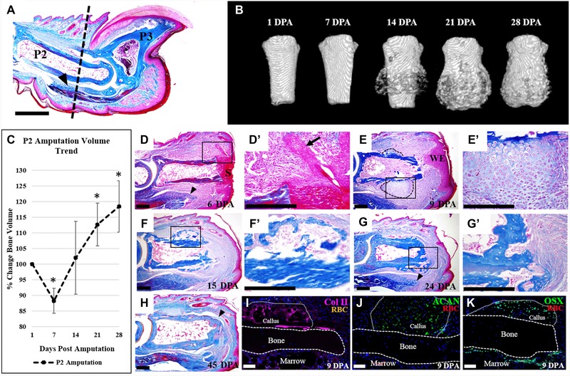
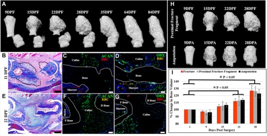
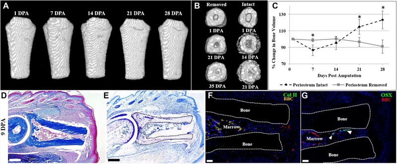
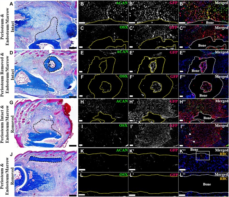
Regeneration of amputated structures is severely limited in humans and mice, with complete regeneration restricted to the distal portion of the terminal phalanx (P3). Here, we investigate the dynamic tissue repair response of the second phalangeal element (P2) post amputation in the adult mouse, and show that the repair response of the amputated bone is similar to the proximal P2 bone fragment in fracture healing. The regeneration-incompetent P2 amputation response is characterized by periosteal endochondral ossification resulting in the deposition of new trabecular bone, corresponding to a significant increase in bone volume; however, this response is not associated with bone lengthening. We show that cells of the periosteum respond to amputation and fracture by contributing both chondrocytes and osteoblasts to the endochondral ossification response. Based on our studies, we suggest that the amputation response represents an attempt at regeneration that ultimately fails due to the lack of a distal organizing influence that is present in fracture healing.

Keywords: Digit; endochondral ossification; fracture; mouse; periosteum; regeneration.


Figures

Figure 1
Figure 1
P2 amputation initiates local endochondral ossification response. (A) Mallory trichrome stained section of an adult mouse hind limb digit. Dashed line indicates the amputation level. (B) μCT reconstructed images show new bone formation initiated by 14 DPA, and continued bone formation at 21 and 28 DPA. (C) Bone volume measurements, normalized to 1 DPA, indicate bone degradation at 7 DPA, resulting in an average bone loss of 12%, followed by a bone growth phase, resulting in an 18% overall increase in bone volume by 28 DPA (t test, ±SEM, *P < 0.005). (D)−(H) Histological sections of digits stained with Mallory trichrome stain. (D), (D′) At 6 DPA wound closure has not completed and wound retraction is evident. (E), (E′) Wound closure is completed by 9 DPA, and cartilaginous growth is evident on the lateral portions of the bone (outlined). (F), (F′) Replacement of the cartilaginous callus with woven bone and marrow cells is apparent by 15 DPA. (G), (G′) By 24 DPA, a bone plug has formed at the distal portion of the stump. (H) Bone remodeling and tendon reattachment to the bone (arrowhead) is evident by 45 DPA. (I)−(K) Immunostaining of 9 DPA samples, counterstained with DAPI. (I) Col II immunostaining indicates cartilaginous tissue along the periosteal surface. (J) Immunostaining for aggrecan (ACAN) confirming cartilaginous tissue is present. (K) Immunostaining for osterix (Osx) reveals osteoblasts localized to the periosteal callus and the endosteum. (A), (D)−(K) Dorsal surface is top, distal to the right. (B) Top is proximal, bottom is distal. S, scab; WE, wound epidermis; RBC, red blood cells. Scale bars: (A) 500 μm; (D)−(H) 200 μm; (I)−(K) 50 μm.
Figure 2
Figure 2
The repair response following amputation is similar to fracture healing. (A), (I) μCT reconstructed images of the fractured P2 digit show periosteal bone formation evident by 15 DPF, with progressive bone growth and eventual merging of the proximal and distal bone fragments by 35 DPF, coinciding with a significant increase in bone volume when normalized to 1 DPF (t test, ±SEM, *P < 0.05). (B)−(D) 11 DPF serial sectioned sample. (B) Mallory trichrome stained section showing cartilaginous growth on the proximal and distal bone segments (outlined). (C) Immunostaining for ACAN confirming cartilaginous cells associated with the periosteum. (D) OSX immunostaining showing osteoblasts localized to the callus and the endosteum/marrow. (E)−(G) 22 DPF serial sectioned sample. (E) Mallory trichrome stained section showing new bone and marrow formation on the proximal bone fragment and cartilaginous bridging of the fracture gap. (F) Immunostaining for ACAN showing cartilage spanning the fracture gap. (G) Immunostaining for OSX showing osteoblasts localized to the proximal bone fragment. (C), (D), (F), (G) Samples counterstained with DAPI. (H) Comparative analysis of μCT reconstructed images showing that the proximal fracture bone fragment is morphologically similar to the amputation stump, with ossification apparent by 15 DPF/DPA, and continued bone formation at 22 and 28 DPF/DPA. (I) Bone volume measurements indicate no statistical significance between the three experimental groups when normalized to the starting bone volume (t test, ±SEM, P < 0.05). (B)−(G) Dorsal surface is top, distal to the right. (A), (H) Top is proximal, bottom is distal. P bone, proximal bone fragment; D bone, distal bone fragment; RBC, red blood cells. Scale bars: (B), (E) 200 μm; (C), (D), (F), (G) 50 μm.
Figure 3
Figure 3
Periosteum removal inhibits cartilaginous callus formation after amputation. (A) μCT reconstructed images of the periosteum‐removed digit showing no discernable morphological change and no significant change in bone volume when normalized to 1 DPA (t test, ±SEM, P < 0.05). (B) Distal end view of μCT reconstructed images of periosteum‐removed digits showing marrow closure by 35 DPA versus marrow closure by 21 DPA in digits with an intact periosteum. (C) Bone volume measurements over 28 days, normalized to 1 DPA, showing no significant volume change in periosteum‐removed digits. This pattern is significantly different from periosteum‐intact digits (t test, ±SEM, *P < 0.05). (D)−(G) Serial sections of a representative periosteum‐removed digit at 9 DPA. (D) Mallory trichrome staining illustrating no circumferential callus formation on the amputated bone. (E) Toluidine blue staining showing cartilage present in the articular region and no cartilaginous staining on the stump bone surface. (F) Col II immunostaining confirming the absence of cartilage formation on the periosteal surface. (G) OSX immunostaining showing no osteoblasts on the periosteal surface and several immunopositive cells in the endosteum/marrow space (arrowheads). (F), (G) Samples counterstained with DAPI. (A) Top is proximal, bottom is distal. (D)−(G) Dorsal surface is top, distal to the right. RBC, red blood cells. Scale bars: (D), (E) 200 μm; (F), (G) 50 μm.
Figure 4
Figure 4
P2 periosteum and endosteum/marrow contribute to wound repair (dorsal surface is top, distal to the right). (A)−(C′′) Serial sections of an intact GFP‐labeled P2 bone grafted into a NOD‐SCID fractured P2 digit, 11 DPF. Representative sample shown. N = 4. (A) Mallory trichrome staining showing callus formation on the grafted bone (outlined). (B)−(B′′) Double immunostaining for ACAN and GFP illustrating cartilage derived from the bone graft (outlined). (C)−(C′′) Double immunostaining for OSX and GFP showing graft‐derived osteoblasts within the periosteal callus and the endosteal/marrow space (graft outlined). Immunohistochemical stained samples counterstained with DAPI. (D)−(F′′) Serial sections of a periosteum‐removed and endosteum/marrow‐intact GFP‐labeled P2 bone grafted into a NOD‐SCID fractured P2 digit, 11 DPF. Representative sample shown. N = 4. (D) Mallory trichrome staining showing no periosteal callus formation on the grafted bone (outlined). (E)−(E′′) Double immunostaining for ACAN and GFP revealing no graft‐derived chondrocytes present on the periosteal surface or within the marrow cavity (graft outlined). (F)−(F′′) Double immunostaining for OSX and GFP showing double‐labeled osteoblasts present within the graft marrow space (outlined). Immunohistochemical stained samples counterstained with DAPI. (G)−(I′′) Serial sections of periosteum‐intact and endosteum/marrow‐removed GFP‐labeled P2 bone grafted into a NOD‐SCID fractured P2 digit, 11 DPF. Representative sample shown. N = 4. (G) Mallory trichrome staining showing graft periosteal callus formation (outlined). (H)−(H′′) Double immunostaining for ACAN and GFP showing the graft‐derived cartilaginous callus (grafted bone outlined). (I)−(I′′) OSX and GFP double immunostaining showing graft‐derived osteoblasts (arrowheads). Immunohistochemical stained samples counterstained with DAPI. (J)−(L′′) Serial sections of a periosteum and endosteum/marrow‐removed GFP‐labeled P2 bone grafted into a NOD‐SCID fractured P2 digit, 11 DPF. Representative sample shown. N = 4. (J) Mallory trichrome staining showing no callus formation on the grafted bone (outlined). (K)−(K′′) Double immunostaining for ACAN and GFP indicating no graft‐derived chondrocytes present (grafted bone outline). (L)−(L′′) Double labeled osteoblasts were not detected by OSX and GFP double immunostaining (grafted bone outlined). (K)−(L′′) Signal indicates red blood cells (RBC). Immunohistochemical stained samples counterstained with DAPI. Scale bars: (A), (D), (G), (J) 200 μm; all immunostaining 50 μm.

References

    1. Agrawal, V. , Johnson, S.A. , Reing, J. , Zhang, L. , Tottey, S. , Wang, G. et al. (2010). Epimorphic regeneration approach to tissue replacement in adult mammals. Proc Natl Acad Sci USA, 107, 3351–3355. - PMC - PubMed
    1. Agrawal, V. , Tottey, S. , Johnson, S.A. , Freund, J.M. , Siu, B.F. & Badylak, S.F. (2011a). Recruitment of progenitor cells by an extracellular matrix cryptic peptide in a mouse model of digit amputation. Tissue Eng Part A, 17, 2435–2443. - PMC - PubMed
    1. Agrawal, V. , Kelly, J. , Tottey, S. , Daly, K.A. , Johnson, S.A. , Siu, B.F. et al. (2011b). An isolated cryptic peptide influences osteogenesis and bone remodeling in an adult mammalian model of digit amputation. Tissue Eng Part A, 17, 3033–3044. - PMC - PubMed
    1. Agrawal, V. , Siu, B.F. , Chao, H. , Hirschi, K.K. , Raborn, E. , Johnson, S.A. et al. (2012). Partial characterization of the Sox2+ cell population in an adult murine model of digit amputation. Tissue Eng Part A, 18, 1454–1463. - PMC - PubMed
    1. Al‐Aql, Z. S. , Alagl, A.S. , Graves, D.T. , Gerstenfeld, L.C. & Einhorn, T. A. (2008). Molecular mechanisms controlling bone formation during fracture healing and distraction osteogenesis. J Dent Res, 87, 107–118. - PMC - PubMed

LinkOut - more resources