

Diagnostic value of H3F3A mutations in giant cell tumour of bone compared to osteoclast-rich mimics
- PMID: 27499898
- PMCID: PMC4858131
- DOI: 10.1002/cjp2.13
Diagnostic value of H3F3A mutations in giant cell tumour of bone compared to osteoclast-rich mimics
Abstract
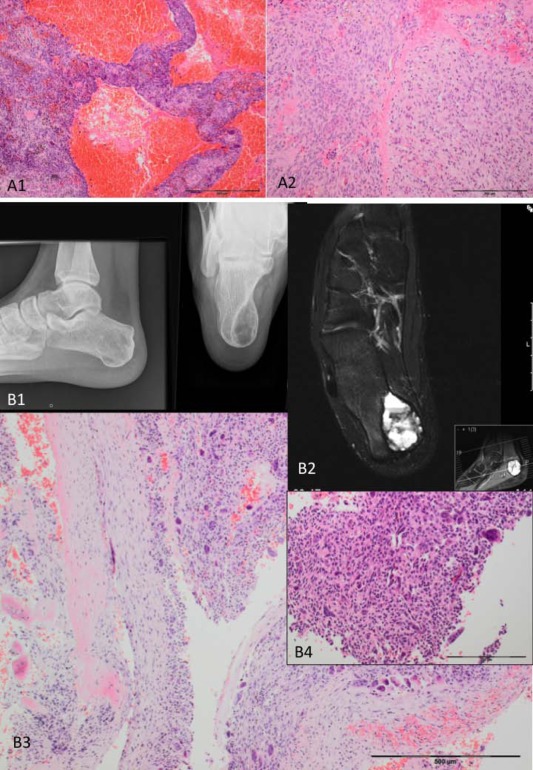
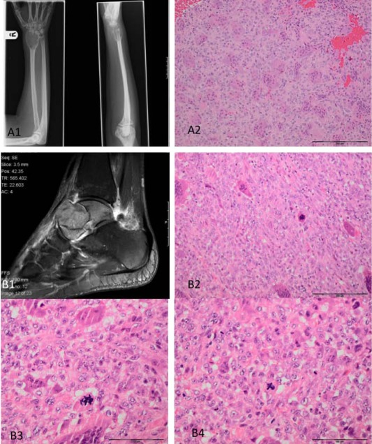
Driver mutations in the two histone 3.3 (H3.3) genes, H3F3A and H3F3B, were recently identified by whole genome sequencing in 95% of chondroblastoma (CB) and by targeted gene sequencing in 92% of giant cell tumour of bone (GCT). Given the high prevalence of these driver mutations, it may be possible to utilise these alterations as diagnostic adjuncts in clinical practice. Here, we explored the spectrum of H3.3 mutations in a wide range and large number of bone tumours (n = 412) to determine if these alterations could be used to distinguish GCT from other osteoclast-rich tumours such as aneurysmal bone cyst, nonossifying fibroma, giant cell granuloma, and osteoclast-rich malignant bone tumours and others. In addition, we explored the driver landscape of GCT through whole genome, exome and targeted sequencing (14 gene panel). We found that H3.3 mutations, namely mutations of glycine 34 in H3F3A, occur in 96% of GCT. We did not find additional driver mutations in GCT, including mutations in IDH1, IDH2, USP6, TP53. The genomes of GCT exhibited few somatic mutations, akin to the picture seen in CB. Overall our observations suggest that the presence of H3F3A p.Gly34 mutations does not entirely exclude malignancy in osteoclast-rich tumours. However, H3F3A p.Gly34 mutations appear to be an almost essential feature of GCT that will aid pathological evaluation of bone tumours, especially when confronted with small needle core biopsies. In the absence of H3F3A p.Gly34 mutations, a diagnosis of GCT should be made with caution.
Keywords: H3F3A; H3F3B; USP6; giant cell granuloma; giant cell tumour of bone; malignant giant cell tumour of bone; solid variant of aneurysmal bone cyst.
Figures
References
-
- Athanasou NA, Bansal M, Forsyth R, et al Giant cell tumour of bone In WHO Classification of Tumours of Soft Tissue and Bone, (4th edn), Fletcher CDM, Bridge JA, Hogendoorn PCW, et al (eds). IARC Press: Lyon, France, 2013; 321–324.
-
- Oliveira AM, Hsi BL, Weremowicz S, et al USP6 (Tre2) fusion oncogenes in aneurysmal bone cyst. Cancer Res 2004; 64: 1920–1923. - PubMed
LinkOut - more resources
Full Text Sources
Other Literature Sources
Research Materials
Miscellaneous
