. 2016 Feb 8;2(2):93-103.
doi: 10.1002/cjp2.38. eCollection 2016 Apr.

Molecular classification of thyroid lesions by combined testing for miRNA gene expression and somatic gene alterations

Affiliations

Molecular classification of thyroid lesions by combined testing for miRNA gene expression and somatic gene alterations

Dennis Wylie et al. J Pathol Clin Res. .

Abstract

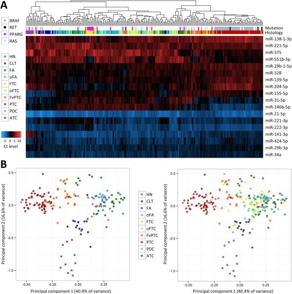
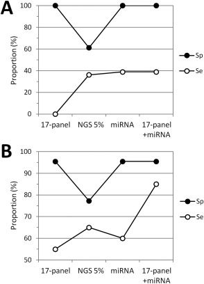
Multiple molecular markers contribute to the pathogenesis of thyroid cancer and can provide valuable information to improve disease diagnosis and patient management. We performed a comprehensive evaluation of miRNA gene expression in diverse thyroid lesions (n = 534) and developed predictive models for the classification of thyroid nodules, alone or in combination with genotyping. Expression profiling by reverse transcription-quantitative polymerase chain reaction in surgical specimens (n = 257) identified specific miRNAs differentially expressed in 17 histopathological categories. Eight supervised machine learning algorithms were trained to discriminate benign from malignant lesions and evaluated for accuracy and robustness. The selected models showed invariant area under the receiver operating characteristic curve (AUC) in cross-validation (0.89), optimal AUC (0.94) in an independent set of preoperative thyroid nodule aspirates (n = 235), and classified 92% of benign lesions as low risk/negative and 92% of malignant lesions as high risk/positive. Surgical and preoperative specimens were further tested for the presence of 17 validated oncogenic gene alterations in the BRAF, RAS, RET or PAX8 genes. The miRNA-based classifiers complemented and significantly improved the diagnostic performance of the 17-mutation panel (p < 0.001 for McNemar's tests). In a subset of resected tissues (n = 54) and in an independent set of thyroid nodules with indeterminate cytology (n = 42), the optimized ThyraMIR Thyroid miRNA Classifier increased diagnostic sensitivity by 30-39% and correctly classified 100% of benign nodules negative by the 17-mutation panel. In contrast, testing with broad targeted next-generation sequencing panels decreased diagnostic specificity by detecting additional mutations of unknown clinical significance in 19-39% of benign lesions. Our results demonstrate that, independent of mutational status, miRNA expression profiles are strongly associated with altered molecular pathways underlying thyroid tumorigenesis. Combined testing for miRNA gene expression and well-established somatic gene alterations is a novel diagnostic strategy that can improve the preoperative diagnosis and surgical management of patients with indeterminate thyroid nodules.

Keywords: diagnosis; gene mutation; gene rearrangement; miRNA; thyroid cancer; thyroid nodule.


Figures

Figure 1
Figure 1
miRNA expression profiling in resected thyroid tissues. (A) Unsupervised hierarchical clustering for histopathological categories represented by at least five independent specimens (n = 248 specimens total). The expression levels of 18 miRNAs as determined by RT‐qPCR are colour‐coded from low expression (high Ct, red) to high expression (low Ct, blue) after mean‐centering normalization. The presence of gene alterations in the BRAF, RAS, PAX8 or RET genes detected with the Luminex platform is colour‐coded at the top of the figure. (B) Principal component (PC) analysis using the same miRNAs and sample set. For clarity, the colour‐coded markers corresponding to oFA, FTC, oFTC and PDC specimens are not displayed in the left panel. Abbreviations: HN, hyperplastic nodule (includes diffuse hyperplasia); CLT, chronic lymphocytic thyroiditis; FA, follicular adenoma; oFA, oncocytic FA; FTC, follicular thyroid carcinoma; oFTC, oncocytic FTC; PTC, papillary thyroid carcinoma (includes conventional and tall cell variants); FvPTC, follicular variant of PTC; PDC, poorly differentiated carcinoma; ATC, anaplastic thyroid carcinoma.
Figure 2
Figure 2
Impact of blood contamination on miRNA expression signatures. (A) Representative examples of miRNA expression levels (Ct) according to the percentage of blood contamination. Nucleic acids from resected thyroid samples (0%) were mixed with nucleic acids from blood samples (100%) to mimic clinical samples contaminated with 10, 25, 50 or 75% of blood. miR‐144‐5p was highly over expressed in blood relative to resected thyroid tissue (ΔCt=Ct 0%Ct 100% >10), which was confirmed by microarray differential analyses (data not shown). (B) Relationship between miRNA expression difference in whole blood relative to resected thyroid tissues determined in Figure 2A (ΔCt, y axis) and the correlation between the Ct of each miRNA with the Ct of miR‐144‐5p measured in 235 preoperative thyroid nodule aspirates (R 2, x axis). In this experiment, the correlation of a given miRNA with miR‐144‐5p in nodule aspirates is taken as a proxy for the degree to which the miRNA's expression measurement is influenced by blood contamination. Abbreviation: FNA, fine‐needle aspiration.
Figure 3
Figure 3
Comprehensive molecular characterization of thyroid lesions by miRNA gene expression and mutation testing. (A) Resected thyroid tissues. The graph shows the diagnostic specificity (Sp) and sensitivity (Se) for the panel of 17 gene alterations detected with the Luminex platform (17‐panel), next‐generation sequencing at 5% allele variant threshold (NGS 5%), the ThyraMIR test (miRNA), and for combination testing with the 17‐mutation panel and ThyraMIR in 54 resected tissues all negative with the 17‐mutation panel (100% specificity, 0% sensitivity). (B) Thyroid nodule aspirates. Same analysis for molecular testing performed in 42 aspirates from nodules with an indeterminate cytology diagnosis of atypia of undetermined significance/follicular lesion of undetermined significance (Bethesda category III) or follicular neoplasm/suspicious for a follicular neoplasm (Bethesda category IV).

References

    1. Hsiao SJ, Nikiforov YE. Molecular approaches to thyroid cancer diagnosis. Endocr Relat Cancer 2014; 21: T301–T313. - PMC - PubMed
    1. Xing M. Molecular pathogenesis and mechanisms of thyroid cancer. Nat Rev Cancer 2013; 13: 184–199. - PMC - PubMed
    1. Xing M, Liu R, Liu X, et al BRAF V600E and TERT promoter mutations cooperatively identify the most aggressive papillary thyroid cancer with highest recurrence. J Clin Oncol 2014; 32: 2718–2726. - PMC - PubMed
    1. Melo M, da Rocha AG, Vinagre J, et al TERT promoter mutations are a major indicator of poor outcome in differentiated thyroid carcinomas. J Clin Endocrinol Metab 2014; 99: E754–E765. - PMC - PubMed
    1. Beaudenon‐Huibregtse S, Alexander EK, Guttler RB, et al Centralized molecular testing for oncogenic gene mutations complements the local cytopathologic diagnosis of thyroid nodules. Thyroid 2014; 24: 1479–1487. - PubMed

LinkOut - more resources