. 2016 Jun 6;2(3):175-86.
doi: 10.1002/cjp2.48. eCollection 2016 Jul.

Rapid and reliable diagnosis of Wilson disease using X-ray fluorescence

Affiliations

Rapid and reliable diagnosis of Wilson disease using X-ray fluorescence

Slávka Kaščáková et al. J Pathol Clin Res. .

Abstract

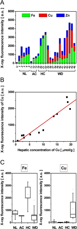
Wilson's disease (WD) is a rare autosomal recessive disease due to mutations of the gene encoding the copper-transporter ATP7B. The diagnosis is hampered by the variability of symptoms induced by copper accumulation, the inconstancy of the pathognomonic signs and the absence of a reliable diagnostic test. We investigated the diagnostic potential of X-ray fluorescence (XRF) that allows quantitative analysis of multiple elements. Studies were performed on animal models using Wistar rats (n = 10) and Long Evans Cinnamon (LEC) rats (n = 11), and on human samples including normal livers (n = 10), alcohol cirrhosis (n = 8), haemochromatosis (n = 10), cholestasis (n = 6) and WD (n = 22). XRF experiments were first performed using synchrotron radiation to address the elemental composition at the cellular level. High-resolution mapping of tissue sections allowed measurement of the intensity and the distribution of copper, iron and zinc while preserving the morphology. Investigations were further conducted using a laboratory X-ray source for irradiating whole pieces of tissue. The sensitivity of XRF was highlighted by the discrimination of LEC rats from wild type even under a regimen using copper deficient food. XRF on whole formalin-fixed paraffin embedded needle biopsies allowed profiling of the elements in a few minutes. The intensity of copper related to iron and zinc significantly discriminated WD from other genetic or chronic liver diseases with 97.6% specificity and 100% sensitivity. This study established a definite diagnosis of Wilson's disease based on XRF. This rapid and versatile method can be easily implemented in a clinical setting.

Keywords: Wilson disease; X‐ray fluorescence spectroscopy; copper; diagnosis.


Figures

Figure 1
Figure 1
SR‐XRF analysis on tissue sections from normal liver and Wilson disease. (A) Tissue sections of 5 μm thickness were stained with HES. Portal tract and centrilobular vein are present on the section of normal liver (patient #3) (upper image). WD liver (patient #46) exhibits nodules surrounded by fibrosis (lower image). Synchrotron radiation XRF spectra were acquired on the area delimited by the black squares. Representative XRF spectra acquired on tissue sections from normal liver and WD showing peaks characteristic of the composite elements. (B) The distribution of Fe (green), Cu (red) and Zn (blue) in the normal liver tissue (left panel) and in the hepatic nodule of WD tissue (right panel). The three component images are overlaid at lower right. The scale bar represents 500 μm, where the image is 2 × 2 mm2 with 3 × 3 μm2 pixels.
Figure 2
Figure 2
Assessment of elemental content by SR‐XRF. (A) The integrated fluorescence intensity from tissue sections shows both the total fluorescence (bar height, y‐axis) and the relative contribution of each of the components (coloured bar section) within 24 human liver samples; normal liver (NL), alcohol‐induced cirrhosis (AC), haemochromatosis (HC) and Wilson disease (WD) are shown alongside and empty Ultralene (U) sample holder. (B) For each WD patient the fluorescence intensity of the Cu was plotted as a function of intrahepatic Cu concentration measured from the same surgical specimen by atomic emission spectrometry (r 2 = 0.95). (C) The X‐ray fluorescence intensities of Fe and Cu within the different liver pathologies: NL (n = 5), AC (n = 3), HC (n = 4) and WD (n = 12).
Figure 3
Figure 3
XRF analysis on whole pieces of unfixed or fixed tissues from animal models. (A) Representative XRF spectra obtained on whole pieces of unfixed tissues from wild type (WT) rat and LEC rat (model of WD), or obtained on tissue embedded in paraffin (FFPE). The time of spectrum acquisition was 30 min. (B) Relative Cu content expressed as ratio of Cu/(Cu + Fe + Zn) measured by XRF in the liver of WT and LEC rat which were fed with normal (13 ppm) or low (1 ppm) content of copper. LEC rats (n = 5) and WT rats (n = 5) received normal food (eg food with 13 mg/L; 13 ppm Cu content); LEC rats (n = 6) and WT rats (n = 5) received food with low Cu content (eg 1 mg/L; 1 ppm). A statistically significant difference was found between the LEC and the WT groups for both intake regimen (p < 0.05).
Figure 4
Figure 4
Diagnosis of WD by XRF on FFPE needle biopsy. (A) Comparison of XRF spectra measured from formalin‐fixed paraffin‐embedded (FFPE) needle biopsy from normal liver and from a patient with WD. The time of spectra acquisition was 30 min. (B) Cu content expressed as ratio of Cu/(Cu + Fe + Zn) measured by XRF within FFPE needle biopsy from normal liver (NL) (n = 5), alcohol‐induced cirrhosis (AC) (n = 5), cholestasis (CD) (n = 6), hemochromatosis (HC) (n = 6) and Wilson disease adult (age >16 years) (WD Adult) (n = 6) and paediatric Wilson disease patients (WD Paediatric) (age ≤16 years) (n = 6); ** p < 0.05. (C) Receiver operating characteristic (ROC) curve for XRF as a diagnostic test for WD on paraffin liver biopsies (red dashed line). The Cu fluorescence intensity from the FFPE needle biopsies from normal liver, alcohol‐induced cirrhosis, chronic cholestasis, haemochromatosis and Wilson disease were used to perform the ROC analysis. Area under the curve (AUC) with 95% confidence interval is AUC = 0.999. Gray line indicates the threshold of significance.
Figure 5
Figure 5
Comparison of copper detection by histochemical rhodanine staining and by XRF on two patients with Wilson disease. For each patient tissue sections of 5 μm thickness were stained with rhodanine (left) (scale bar: 1 mm) and XRF spectra were acquired (right) in 30 min. (A) Patient #55 represents positivity in Cu detection by histochemical rhodanine staining as well as by XRF. (B) Patient #53 shows false negativity for Cu deposits by rhodanine staining whereas XRF allowed the detection of high Cu intensity in the tissue.

References

    1. Tao TY, Gitlin JD. Hepatic copper metabolism: insights from genetic disease. Hepatology 2003; 37: 1241–1247. - PubMed
    1. Ala A, Walker AP, Ashkan K, et al Wilson's disease. Lancet 2007; 369: 397–408. - PubMed
    1. Coffey AJ, Durkie M, Hague S, et al A genetic study of Wilson's disease in the United Kingdom. Brain 2013; 136: 1476–1487. - PMC - PubMed
    1. Dziezyc K, Litwin T, Chabik G, et al Families with Wilson's disease in subsequent generations: clinical and genetic analysis. Mov Disord 2014; 29: 1828–1832. - PubMed
    1. Schilsky M, Tavill AS. Wilson disease In Disease of the Liver 2003 (9th edn). Lippicott Williams & Wilkins: Philadelphia, 2003; 1169–1186.

LinkOut - more resources