Subarachnoid hemorrhage caused by an undifferentiated sarcoma of the sellar region
- PMID: 27500006
- PMCID: PMC4960927
- DOI: 10.4103/2152-7806.185775
Subarachnoid hemorrhage caused by an undifferentiated sarcoma of the sellar region
Abstract
Background: It is rare for patients with pituitary apoplexy to exhibit concomitant subarachnoid hemorrhage (SAH). Only a handful of patients with pituitary apoplexy have developed such hemorrhagic complications, and histopathological examination revealed pituitary adenoma as the cause of SAH.
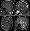
Case report: A previously healthy 35-year-old woman was brought to our institution after complaining of severe headache and left monocular blindness. Brain computed tomography showed a diffuse SAH with a central low density. Subsequently, the brain magnetic resonance imaging revealed an intrasellar mass with heterogeneous contrast enhancement. The patient was presumptively diagnosed with SAH secondary to hemorrhagic pituitary adenoma and underwent transcranial surgery to remove both the tumor and subarachnoid clot. A histological evaluation of the surgical specimen revealed malignant cells with strong predilection for vascular invasion. Following immunohistochemical evaluation, the tumor was negative for the majority of tumor markers and was positive only for vimentin and p53; thus, a diagnosis of undifferentiated sarcoma was established.
Conclusions: This case was informative in the respect that tumors other than pituitary adenoma should be included in the differential diagnosis of patients with pituitary apoplexy.
Keywords: Pituitary apoplexy; sella turcica; subarachnoid hemorrhage; undifferentiated sarcoma.
Figures


Similar articles
-
[A case of pituitary adenoma progressing to pituitary apoplexy on the occasion of cerebral angiography].No Shinkei Geka. 1996 May;24(5):475-9. No Shinkei Geka. 1996. PMID: 8692376 Review. Japanese.
-
Ruptured aneurysm-induced pituitary apoplexy: illustrative case.J Neurosurg Case Lessons. 2021 Jun 28;1(26):CASE21169. doi: 10.3171/CASE21169. eCollection 2021 Jun 28. J Neurosurg Case Lessons. 2021. PMID: 35854902 Free PMC article.
-
Post-Traumatic Pituitary Tumor Apoplexy After Closed Head Injury: Case Report and Review of the Literature.World Neurosurg. 2018 Dec;120:331-335. doi: 10.1016/j.wneu.2018.08.238. Epub 2018 Sep 10. World Neurosurg. 2018. PMID: 30213676
-
Pituitary adenoma apoplexy caused by rupture of an anterior communicating artery aneurysm: case report and literature review.World J Surg Oncol. 2015 Jul 30;13:228. doi: 10.1186/s12957-015-0653-z. World J Surg Oncol. 2015. PMID: 26220796 Free PMC article. Review.
-
Subarachnoid hemorrhage with normal cerebral angiography: a prospective study on sellar abnormalities and pituitary function.Neurosurgery. 1986 Dec;19(6):1012-5. doi: 10.1227/00006123-198612000-00018. Neurosurgery. 1986. PMID: 3808231
Cited by
-
Sarcomas of the sellar region: a systematic review.Pituitary. 2021 Feb;24(1):117-129. doi: 10.1007/s11102-020-01073-9. Pituitary. 2021. PMID: 32785833
References
-
- Alpert TE, Hahn SS, Chung CT, Bogart JA, Hodge CJ, Montgomery C. Successful treatment of spindle cell sarcoma of the sella turcica.Case report. J Neurosurg. 2002;97(5 Suppl):438–40. - PubMed
-
- Arita K, Sugiyama K, Tominaga A, Yamasaki F. Intrasellar rhabdomyosarcoma: Case report. Neurosurgery. 2001;48:677–80. - PubMed
-
- Capatina C, Inder W, Karavitaki N, Wass JA. Management of endocrine disease: Pituitary tumour apoplexy. Eur J Endocrinol. 2015;172:R179–90. - PubMed
-
- Glezer A, Bronstein MD. Pituitary apoplexy: Pathophysiology, diagnosis and management. Arch Endocrinol Metab. 2015;59:259–64. - PubMed
-
- Inamasu J, Hori S, Sekine K, Aikawa N. Pituitary apoplexy without ocular/visual symptoms. Am J Emerg Med. 2001;19:88–90. - PubMed
Publication types
LinkOut - more resources
Full Text Sources
Other Literature Sources
Research Materials
Miscellaneous

