Review
. 2016 Aug;6(4):396-408.
doi: 10.21037/cdt.2016.06.01.

Pathology of coronary atherosclerosis and thrombosis

Affiliations
Review

Pathology of coronary atherosclerosis and thrombosis

Fumiyuki Otsuka et al. Cardiovasc Diagn Ther. 2016 Aug.

Abstract

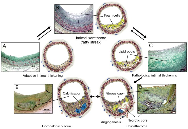
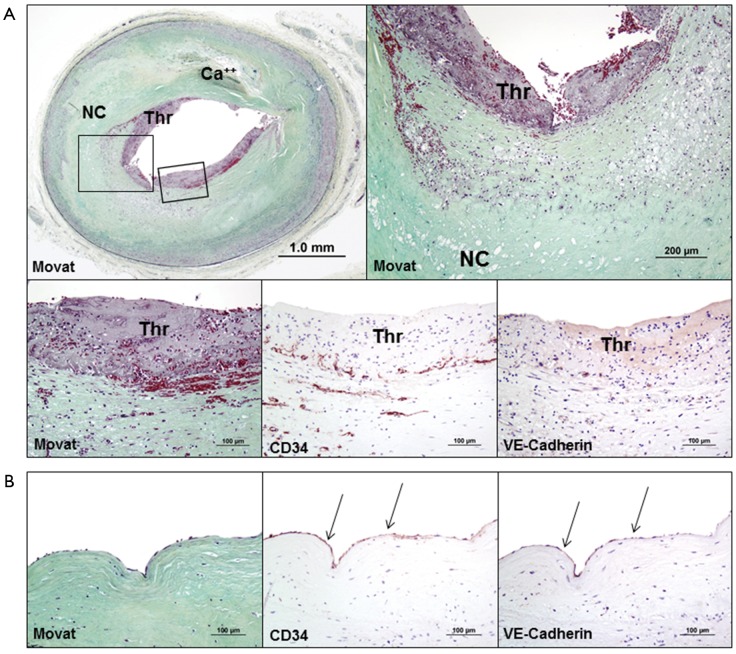
The process of early atherosclerotic plaque progression is characterized by the development of pathologic intimal thickening (PIT) with lipid pool that may transform into the necrotic core to form fibroatheroma, where infiltration of foamy macrophages plays a crucial role. The expansion of the necrotic core is also attributable to intraplaque hemorrhage. Thin-cap fibroatheroma (TCFA) is characterized by a relatively large necrotic core with an overlying thin fibrous cap measuring <65 µm typically containing numerous macrophages, and is considered to be the precursor lesion of plaque rupture which is the most common cause of coronary thrombosis. The second common cause of acute thrombosis is plaque erosion, while calcified nodules is known to be the least frequent cause of coronary thrombosis. Coronary thrombosis can occur without symptoms to form healed lesions, which contributes to an increase in plaque burden and luminal narrowing. The process of plaque progression is generally accompanied by the progression of calcification. An understanding of the histomorphological characteristics of coronary plaques should provide important insights into the pathogenesis, diagnosis, and treatment of atherosclerotic coronary disease for both basic and clinical researchers as well as for clinicians.

Keywords: Atherosclerosis; coronary disease; pathology; thrombosis.


Conflict of interest statement

Conflicts of Interest: The authors have no conflict of interests to declare.

Figures

Figure 1
Figure 1
Lesion types of atherosclerosis and a proposed sequence of their development. (A) Adaptive intimal thickening (AIT) characterized by smooth muscle cell accumulation within the intima; (B) intimal xanthoma corresponding to the accumulation of foamy macrophages within the intima. Pathological intimal thickening in (C) denotes the accumulation of extracellular lipid pools in the absence of apparent necrosis; (D) fibroatheroma indicating the presence of a necrotic core. The necrotic core and surrounding tissue may eventually be calcified, which forms fibrocalcific plaque shown in (E). Because some of the advanced lesion types (fibroatheromas and fibrocalcific plaques) evolve simultaneously in life, their interrelationships are difficult to resolve in autopsy studies. Movat pentachrome stain. Reproduced with permission from Bentzon JF, et al. Circ Res 2014;114:1852-66 (3).
Figure 2
Figure 2
Representative histologic sections showing pathologic intimal thickening (PIT) without macrophage (mac) infiltration, PIT with macrophages, early fibroatheroma (EFA), and late fibroatheroma (LFA) in human coronary plaques. Movat pentachrome stains show lipid pool (LP) with or without macrophage infiltration in PIT and necrotic core (NC) formation in EFA and LFA. Plaque progression from PIT to LFA is accompanied by an increase in macrophage infiltration which is confirmed by immunohistochemical stains for CD68. Immunohistochemical identification of extracellular matrix molecules hyaluronan and proteoglycan versican show intense staining for hyaluronan in LPs of PIT, whereas early NC shows partial loss of staining and late NC exhibits almost complete loss of hyaluronan. Gradual decrease in versican was also noted from PIT without macrophages to LFA where the staining was almost absent in late NC. Reproduced with permission from Otsuka F, et al. Atherosclerosis 2015;241:772-82 (8).
Figure 3
Figure 3
Representative histologic sections showing thin-cap fibroatheroma (A) and plaque rupture (B) in human coronary plaques. (A) Thin-cap fibroatheroma (TCFA) with extensive intraplaque hemorrhage within the necrotic core (NC). High power histologic images (Movat pentachrome stains) highlight thin fibrous cap heavily infiltrated by macrophages which are confirmed by immunohistochemistry for CD68 (arrows); (B) plaque rupture with acute luminal thrombus (Thr) and underlying large necrotic core. Arrows in a low power image [hematoxylin and eosin (H&E) stain] indicate the site of fibrous cap disruption. A high power image (Movat pentachrome stain) highlights disrupted fibrous cap with microcalcification and overlying luminal thrombus. Reproduced with permission from Otsuka F, et al. Nat Rev Cardiol 2014;11:379-89 (15), Otsuka F, et al. Atherosclerosis 2013;229:34-7 (16), and Otsuka F, et al. Arterioscler Thromb Vasc Biol 2014;34:724-36 (17).
Figure 4
Figure 4
Representative histologic sections showing progression of coronary calcification in human coronary plaques, obtained from non-decalcified arterial segments (A,B) and decalcified segments (C-E). (A) Pathological intimal thickening (PIT) characterized by lipid pool (LP) showing early microcalcification (≥0.5 µm, typically <15 µm in diameter) likely resulting from smooth muscle cell apoptosis, and calcification is detected by von Kossa staining within the LP (corresponding with a boxed area in the Movat image); (B) early necrotic core (NC) not only lacks SMCs but also is infiltrated by macrophages which eventually undergo apoptosis and calcification, which is observed as relatively larger punctate (≥15 µm) areas of calcification by von Kossa staining; (C) The degree of calcification in NC typically increases toward the medial wall where fragmented calcifications can be seen; (D) calcification generally progress into the surrounding area of the NC, which leads to the development of sheets of calcification; (E) nodular calcification may occur within the plaque in the absence of luminal thrombus and is characterized by breaks in calcified plates with fragments of calcium separated by fibrin. Reproduced with permission from Otsuka F, et al. Arterioscler Thromb Vasc Biol 2014;34:724-36 (17).
Figure 5
Figure 5
Representative histologic sections showing plaque erosion at the culprit site with acute thrombus (A) and at the adjacent lesion without thrombus (B) in human coronary plaques. (A) A low power histologic image (Movat pentachrome stain) shows plaque erosion with non-occlusive platelet rich thrombi (Thr) and underlying fibroatheroma with early necrotic core (NC) and focal mild calcification (Ca++). High power images highlight luminal thrombus and underlying early NC with foamy macrophages, along with luminal surface lacking the endothelium, which is in contact with acute thrombus. Lack of endothelium at luminal surface is confirmed by immunostaining for two endothelial markers: CD34 and VE-Cadherin; (B) a high power image of an adjacent section shows normal endothelialized surface, which is confirmed by immunostaining for CD34 and VE-Cadherin (arrows). Reproduced with permission from Otsuka F, et al. Nat Rev Cardiol 2014;11:379-89 (15).
Figure 6
Figure 6
Representative histologic sections showing healed plaque rupture (A) and chronic total occlusion (B) in human coronary plaques. (A) A low power histologic image (Movat pentachrome stain) shows healed plaque rupture and underlying necrotic core (NC) with extensive hemorrhage. A high power image (Movat pentachrome stain) shows numerous smooth muscle cells within the newly formed proteoglycan-rich neointima (black double arrows close to the luminal surface), with clear demarcation from the underlying old collagen-rich fibrous cap. In a high-power image of a Sirius-red-stained section (taken with polarized light), dense (type I) collagen forms a fibrous cap seen as a reddish-yellow region, and is overlaid with newer (type III) collagen detected as a greenish area; (B) silent occlusive thrombosis results in the development of chronic total occlusion which typically consists of organized thrombus with numerous micro-vessels. Reproduced with permission from Otsuka F, et al. Nat Rev Cardiol 2014;11:379-89 (15) and Sakakura K, et al. Eur Heart J 2014;35:1683-93 (43).

References

    1. Mozaffarian D, Benjamin EJ, Go AS, et al. Heart disease and stroke statistics--2015 update: a report from the American Heart Association. Circulation 2015;131:e29-322. 10.1161/CIR.0000000000000152 - DOI - PubMed
    1. Davies MJ, Thomas A. Thrombosis and acute coronary-artery lesions in sudden cardiac ischemic death. N Engl J Med 1984;310:1137-40. 10.1056/NEJM198405033101801 - DOI - PubMed
    1. Bentzon JF, Otsuka F, Virmani R, et al. Mechanisms of plaque formation and rupture. Circ Res 2014;114:1852-66. 10.1161/CIRCRESAHA.114.302721 - DOI - PubMed
    1. Stary HC, Chandler AB, Glagov S, et al. A definition of initial, fatty streak, and intermediate lesions of atherosclerosis. A report from the committee on vascular lesions of the council on arteriosclerosis, american heart association. Circulation 1994;89:2462-78. 10.1161/01.CIR.89.5.2462 - DOI - PubMed
    1. Virmani R, Kolodgie FD, Burke AP, et al. Lessons from sudden coronary death: A comprehensive morphological classification scheme for atherosclerotic lesions. Arterioscler Thromb Vasc Biol 2000;20:1262-75. 10.1161/01.ATV.20.5.1262 - DOI - PubMed