. 2016 Aug 8;12(8):e1006215.
doi: 10.1371/journal.pgen.1006215. eCollection 2016 Aug.

Control of Oocyte Reawakening by Kit

Affiliations

Control of Oocyte Reawakening by Kit

Hatice Duygu Saatcioglu et al. PLoS Genet. .

Abstract

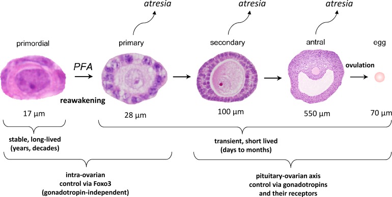
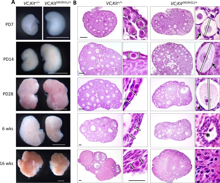
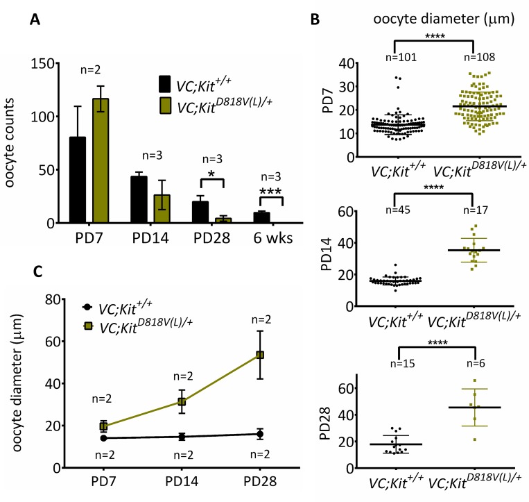
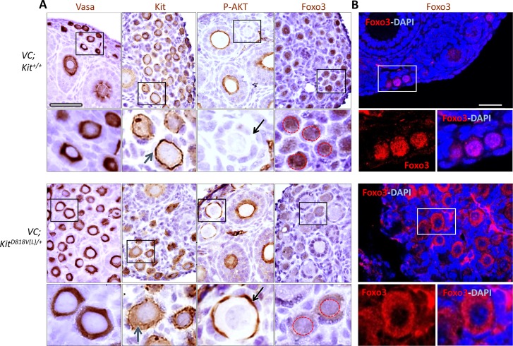
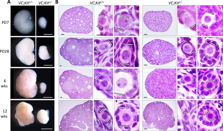
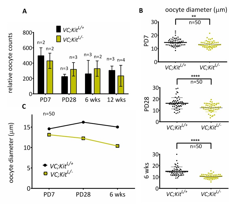
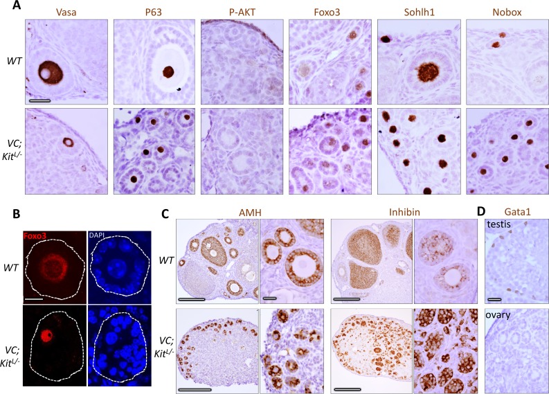
In mammals, females are born with finite numbers of oocytes stockpiled as primordial follicles. Oocytes are "reawakened" via an ovarian-intrinsic process that initiates their growth. The forkhead transcription factor Foxo3 controls reawakening downstream of PI3K-AKT signaling. However, the identity of the presumptive upstream cell surface receptor controlling the PI3K-AKT-Foxo3 axis has been questioned. Here we show that the receptor tyrosine kinase Kit controls reawakening. Oocyte-specific expression of a novel constitutively-active KitD818V allele resulted in female sterility and ovarian failure due to global oocyte reawakening. To confirm this result, we engineered a novel loss-of-function allele, KitL. Kit inactivation within oocytes also led to premature ovarian failure, albeit via a contrasting phenotype. Despite normal initial complements of primordial follicles, oocytes remained dormant with arrested oocyte maturation. Foxo3 protein localization in the nucleus versus cytoplasm explained both mutant phenotypes. These genetic studies provide formal genetic proof that Kit controls oocyte reawakening, focusing future investigations into the causes of primary ovarian insufficiency and ovarian aging.


Conflict of interest statement

The authors have declared that no competing interests exist.

Figures

Fig 1
Fig 1. Stages of follicular maturation.
Mouse follicles are depicted (not to scale: approximate follicle sizes including granulosa cells are shown below each follicle). Follicle growth is normally asynchronous, such that the adult ovary contains follicles at all stages of development. However, the vast majority of follicles at any time are primordial, with only a very small percentage of follicles actively growing. Follicle reawakening, sometimes called initiation or activation, is the regular, metered process by which individual primordial follicles are continually selected to begin the elaborate program of follicle maturation that culminates in ovulation. The earliest morphologic hallmark of reawakening is increased oocyte size followed by a change in granulosa cells from a flattened to a cuboidal shape, and then by granulosa cell proliferation. Whereas primordial follicles are long-lived, growing follicles are transient and short-lived, because growing follicles that do not progress to the next stage undergo atresia. Primary follicles are defined as follicles that have initiated oocyte growth and undergone a transition from flattened to cuboidal granulosa cells but have only one layer of granulosa cells. Secondary follicles have two layers of granulosa cells, and antral follicles are large follicles with a chamber (antrum) in preparation for ovulation.
Fig 2
Fig 2. Global oocyte reawakening phenotype following conditional Kit activation within oocytes.
(A) VC; KitD818V(L)/+ ovaries were larger than sibling controls up to PD28; scale bars = 1 mm. (B) Histological analyses (H&E-stained sections) revealed significant oocyte enlargement (white lines) and flattened granulosa cells (arrows) in many of the activated follicles (PD7-28). Numerous preantral and early antral follicles are present at PD28. By 6 weeks there was some oocyte death, follicle atresia and empty follicles (asterisk); by 16 weeks there was a complete absence of follicles with the ovary consisting of a luteinized stroma. These features indicate a classic global reawakening phenotype. Scale bars = 100 μm in large panels (same for control and experimental sections); 25 μm for small panels (all at the same magnification). Pictures are representative of at least three animals analyzed per genotype/time-point.
Fig 3
Fig 3. Quantitative analyses of oocyte phenotypes following conditional Kit activation within oocytes.
(A) Primordial/primary oocyte numbers of VC; KitD818V(L)/+ and control ovaries (n≥2 animals per genotype per timepoint; *p<0.05; ***p<0.001, unpaired student t test, error bars = S.E.M. (B) Oocyte diameters (micrometers) of early oocytes in VC; KitD818V(L)/+ and VC; +/+ mice; ****p<0.0001, n≥6 unpaired student t test. Error bars are S.E.M. (C) Average primordial/primary oocyte diameters in VC; KitD818V(L)/+ and VC; +/+ ovaries; at least 6 oocytes were measured from two different animals per genotype/time-point.
Fig 4
Fig 4. Kit activation promotes oocyte reawakening through AKT-Foxo3 axis.
(A) Immunohistochemistry for markers as shown; slides counterstained with hematoxylin. Lower panels are higher magnifications of areas shown in black rectangles. Blue arrows, Kit localization in experimental and control oocytes. Note membrane-bound staining in the control vs. greater cytoplasmic staining in VC; KitD818V(L)/+ oocytes. Black arrows, oocyte membrane of primordial/primary follicles in control vs. mutant; only mutant oocytes were positive for P-AKT. Red dashed circles in Foxo3 panels demarcate nuclei to highlight nuclear to cytoplasmic export in the mutant. Scale bar = 25 μm, all panels at same magnification. (B) Foxo3 localization by confocal microscopy/immunofluorescence of control and experimental ovaries at PD14. Scale bar = 33 μm for both large panels. Lower panels are higher magnifications of areas in white rectangles.
Fig 5
Fig 5. Primordial follicle arrest phenotype following Kit inactivation in oocytes.
(A) VC; KitL/- ovaries were minute relative to VC; KitL/+ sibling controls at all timepoints analyzed; scale bar = 1 mm. (B) Histological analyses (H&E-stained sections) revealed abundant primordial follicles with a complete failure of oocyte growth and absence of advanced follicles in VC; KitL/- ovaries. White lines demarcate oocyte diameters. Scale bars = 50 μm (large panels) or 10 μm (small panels). Pictures are representative of ≥4 ovaries from ≥2 different animals per timepoint.
Fig 6
Fig 6. Quantitative analyses of oocyte phenotypes following Kit inactivation in oocytes.
(A) Relative oocyte numbers at PD7 to 12 wks; n = 3 animals per genotype per timepoint except PD7 and 6 weeks experimental (n = 2 animals, total of 4 ovaries analyzed per genotype), error bars = S.E.M. Note that there is no significant oocyte loss up to 12 weeks (p = 0.4475 at 12 weeks, unpaired student t-test). (B) Oocyte diameters (micrometers) of early (primordial/primary) oocytes; **p<0.01, ****p<0.0001; unpaired student t test; n = 50 per genotype. (C) Average oocyte diameters, n = 50 oocytes per genotype per timepoint.
Fig 7
Fig 7. Marker studies of Kit-deficient oocytes are consistent with specific defect in oocyte reawakening via Foxo3.
(A) Immunohistochemistry for markers as shown at 6 weeks of age; slides counterstained with hematoxylin. Note absence of P-AKT (in contrast to D818V mutant) and constitutively nuclear localization of Foxo3. Scale bars = 25 μm; all panels at same magnification. (B) Immunofluorescence detection of Foxo3 protein; slides counterstained with DAPI. Foxo3 undergoes nuclear to cytoplasmic export in wild-type primary follicles but is constitutively nuclear in the mutant. Scale bar = 10 μm, same for all panels. (C) Analysis of granulosa cells in wild-type and VC; KitL/- ovaries. Immunohistochemistry for markers as shown at 6 wks of age; slides counterstained with hematoxylin. Scale bars = 200 μm in large panels, or 25 μm in small panels; all small panels at same magnification. (D) Absence of Gata1-positive cells in aberrant follicles in VC; KitL/- ovaries. Wild-type testis is shown as a positive control (Gata1 is expressed in Sertoli cells). Scale bar = 25 μm; both panels at same magnification.

Similar articles

Cited by

References

    1. Zhang H., et al., Life-long in vivo cell-lineage tracing shows that no oogenesis originates from putative germline stem cells in adult mice. Proc Natl Acad Sci U S A, 2014. 111(50): p. 17983–8. 10.1073/pnas.1421047111 - DOI - PMC - PubMed
    1. McGee E.A. and Hsueh A.J., Initial and cyclic recruitment of ovarian follicles. Endocr Rev, 2000. 21(2): p. 200–14. - PubMed
    1. Sullivan S.D. and Castrillon D.H., Insights into Primary Ovarian Insufficiency through Genetically Engineered Mouse Models. Semin Reprod Med, 2011. 29(4): p. 283–98. 10.1055/s-0031-1280914 - DOI - PubMed
    1. Lintern-Moore S. and Moore G.P., The initiation of follicle and oocyte growth in the mouse ovary. Biol Reprod, 1979. 20(4): p. 773–8. - PubMed
    1. Matzuk M.M., et al., Intercellular communication in the mammalian ovary: oocytes carry the conversation. Science, 2002. 296(5576): p. 2178–80. - PubMed

Publication types

MeSH terms

Substances