. 2016 Aug 8;10(8):e0004893.
doi: 10.1371/journal.pntd.0004893. eCollection 2016 Aug.

Cysteine and Aspartyl Proteases Contribute to Protein Digestion in the Gut of Freshwater Planaria

Affiliations

Cysteine and Aspartyl Proteases Contribute to Protein Digestion in the Gut of Freshwater Planaria

Louise S Goupil et al. PLoS Negl Trop Dis. .

Abstract

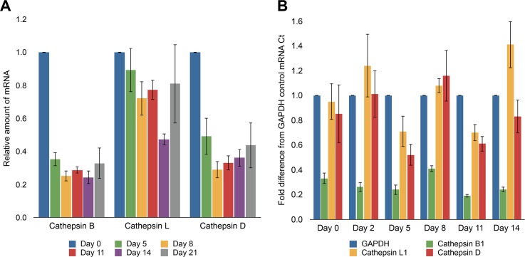
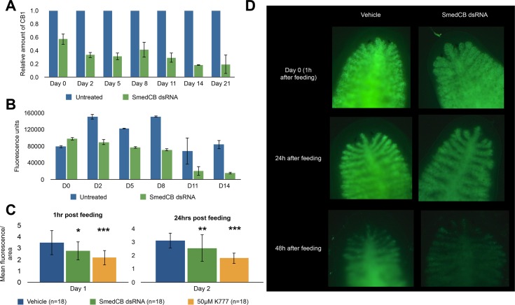
Proteases perform numerous vital functions in flatworms, many of which are likely to be conserved throughout the phylum Platyhelminthes. Within this phylum are several parasitic worms that are often poorly characterized due to their complex life-cycles and lack of responsiveness to genetic manipulation. The flatworm Schmidtea mediterranea, or planaria, is an ideal model organism to study the complex role of protein digestion due to its simple life cycle and amenability to techniques like RNA interference (RNAi). In this study, we were interested in deconvoluting the digestive protease system that exists in the planarian gut. To do this, we developed an alcohol-induced regurgitation technique to enrich for the gut enzymes in S. mediterranea. Using a panel of fluorescent substrates, we show that this treatment produces a sharp increase in proteolytic activity. These enzymes have broad yet diverse substrate specificity profiles. Proteomic analysis of the gut contents revealed the presence of cysteine and metallo-proteases. However, treatment with class-specific inhibitors showed that aspartyl and cysteine proteases are responsible for the majority of protein digestion. Specific RNAi knockdown of the cathepsin B-like cysteine protease (SmedCB) reduced protein degradation in vivo. Immunohistochemistry and whole-mount in situ hybridization (WISH) confirmed that the full-length and active forms of SmedCB are found in secretory cells surrounding the planaria intestinal lumen. Finally, we show that the knockdown of SmedCB reduces the speed of tissue regeneration. Defining the roles of proteases in planaria can provide insight to functions of conserved proteases in parasitic flatworms, potentially uncovering drug targets in parasites.


Conflict of interest statement

The authors have declared that no competing interests exist.

Figures

Fig 1
Fig 1. Incubation of worm regurgitant with fluorescent substrates and MSP-MS reveals highest activity at low pH and is dependent on aspartyl, cysteine, and metalloproteases.
(A) Replicates of S. mediterranea regurgitant were incubated with a library of fluorescent substrates at pH 3.5, 5.5, and 7.5. Each sample showed the highest rate of cleavage at pH 3.5. No detectable activity was found in samples without regurgitant. Fluorescence was measured in relative fluorescence units (RFU) per second. (B) The regurgitant was incubated with peptide substrates for MSP-MS and the total number of cleavage sites was detected after 15, 60, 240, and 1200 minutes of incubation with the peptide mixture at pH 3.5, 5.5, and 7.5. The largest number of cleavages occurred at pH 3.5. (C) An iceLogo generated from the pattern of cleavage events after 60 minutes reveals the specificity of protease activity at pH 3.5, 5.5, and 7.5. Amino acids that are most frequently observed at each position (P4-P4’) are shown above the axis, while less frequently observed amino acids are shown below. (D) The pooled samples of regurgitant were assayed with the same fluorescent library in the presence of several inhibitors, including pepstatin (aspartyl protease inhibitor), E-64 (cysteine protease inhibitor), 1,10-phenanthroline (metalloprotease inhibitor), and AEBSF (serine protease inhibitor). While the effect of inhibition of astpartyl, cysteine, and metalloproteases varied according to pH, inhibition of serine proteases had no effect on the overall amount of activity detected at any pH.
Fig 2
Fig 2. Inhibition of cysteine, aspartyl, but not metalloproteases, inhibits planaria digestion.
(A) Animals were treated with 50μM protease-specific chemical inhibitors or dsRNA prior to feeding with rhodamine-labeled bovine serum albumin (RhBSA) and imaging. Fluorescence, due to cleavage of quenched albumin, was imaged (B). These representative images show a decrease in fluorescence in worms treated with K11777. Inhibition of aspartyl proteases by pepstatin as seen in (C) also decreased digestion. Combined inhibition of cathepsin B, L, and D completely reduced fluorescence. In contrast, inhibition of metalloproteases with bestatin and EDTA (D) did not reduce fluorescence. These data are quantified in (E) for K11777 and pepstatin and show that the reduction in fluorescence is significant in the presence of these inhibitors. (F) Treatment with metalloprotease inhibitors bestatin and EDTA did not lead to a significant change in fluorescence.
Fig 3
Fig 3. S. mediterranea cathepsin protease mRNA is reduced after starvation while dsRNA treatment with SmedCB is specific to cathepsin B.
(A) RNA was extracted from three sets of 10 to 15 worms starved for three weeks. mRNA levels for three major cathepsin protease genes (B, L, and D) were quantified using RTPCR; levels were compared to recently fed worms (day 0) to measure the relative change in transcript levels over time. Starved worms demonstrated a 65–75% reduction in cathepsin B mRNA levels and 50–65% reduction in cathepsin D mRNA. Cathepsin L mRNA was reduced, on average, 20% with the exception of day 14, which had a 50% reduction. (B) Worms injected with ~100ng of SmedCB dsRNA for three consecutive days, followed by amputation (day 0) and three weeks of starvation, showed a marked reduction of SmedCB mRNA when analyzed via RTPCR using GAPDH mRNA levels as a baseline control. SmedCB levels decreased by ~80% after two weeks. Cathepsin L and D showed some fluctuation in mRNA levels, but remained within 50% of baseline levels and there was no continued trend observed over time. This indicates that fluctuations in levels are due to variations in worm populations chosen for RNA extraction rather than significant off-target effects of SmedCB dsRNA.
Fig 4
Fig 4. SmedCB RNAi treatment reduces cathepsin B mRNA levels and proteolytic activity, inhibiting digestion.
A) Worms injected with ~100ng of SmedCB dsRNA for three consecutive days, followed by amputation (day 0) and three weeks of starvation, showed a marked reduction of SmedCB mRNA when analyzed via RTPCR in comparison to untreated worms. The initial three days of injection prior to day 0 causes a 45% reduction that increases to over 80% after two weeks of starvation during regeneration. SmedCB dsRNA treated worms also showed a marked reduction of SmedCB protease activity via Z-RR-AMC cleavage (B). Treated worms exhibited between 50–75% less fluorescence from Z-RR-AMC cleavage than untreated worms over two weeks. C) Fluorescence from digestion of Rh-BSA is decreased in worms treated with SmedCB dsRNA. This decrease is less dramatic than treatment with the cathepsin B and cathepsin L inhibitor K11777, but is statistically significant. Representative images of this fluorescence can be observed in D.
Fig 5
Fig 5. Total fluorescence of Z-RR-AMC and Z-FR-AMC cleavage by planaria lysate is reduced by inhibition of cathepsin activity.
(A) Whole worm lysate was pre-incubated with 50μM E-64, a pan-cysteine protease inhibitor, before assaying with the fluorescent probe Z-RR-AMC. Pretreatment with E-64 reduced fluorescence by 93%. Inhibition of cathepsin B protease alone by CA-074 caused a 71% decrease in activity, indicating that the majority of proteolytic cleavage is caused by cathepsin B-like proteases. (B) Similar effects were observed when Z-FR-AMC, a probe cleaved by both cathepsin B and cathepsin L proteases, was used.
Fig 6
Fig 6. Immunofluorescence labeling and electron microscopy of zymogen and catalytic SmedCB shows strong labeling in cells lining the intestinal lumen.
A) Several antibodies were generated against Schmidtea cathepsin B, including one against the catalytic region only (underlined region), and one against the zymogen tag and catalytic region (a combination of the green and pink peptides). (B) Western blot with antibody used to localize SmedCB at 1:5,000. Lane 1, recombinant prep of SmedCB from E. coli. Band at 70kDa is a dimer of SmedCB, lower band at 35kDa is zymogen SmedCB. Lanes 2–5 contain planaria lysate at 15μL, 7.5 μL, 5μL, and 15μL, respectively. Higher band (~37kDa) is zymogen, lower band (~27kDa) is catalytic domain. Lanes 1–4 use the mouse-derived antibody underlined in A, while lane 5 was blotted against the rabbit-derived antibody from the pink and green peptides in A. (C) Labeling of paraffin-embedded S. mediterranea cross sections of worms starved for one week with (1:100) anti-SmedCB zymogen pro-peptide antibody showed strongest signal in the cells lining the intestinal lumen. Comparison with the negative control (D) anti-rabbit secondary (1:100) only shows that the labeling on the border of the planaria cross sections is due to drying artifact. This pattern is also seen in worms with the anti-SmedCB catalytic domain only antibody (1:100) (E). The negative control (F) with anti-mouse secondary only (1:100), shows labeling only on the edges of the cross sections due to drying artifact. Worms were also starved for two weeks prior to labeling (G) as opposed to one week in C and E. SmedCB labeling remained the same. (H) Cross sections of S. mediterranea worms (starved one week) were labeled with (1:100) anti-SmedCB zymogen pro-peptide antibody. Intestinal lumen seen as white space in the center of the image. (I) Immuno-gold labeling of antibody target, visualized as black dots, shows vesicle-like structures are heavily labeled. Other tissue, like the rounded, gray lipid droplets in (J) did not show labeling.
Fig 7
Fig 7. Whole-mount in situ hybridization of SmedCB shows labeling throughout the worm gut in intact and regenerating animals.
An in situ DIG-12-UTP riboprobe against SmedCB shows labeling throughout the branched intestine. This pattern is observed in worms fixed four days after feeding (A) and eight days after feeding (B). For regenerating worms, animals were fixed three, five, or seven days after amputation. Some regenerating animals have strong labeling near the blastema (newly regenerated tissue, circled in red) as well as the newly-forming gut. Amputated heads (C) have labeling in the blastema up to seven days after amputation, while pharynxes (D) only show labeling near the blastema for three days. Tails (E) exhibit an intermediate phenotype with blastema labeled five days after amputation. All animals were treated with 1:100 dilution of the same riboprobe.
Fig 8
Fig 8. SmedCB shows a transient spike of protease activity following digestion and amputation, while chemical inhibition of both cathepsin B and L leads to a significant decrease in regeneration.
(A) Whole worm lysate from S. mediterranea was labeled with the activity-based probe DCG-04 and resolved using SDS-PAGE. Western blot against the biotin tag of DCG-04 was used to analyze activity levels. S. mediterranea lysate showed an increase in cathepsin B labeling following digestion (day 4–6) before returning to base levels by day 14. B) When S. mediterranea worms were amputated following feeding, the increase in DCG-04 signal extended to day 8 before returning to baseline. The large upper band was quantified as SmedCB activity; five micrograms of total protein was used in each reaction with 10μM DCG-04. (C) Worms treated with SmedCB dsRNA did not show any significant defects in regeneration compared to untreated controls, but treatment with K11777 (D) showed a highly significant decrease in growth compared to vehicle worms. This effect was dose dependent. Relative changes in area are reported to account for any differences in initial size of worms selected. A negative value indicates that fragments shrank over time (heads), while positive values show relative growth. K11777 treated worms, on average, grew less (pharynxes, tails) and shrank more (heads), than untreated worms. These results were highly significant at 40 and 50μM treatment in all fragments, but not significant at lower concentrations.

Similar articles

Cited by

References

    1. Morgan T.H. Experimental studies of the regeneration of Planaria maculata. Arch. Entwm. Org. 1898; 7: 364–397.
    1. Reddien PW, Sánchez Alvarado A. Fundamentals of planarian regeneration. Annu. Rev. Cell. Dev. Biol. 2004; 20: 725–757. - PubMed
    1. Reddien P.W., Bermange A.L., Murfitt K.J., Jennings J.R., Sánchez Alvarado A. Identification of genes needed for regeneration, stem cell function, and tissue homeostasis by systematic gene perturbation in planaria. Cell. 2005; 8: 635–649. - PMC - PubMed
    1. Sánchez Alvarado A., Newmark P.A., Robb S. M., Juste R. The Schmidtea mediterranea database as a molecular resource for studying platyhelminthes, stem cells and regeneration. Development. 2002; 129: 5659–5665. - PubMed
    1. Collins J. J., Newmark P. A. It’s no fluke: the planarian as a model for understanding schistosomes. PLoS Pathog. 2013; 9:(7) e1003396 10.1371/journal.ppat.1003396 - DOI - PMC - PubMed

Publication types