. 2016 Aug 25;536(7617):464-8.
doi: 10.1038/nature19312. Epub 2016 Aug 8.

The prion protein is an agonistic ligand of the G protein-coupled receptor Adgrg6

The prion protein is an agonistic ligand of the G protein-coupled receptor Adgrg6

Alexander Küffer et al. Nature. .

Abstract

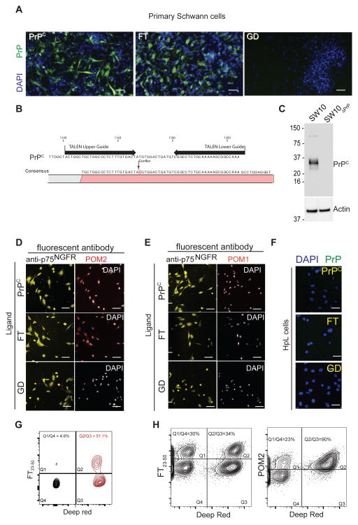
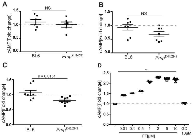
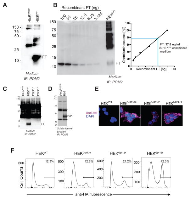
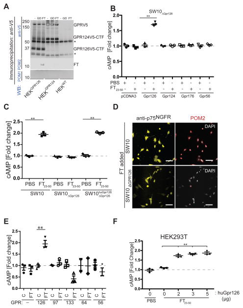
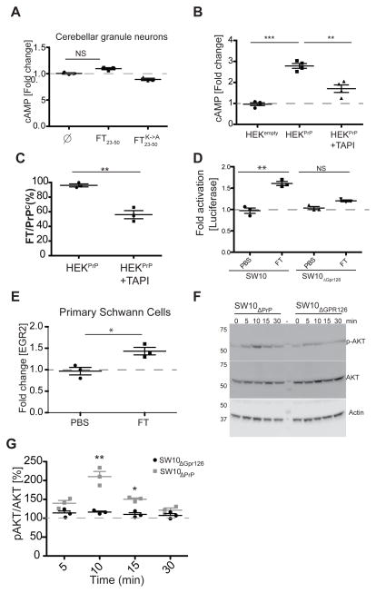
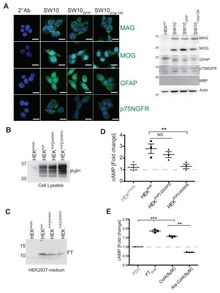
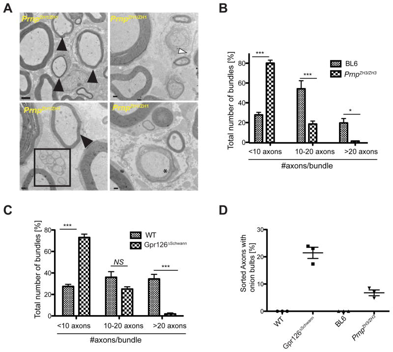
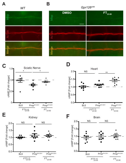
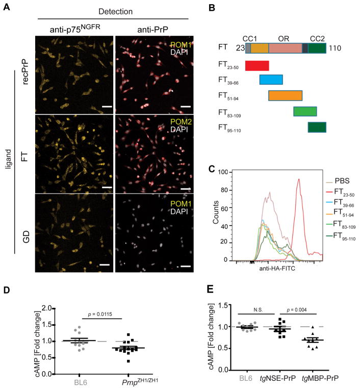
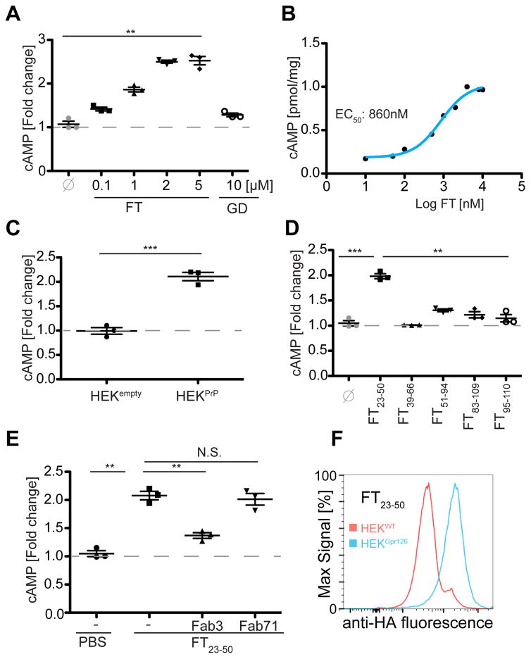
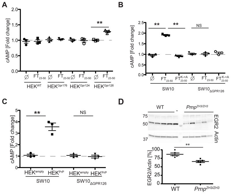
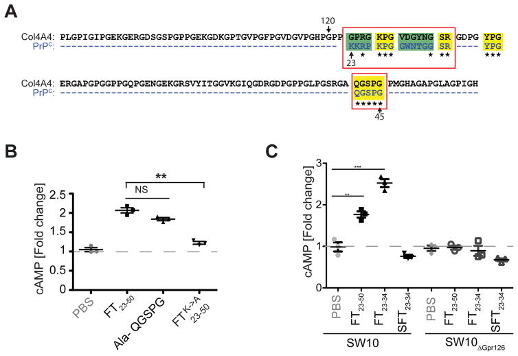
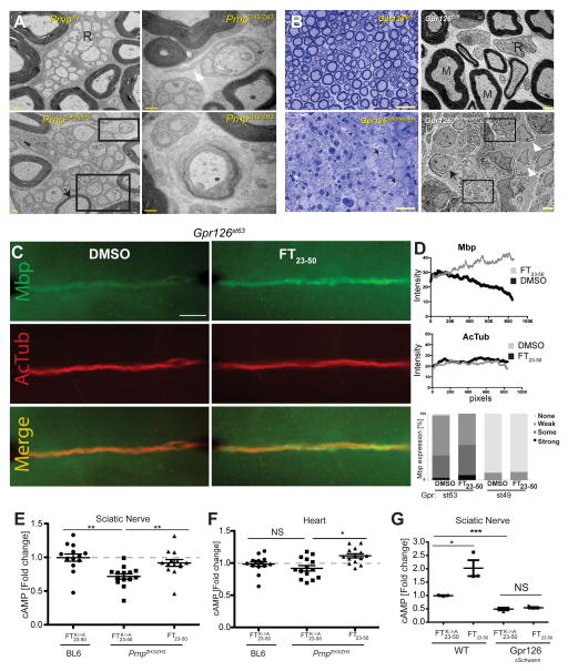
Ablation of the cellular prion protein PrP(C) leads to a chronic demyelinating polyneuropathy affecting Schwann cells. Neuron-restricted expression of PrP(C) prevents the disease, suggesting that PrP(C) acts in trans through an unidentified Schwann cell receptor. Here we show that the cAMP concentration in sciatic nerves from PrP(C)-deficient mice is reduced, suggesting that PrP(C) acts via a G protein-coupled receptor (GPCR). The amino-terminal flexible tail (residues 23-120) of PrP(C) triggered a concentration-dependent increase in cAMP in primary Schwann cells, in the Schwann cell line SW10, and in HEK293T cells overexpressing the GPCR Adgrg6 (also known as Gpr126). By contrast, naive HEK293T cells and HEK293T cells expressing several other GPCRs did not react to the flexible tail, and ablation of Gpr126 from SW10 cells abolished the flexible tail-induced cAMP response. The flexible tail contains a polycationic cluster (KKRPKPG) similar to the GPRGKPG motif of the Gpr126 agonist type-IV collagen. A KKRPKPG-containing PrPC-derived peptide (FT(23-50)) sufficed to induce a Gpr126-dependent cAMP response in cells and mice, and improved myelination in hypomorphic gpr126 mutant zebrafish (Danio rerio). Substitution of the cationic residues with alanines abolished the biological activity of both FT(23-50) and the equivalent type-IV collagen peptide. We conclude that PrP(C) promotes myelin homeostasis through flexible tail-mediated Gpr126 agonism. As well as clarifying the physiological role of PrP(C), these observations are relevant to the pathogenesis of demyelinating polyneuropathies--common debilitating diseases for which there are limited therapeutic options.


Conflict of interest statement

Competing Financial Interests: None

Figures

Extended Data Figure 1
A: Primary Schwann cells were isolated from the sciatic nerves of PrnpZH1/ZH1 mice and grown on coverslips. Cells were exposed for 20 min to recombinant PrPC, FT, or GD (2μM), fixed, and stained with POM2 (FT, PrPC) or POM1 (GD). Antibodies were visualized in the green channel and nuclei were stained with DAPI (blue). PrPC and FT, but not GD, adhered to the cells. Scale bars: 25μm B: Schematic representation of the target region for transcription activator-like endonucleases (TALEN) in the Prnp gene. Target guides are indicated by arrows. Gene editing resulted in a deletion leading to a frame shift in the PrPC coding sequence (designated as conflict in the figure) and a premature stop codon identified by sequencing. C: Wild-type SW10 cells and a subclone isolated after treatment with TALEN (termed SW10ΔPrP) were probed by Western blotting using anti-PrP antibody (POM1). SW10ΔPrP showed complete abrogation of PrPC expression and was used for further experiments. Levels of actin on the same membrane were monitored to confirm equal loading of cell lysates onto the gel. For uncropped gels see Supplementary Information File 1. D–E: SW10ΔPrP cells were treated with full-length recombinant (PrPC, residues 23-231), flexible tail (FT, 23-110), or globular domain (GD, 121-231). PrP epitopes were detected with POM2 (D) or POM1 (E) (red). Grey: DAPI. As expected, FT was detected only by POM2. Cells were also labeled with antibodies to the p75 nerve-growth factor receptor (yellow), a Schwann cell marker. PrPC and FT, but not GD, adhered to Schwann cells. Scale bar: 26 μm. F: the PrPC-deficient cell line HpL was treated with recombinant PrPC, FT, and GD as in panel A. None of the recombinant proteins adhered to HpL cells. Scale bars: 20 μm. G: SW10 cells were trypsinized, washed, and mixed with non-trypsinized SW10 cells labeled with Deep Red cell tracker. Cells were incubated with HA-tagged peptide FT23-50, and binding was visualized by flow cytometry. The Deep red signal (abscissa) was used to differentiate trypsinized from non-trypsinized cells. 51% of untreated cells, but only 5% of trypsinized cells, became decorated by FT23-50-HA, indicating that FT23-50 reacted with trypsin-sensitive surface molecules. H: SW10 cells were digested (30 min) with phosphatidylinositol phospholipase C (PI-PLC, 0.5 units), washed, and incubated with FT23-50-HA along with undigested Deep-Red labeled cells (left panel). The proportion of binders in the digested (34%) and the undigested samples (30%) was similar, indicating that the FT23-50 receptor was neither PrPC itself nor any other GPI-linked protein. To monitor the efficiency of PI-PLC treatment, we assessed POM2 binding to PrPC on both treated and untreated cells (right panel). POM2 binding was significantly decreased in PI-PLC treated cells (23%) compared to untreated cells (90%). Panels depict biologically independent triplicates; unpaired Student’s t-test was used for statistical analysis.
Extended Data Figure 2
A–B: cAMP was measured in sciatic nerves isolated from 4-day old (A) or 4-week old (B) BL6 and PrnpZH1/ZH1 mice. No difference was observed in cAMP levels in 4-day old mice, whereas 4-week old PrnpZH1/ZH1 mice displayed a trend towards decreased cAMP levels. C: Sciatic nerve from 10-week old PrnpZH3/ZH3 mice showed a significant decrease in cAMP (p= 0.0151). D: PrPC expression by neurons (tgNSE-PrP), but not by Schwann cells (tgMBP-PrP), restored cAMP levels in sciatic nerves of 10-16 week-old mice. Each dot represents one individual mouse. E: SW10 cells were seeded in 6 well plates and treated (20’) with recombinant FT or GD (10 μM). Addition of FT, but not of GD, resulted in a concentration-dependent intracellular cAMP increase. F: Primary PrnpZH1/ZH1 Schwann cells were treated with FT (20’), and cAMP concentrations were determined by immunoassay. A dose-response curve was interpolated. Data are representative of three biologically independent experiments and statistical significance was evaluated by unpaired Student’s t-Tests.
Extended Data Figure 3
A: HEK293T cells were transfected with an empty vector (HEKempty) or a plasmid expressing murine PrPC (HEKPrP). Cell medium was collected 48h after transfection and subjected to immunoprecipitation with monoclonal antibody POM2 (against PrPC), followed by western blotting using biotinylated POM2 and streptavidin-HRP. FT was observed only in the medium from HEKPrP cells. B: FT released into the conditioned medium of HEKPrP was immunoprecipitated using POM2 and visualized by Western blotting with biotinylated POM2. Dilutions of recombinant FT (3.125 – 100 ng) were used for calibration, and the concentration of FT released into the medium was estimated at 37 ng/ml. C: Conditioned medium from primary BL6 Schwann cells cultures (PSCBL6) was subjected to immunoprecipitation with antibody POM2 (against PrPC) followed by Western blotting with POM2. For control, we used conditioned medium from HEK cells transfected with a non-coding plasmid (HEKempty) or a with a plasmid encoding murine PrPC (HEKPrP). FT was only detected in conditioned medium from HEKPrP cells (lane 2) but not in conditioned medium from two independent PSCBL6 cultures (lanes 3–4). Asterisks denote immunoglobulins detected by the secondary antibody. D: Sciatic nerves lysates obtained from 10 week old PrnpZH1/ZH1 and BL6 mice and subjected to immunoprecipitation with POM2 antibody followed by Western blotting with POM2. Full length PrPC, but no FT, was detectable in the immunoprecipitates from BL6 mice. E: Wild-type HEK293T cells (HEKWT) or HEK293T cells overexpressing various GPCRs bearing V5 epitope tags (HEKGpr126, HEKGpr124, and HEKGpr176) were grown on coverslips and stained with anti V5 antibody (detecting tagged GPCRs; magenta). Nuclei were stained with DAPI (blue). Staining revealed cell surface expression of all transfected GPCRs. Scale bar: 8μm. F: HA-tagged FT23-50 peptide (2μM) was added to wild-type HEK293T (HEKWT) cells or to HEKGPR126 cells, labeled with anti-HA antibody, and subjected to cytofluorimetry. Overexpression of Gpr126 increased the binding of FT23-50. G: Binding of HA-tagged FT23-50 to HEKGPR126 cells (right panel, monitored by cytofluorimetry) was conspicuously increased over that of wild-type, Gpr176, and Gpr124-overexpressing HEK293T cells. Data are representative of three biologically independent experiments; statistical significance was evaluated by unpaired Student’s t-test.
Extended Data Figure 4
A: HEK293T, HEKGPR124 and HEKGPR126 cells were exposed (20 min) to recombinant FT, GD (2μM), or PBS, and subjected to immunoprecipitation using the anti-V5 antibody, followed by Western blotting using POM2, anti-V5 or POM1. Anti-V5 detected both full-length Gpr126, Gpr124 (denoted as GprV5 for both proteins) and the respective C-terminal fragments (Gpr126V5-CTF, Gpr124V5-CTF). POM2 revealed a band corresponding to the FT (lane 3) that co-precipitated with GPR126. POM1 indicated that GD did not bind. Lanes 1, 2 and 3: HEKGPR126 cells treated with PBS, GD and FT. Lanes 4, 5 and 6: HEKGPR124 cells treated with PBS, GD and FT. Lanes 7, 8 and 9: HEK293T cells treated with PBS, GD and FT Asterisks: immunoglobulin heavy and light chains. For uncropped gels see Supplementary Information File 1. B: SW10ΔGpr126 cells plated at a density of 100’000 cells/well in 6-well plates were transfected with control plasmid (pCDNA3) or plasmids encoding various GPCRs (Gpr126, 124, 176, and 56) bearing C-terminal V5 tags. Only cells transfected with pCGpr126-V5 showed a cAMP response to FT23-50 48h post transfection. PBS treatment was used for control. C: Intracellular cAMP responses to FT treatment (2 μM, 20’) in SW10 and SW10ΔGpr126 cells, as well as SW10ΔGpr126huGpr126 cells expressing V5-tagged human Gpr126 (pCGpr126-V5). A significant increase in cAMP was observed in SW10 cells, whereas SW10ΔGpr126 showed no change. In contrast, SW10ΔGpr126huGpr126 cells showed a significant cAMP increase, indicative of successful complementation. D: SW10 and SW10ΔGpr126 cells were incubated (20 min) with conditioned medium from HEKempty or HEKPrP cells.. HEKPrP-conditioned medium induced a robust cAMP spike in SW10, but not SW10ΔGpr126 cells. E: SW10 and SW10ΔGpr126 cells were grown on coverslips for 24h and exposed to recombinant FT (2 μM, 20 min). Cells were stained with POM2 (detecting FT; red, DAPI-stained nuclei: grey) and antibodies to p75NGFR (yellow). Deletion of Gpr126 largely suppressed FT binding. Scale bar: 26 μm. F: HEK293(H) cell lines were transfected with plasmids expressing different adhesion GPCRs (Gpr: 97, 133, 64, 56), followed by selection of cells expressing the receptor in presence of geneticin. GPCR expressing cells and HEKGpr126 cells were then treated with either FT23-50 or FT23-50KA for control (FT and C, respectively). Only cells expressing Gpr126 responded to FT23-50 with a cAMP spike. Interestingly, cells expressing Gpr133 reacted with a decrease in cAMP levels. Data are representative of three biologically independent experiments; statistical significance was evaluated by unpaired Student’s t-test.
Extended Data Figure 5
A: HEK293T cells were transfected with increasing amounts of human Gpr126 plasmid (2–5μg/well of a 6 well plate). 48h post transfection cells were treated with FT23-50 or PBS as a control. Increasing amount of Gpr126 cDNA did not result in amplification of the cAMP signal. B: Primary PrnpZH1/ZH1 cerebellar granule neuron cultures were seeded in 6-well plates at a density of 5x10⁵ cells and treated with FT23-50, FT23-50KA, or PBS. No alterations in the levels of cAMP were noticed. C: SW10 cells were exposed to conditioned medium from HEK cells that had been transfected with empty vector (HEKempty) or a PrPC expression vector (HEKPrP). HEKPrP were optionally treated with 100 μM of the TAPI-2 protease inhibitor for 24h before harvesting the medium. TAPI-2 treatment resulted in reduced cAMP induction, suggesting that impaired proteolytic cleavage of the FT from PrPC resulted in decreased signaling. D: Quantification of FT released into the medium relative to the total amount of PrPC in lysates by Western blotting. The spent medium of HEKPrP cells treated with TAPI-2 contained less FT. E: SW10 and SW10ΔGpr126 cells were transfected with an Egr2-controlled firefly luciferase reporter and treated with recombinant FT (2μM) or PBS (24 hrs). Ordinate: luciferase expression normalized to a renilla luciferase control (n=3; *: p<0.05; t-test). Luciferase activity was observed only in SW10 cells stimulated with FT but not in SW10ΔGpr126 cells. F: Primary Schwann cells were exposed to recombinant FT (2 μM, 1h) or PBS. Egr2 mRNA expression was measured by quantitative RT-PCR and normalized against a panel of housekeeping genes. For uncropped gels see Supplementary Information File 1. G: SW10ΔPrP and SW10ΔGpr126 cells were grown in 6-well plates, exposed to recombinant FT (≤30 min), and analyzed by Western blotting (left). Densitometry (right) showed increased phospho-AKT/AKT ratio in SW10ΔPrP cells, but not in SW10ΔGpr126 cells. Data are representative of three biologically independent experiments; statistical significance was evaluated by unpaired Student’s t-test.
Extended Data Figure 6
A: SW10, SW10ΔPrP and SW10ΔGpr126 cells were grown on coverslips and stained with antibodies against Myelin associated glycoprotein (MAG), Myelin oligodendrocyte glycoprotein (MOG), glial fibrillary acidic protein (GFAP) and p75 nerve growth factor receptor (p75NGFR) (left panel, all green; DAPI-stained nuclei: blue). Cells labeled with secondary antibody alone (2° Ab) were used as control to determine unspecific staining. Scale bars: 10μm. Expression in all cell lines was confirmed by western blotting (right panel). Lysate from HEK293T wild-type cells (HEKWT) was used as control. All proteins except Myelin basic protein (MBP) were expressed in SW10 cells and its derivatives. For uncropped gels see Supplementary Information File 2. B: Western blot (developed with POM2) of HEK 293T cells transfected with expression plasmids for wild-type murine PrPC or for PrPC bearing lysine-to-alanine substitutions in the KKRPK and QGSPG motifs (lanes 3 and 4, respectively). The mutations did not affect the biogenesis and processing of PrPC C: Western blot of the medium collected from the cells shown in panel B. FT fragments bearing the mutations were released into the medium similarly to wild-type FT. For uncropped gels see Supplementary Information File 1. D: SW10ΔPrP cells were treated with conditioned medium from HEK293T cells transfected with an empty vector (HEKempty), with PrPC (HEKPrP), or with full-length PrPC versions in which the QGSPG (HEKPrPΣQGSPG) or KKRPK (HEKPrPΣKKRPK) motifs were substituted (Σ) with alanines. The charge neutralization within the KKRPK motif abrogated the cAMP induction. E: SW10ΔPrP cells were treated with FT23-50 (2 μM) or a Col4-derived 21-meric synthetic peptide containing either the GPRGKPG domain or its alanine-substituted variant (AAAGAAG; both 8 μM). Both FT23-50 and the native Col4 peptide, but not the alanine-substituted peptide (Ala-Col4), induced cAMP. Data are representative of three biologically independent experiments; statistical significance was evaluated by unpaired Student’s t-test.
Extended Data Figure 7
A: Transmission electron micrographs of 14 month-old PrnpZH1/ZH1 sciatic nerves (ZH1). Black arrowhead: thinly myelinated axons; white arrowhead: abnormal cytoplasmic Schwann cell protrusions; boxes: loss of axon-Schwann cell interactions; asterisk: initial onion bulb formation. Scale bar: 2 μm in upper left panel; 500 nm in all other panels. B–C: Quantification of unmyelinated axons in Remak bundles was performed manually by counting number of axons in the bundles from electron microscopy images (1500x magnification, 10 images per mouse were analyzed and three mice per genotype were used in total). The bundles were further sorted into three categories; <10 axons, 10–20 axons and >20axons/bundle. Comparisons were performed between either BL6 and PrnpZH3/ZH3 (B) or Gpr126fl/fl (WT) and DhhCre::Gpr126fl/fl (Gpr126ΔSchwann) mice (all mice were 13 month old in age). Both PrnpZH3/ZH3 and Gpr126ΔSchwann mice showed a similar inclination towards decrease in number of axons per bundle. Statistical significance was established by performing a two-way annova with Bonferroni correction. D: Onion bulb like structures were quantified from electron microscopy images (1500x magnification, 10 images per mouse were analyzed and three mice per genotype were used in total) of either BL6 and PrnpZH3/ZH3 or Gpr126fl/fl (WT) and DhhCre::Gpr126fl/fl (Gpr126ΔSchwann) mice. These onion bulb like structures were prevalent only in PrnpZH3/ZH3 and Gpr126ΔSchwann mice, with Gpr126ΔSchwann exhibiting more.
Extended Data Figure 8
A: Immunofluorescence for myelin basic protein (Mbp; green) in the posterior lateral line nerve of wild-type zebrafish larvae. AcTub: acetylated tubulin (red) labels the axons. Scale bar = 20 μm. B: gpr126st49 hypomorphic mutant larvae were treated with vehicle (DMSO) or FT23-50 (20 μM) at 50–55 hours post fertilization (hpf) and immunostained at 5 days post fertilization (dpf) for myelin basic protein (Mbp, green). AcTub: acetylated tubulin (red) labeling axons. Scale bar = 20 μm. FT23-50 treatment did not alter Mbp immunofluorescence. C: FT23-50 or FT23-50KA was intravenously administered to PrnpZH1/ZH1 and BL6 mice (600 μg/mouse, 20 min). After FT23-50 injection, cAMP levels in PrnpZH1/ZH1 mice increased to levels approaching those of BL6 mice. Each dot represents an individual animal. D: cAMP spiked also in hearts of FT23-50 injected but not in FT23-50KA injected mice. *: p<0.05; **: p<0.01. C–D: FT23-50 or FT23-50KA was injected intravenously into 10–16-week old PrnpZH1/ZH1 or BL6 mice (600 μg/animal, 20 minutes). cAMP levels in kidneys (E) and brain (F) showed no significant changes. Each dot represented an individual animal; statistical significance was evaluated by unpaired Student’s t-test.
Figure 1
Figure 1. Schwann cells selectively bind the flexible tail (FT) of PrPC
A: Prnp-ablated SW10 cells (SW10ΔPrP) were exposed to recombinant PrPC, FT, or GD (“ligand”). PrPC and FT, but not GD, adhered to Schwann cells (red: POM1 and POM2 antibodies; grey: DAPI). p75NGFR antibodies identified Schwann cells. Scale bar: 26 μM. B: FT-derived peptides and PrPC domains. CC1 and CC2: charge clusters 1 and 2. OR: octapeptide repeats. Peptides are color-coded as in panel C. C: SW10ΔPrP cells were exposed to FT-derived peptides (2 μM; 20 min) carrying a carboxy-terminal hemagglutinin tag (HA). Flow cytometry showed strong binding by peptide FT23-50. D: PrnpZH1/ZH1 sciatic nerves displayed lower cAMP than wild-type BL6 nerves. Dots: individual mice (11–15 mice/group). N.S.: non-significant. Error bars: standard error of the mean. Unpaired Student’s t test was used for statistical analysis. Data (A, C) are representative of 3 biological replicates.
Figure 2
Figure 2. The FT fragment elicits a concentration-dependent cAMP response
A: Primary PrnpZH1/ZH1 Schwann cells were treated (20’) with increasing concentrations of recombinant FT or with 10 μM GD. ∅: untreated cells. cAMP levels were determined in cell lysates (5x10⁵ cells/assay). Addition of FT, but not of GD, induced a concentration-dependent cAMP response in Schwann cells. Here and henceforth: *: p<0.05; **: p<0.01; ***: p<0.001. B: cAMP concentrations in primary PrnpZH1/ZH1 Schwann cell cultures exposed to medium conditioned by HEK293T cells overexpressing wild-type murine PrPC (HEKPrP, right) or a non-coding vector (HEKempty, left) as control. FT-containing medium resulted in cAMP induction. C: Synthetic peptides (27-44 residues) were added to SW10ΔPrP cells (2 μM each, 20 min). Only FT23-50 induced cAMP. D: FT23-50 was preincubated with a two-fold molar excess of miniantibodies Fab3 or Fab71, and added to SW10ΔPrP cells (20 min). Preincubation with Fab3, but not with Fab71, significantly quenched the FT-dependent cAMP spike. Panels depict independent triplicates; unpaired Student’s t-test was used for statistical analysis.
Figure 3
Figure 3. FT-dependent cAMP signaling in Gpr126-ablated Schwann cells
A: Intracellular cAMP in wild-type HEK293T cells and in Gpr176, Gpr124 and Gpr126 overexpressors exposed to FT23-50 (0.5 μM, 20 min). Only HEKGpr126 cells showed a cAMP increase. B: Wild-type (left) and Gpr126-ablated (right) SW10 cells were exposed to FT23-50 (2μM, 20 min). SW10 cells, but not SW10ΔGpr126 cells, respond to FT23-50 with a cAMP spike. Moreover, SW10 cells did not respond to alanine-substituted FT23-50(FT23-50KA) C: Protein was isolated from wild-type or PrnpZH3/ZH3 sciatic nerves (13-week old female mice) and Western blots were probed for EGR2 and actin. Densitometry (below) showed reduced Egr2 in PrnpZH3/ZH3 nerves (p=0.028). For uncropped gels see Supplementary Information File 1. Panels depict independent triplicates; unpaired Student’s t-test was used for analysis.
Figure 4
Figure 4. FT and collagen-IV share a cAMP-inducing domain
A: Sequence alignment revealed two regions of similarity between the FT and Col4 (red boxes). Yellow and green shades represent high and moderate similarity, respectively. Dotted line: non-homologous residues. B: SW10ΔPrP cells were treated with synthetic FT23-50 or modified version of FT23-50 in which the KKRPK or QGSPG motifs were replaced with alanines (2μM, 20’). Alanine substitution of KKRPK (peptide FT23-50KA), but not of QGSPG, abrogated cAMP induction. C: SW10ΔPrP and SW10ΔGpr126 cells were exposed (2μM, 20’) to the synthetic peptides FT23-50, FT23-34 or SFT23-34 (peptide containing scrambled amino acid sequence of FT23-34). FT23-50 and FT23-34 induced cAMP in SW10ΔPrP cells but not SW10ΔGpr126 cells. Panels depict independent triplicates; unpaired Student’s t-test was used for analysis.
Figure 5
Figure 5. Myelinotrophic effect of FT in zebrafish and mice
A: Transmission electron micrographs from 14 month old wild-type BL6 and PrnpZH3/ZH3 sciatic nerves (N=3/4 animals). Thinly myelinated axons (arrowhead), loss of axon-Schwann cell interaction (boxes), abnormal cytoplasmic Schwann cell protrusions (white arrowhead) and initial onion bulb formation (asterisk) were observed in PrnpZH3/ZH3 mice. Scale bars: 500 nm. B: Neuropathic phenotype of one-year-old DhhCre;Gpr126fl/fl mutant mouse nerves. Left panels: toluidine blue-stained sections of sciatic nerves from control Gpr126fl/fl (phenotypically wild-type) and DhhCre::Gpr126fl/fl (Gpr126ΔSchwann) mice. Gpr126fl/fl nerves were well myelinated (N = 3/3 animals), whereas Gpr126ΔSchwann nerves exhibited myelin loss with readily apparent onion bulb-like structures (arrowheads) (N = 3/3 animals). Right panels: Transmission electron micrographs from Gpr126fl/fl and Gpr126ΔSchwann sciatic nerves. Myelinated axons (“M”) and Remak bundles (“R”) were found in Gpr126fl/fl sciatic nerves (N = 3/3 animals). Numerous defects were observed in Gpr126ΔSchwann sciatic nerves (N = 3/3 animals) including onion bulbs (black arrowheads), abnormal cytoplasmic protrusions (white arrows), and loss of axon-Schwann cell interactions (boxes) similar to PrnpZH3/ZH3 mice. Scale bars = 20 μm (a–b), 2 μm (c–d). C: gpr126st63 hypomorphic mutant larvae were treated with vehicle (DMSO) or FT23-50 (20 μM) at 50–55 hours post fertilization (hpf) and posterior lateral line nerve was immunostained at 5 days post fertilization (dpf) for myelin basic protein (Mbp, green). AcTub: acetylated tubulin (red) labeling axons. Scale bar = 20 μm. The intensity of immunofluorescence was assessed by morphometry (right graphs). FT treatment enhanced Mbp immunofluorescence without affecting AcTub. D: Mbp expression was scored in larvae treated with FT23-50 or vehicle (DMSO). FT23-50 treatment resulted in a higher proportion of rescued (“some” and “strong”) MBP expression in gpr126st63 (53% vs. 34%), but not in gpr126st49 larvae (p < 0.05, Fisher’s two-tailed exact test). N.S. = not significant. N ≥ 25 larvae per replicate treatment. E: PrnpZH3/ZH3 and BL6 mice were intravenously exposed to either FT23-50 or its non-charged analogue FT23-50KA (600 μg/animal, 20 min). PrnpZH3/ZH3 sciatic nerves showed a significant cAMP increase after injection of FT23-50, but not of FT23-50KA. FT23-50-treated PrnpZH3/ZH3 sciatic nerves reached cAMP levels similar to those of BL/6 mice injected with FT23-50KA. Each dot represents an individual animal. F: Heart cAMP levels were also increased in FT23-50-injected mice.. G: Control Gpr126fl/fl (WT) and DhhCre::Gpr126fl/fl mutant (Gpr126ΔSchwann) mice were intravenously injected with either FT23-50 or uncharged FT23-50(FT23-50KA) (600 μg/animal). Sciatic nerves were isolated 20 min post injection. FT23-50 elicited a significant cAMP increase in WT mice, but not in Gpr126ΔSchwann mice. FT23-50KA injection did not alter cAMP levels (N=3). Unpaired Student’s t-test was used for analysis.

Similar articles

Cited by

References

    1. Bremer J, et al. Axonal prion protein is required for peripheral myelin maintenance. Nat Neurosci. 2010;13:310–318. - PubMed
    1. Paavola KJ, Sidik H, Zuchero JB, Eckart M, Talbot WS. Type IV collagen is an activating ligand for the adhesion G protein-coupled receptor GPR126. Sci Signal. 2014;7:ra76. - PMC - PubMed
    1. Bueler H, et al. Normal development and behaviour of mice lacking the neuronal cell-surface PrP protein. Nature. 1992;356:577–582. - PubMed
    1. Polymenidou M, et al. The POM monoclonals: a comprehensive set of antibodies to non-overlapping prion protein epitopes. PLoS One. 2008;3:e3872. - PMC - PubMed
    1. Kuwahara C, et al. Prions prevent neuronal cell-line death. Nature. 1999;400:225–226. - PubMed

Publication types

MeSH terms