. 2016 Oct;157(10):3788-3799.
doi: 10.1210/en.2016-1328. Epub 2016 Aug 8.

Chronically Increased Amino Acids Improve Insulin Secretion, Pancreatic Vascularity, and Islet Size in Growth-Restricted Fetal Sheep

Affiliations

Chronically Increased Amino Acids Improve Insulin Secretion, Pancreatic Vascularity, and Islet Size in Growth-Restricted Fetal Sheep

Laura D Brown et al. Endocrinology. 2016 Oct.

Abstract

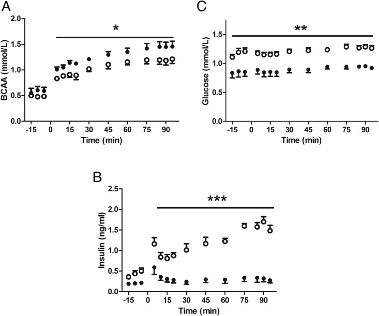
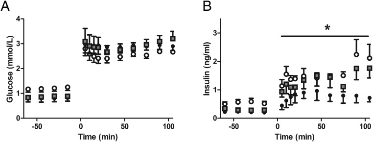
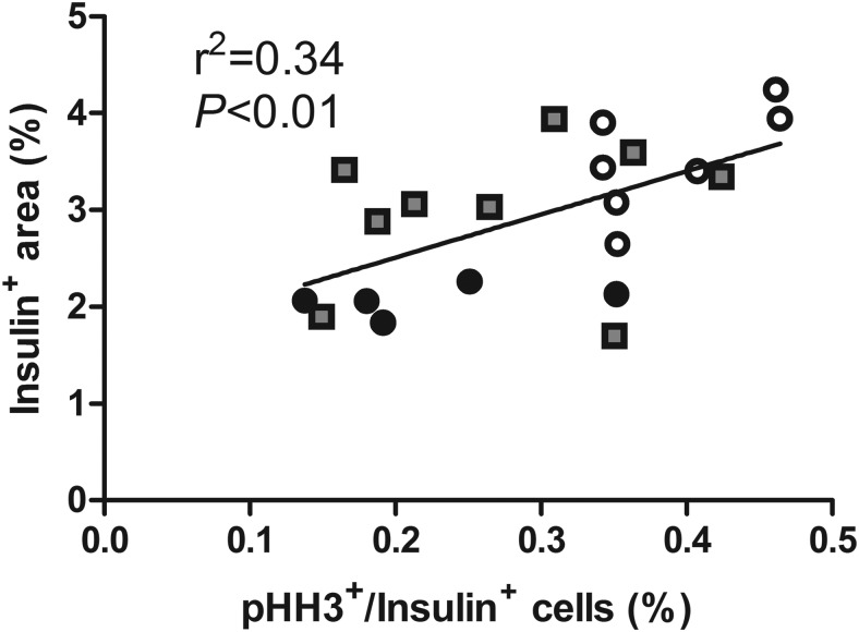
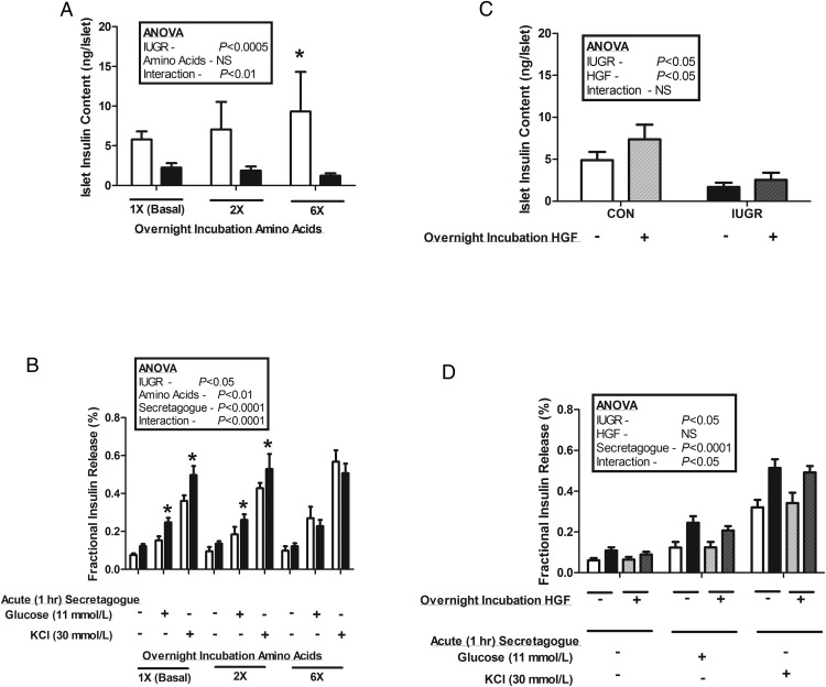
Placental insufficiency is associated with reduced supply of amino acids to the fetus and leads to intrauterine growth restriction (IUGR). IUGR fetuses are characterized by lower glucose-stimulated insulin secretion, smaller pancreatic islets with less β-cells, and impaired pancreatic vascularity. To test whether supplemental amino acids infused into the IUGR fetus could improve these complications of IUGR we used acute (hours) and chronic (11 d) direct fetal amino acid infusions into a sheep model of placental insufficiency and IUGR near the end of gestation. IUGR fetuses had attenuated acute amino acid-stimulated insulin secretion compared with control fetuses. These results were confirmed in isolated IUGR pancreatic islets. After the chronic fetal amino acid infusion, fetal glucose-stimulated insulin secretion and islet size were restored to control values. These changes were associated with normalization of fetal pancreatic vascularity and higher fetal pancreatic vascular endothelial growth factor A protein concentrations. These results demonstrate that decreased fetal amino acid supply contributes to the pathogenesis of pancreatic islet defects in IUGR. Moreover, the results show that pancreatic islets in IUGR fetuses retain their ability to respond to increased amino acids near the end of gestation after chronic fetal growth restriction.


Figures

Figure 1.
Figure 1.
IUGR fetuses have attenuated acute in vivo AASIS. CON (open circles, n = 5) and IUGR (closed circles, n = 5) underwent a primed, continuous, constant-rate hyperaminoacidemic clamp with a direct fetal infusion of 10% TrophAmine (wt/vol) starting at time 0. A, BCAA concentrations increased in both groups and were higher in the IUGR fetuses compared with CONs (*, P < .05). B, IUGR fetuses had significantly lower fetal arterial plasma insulin concentrations during the clamp (***, P < .0001). C, Fetal arterial plasma glucose concentrations increased slightly in both groups (P < .005) but were consistently lower in the IUGR-SAL fetuses (**, P < .0001). Mean ± SE fetal arterial plasma concentrations are plotted and statistics are by mixed models ANOVA.
Figure 2.
Figure 2.
In vivo GSIS is normalized in IUGR+AA fetuses at the end of the chronic amino acid infusion period. Mean ± SE fetal arterial plasma glucose (A) and insulin (B) concentrations are shown relative to the start of a primed continuous variable-rate fetal hyperglycemic clamp (time = 0 min) in CONs (open circles, n = 6), IUGR (closed circles, n = 6), and IUGR+AA (gray squares, n = 5); *, significantly lower insulin concentrations in IUGR compared with both CON and IUGR+AA (P < .05) by mixed models ANOVA.
Figure 3.
Figure 3.
Pancreatic insulin+ area is correlated with the percentage of β-cells in mitosis. Fetal pancreatic sections from CONs (open circles, n = 7), IUGR (closed circles, n = 6), and IUGR+AA (gray squares, n = 9) were immunostained for insulin to define β-cells and pHH3 to define cells in mitosis. A linear association was found between the insulin+ area (y-axis) and the percentage of β-cells in mitosis (x-axis) by linear regression for all fetuses combined (P < .01).
Figure 4.
Figure 4.
In vitro pancreatic islet insulin secretion. Pancreatic islets were isolated from CON (white bars, n = 10) and IUGR (black bars, n = 16) fetuses and incubated overnight with or without additional amino acids (2× or 6×) or HGF (100 ng/mL). A, Insulin content was lower in IUGR islets than CON islets (P < .0005). CON islet insulin content increased with increasing overnight amino acid supplementation (*, relative to basal conditions, P < .005), an effect not seen in IUGR islets. B, Overnight amino acid supplementation had no effect on IUGR islet glucose and potassium-stimulated insulin secretion. However, CON islets had a dose-response increase in glucose and potassium-stimulated insulin secretion after overnight incubation with supplemental amino acids (P < .05); *, significantly more fractional insulin release from IUGR islets compared with CON islets incubated in the same conditions (P < .05). C, Overnight incubation with HGF resulted in higher insulin contents of both CON and IUGR islets (P < .05). D, Overnight incubation in HGF had no effect on CON or IUGR islet glucose or potassium-stimulated insulin secretion. Data are presented as mean ± SE, and statistics are by mixed models ANOVA.
Figure 5.
Figure 5.
Large vessel endothelial cell HGF mRNA is increased with in vitro supplemental amino acids, leucine, and VEGFA. Large vessel endothelial cells from normal fetal sheep (n = 3) were incubated for 72 hours in basal conditions or with indicated supplementation (amino acids [2× or 6×], leucine [2.4 mmol/L], VEGFA [50 ng/mL]). HGF mRNA was then measured and normalized to basal conditions. For each incubation condition, cells derived from all 3 animals were tested in replicates of 1–3; *, significant difference from basal conditions (P < .005). Data are presented as mean ± SE, and statistics are by mixed models ANOVA after log transformation.

References

    1. Van Assche FA, De Prins F, Aerts L, Verjans M. The endocrine pancreas in small-for-dates infants. Br J Obstet Gynaecol. 1977;84:751–753. - PubMed
    1. Nicolini U, Hubinont C, Santolaya J, Fisk NM, Rodeck CH. Effects of fetal intravenous glucose challenge in normal and growth retarded fetuses. Horm Metab Res. 1990;22:426–430. - PubMed
    1. Thorn SR, Rozance PJ, Brown LD, Hay WW., Jr The intrauterine growth restriction phenotype: fetal adaptations and potential implications for later life insulin resistance and diabetes. Semin Reprod Med. 2011;29:225–236. - PMC - PubMed
    1. Economides DL, Nicolaides KH, Linton EA, Perry LA, Chard T. Plasma cortisol and adrenocorticotropin in appropriate and small for gestational age fetuses. Fetal Ther. 1988;3:158–164. - PubMed
    1. Greenough A, Nicolaides KH, Lagercrantz H. Human fetal sympathoadrenal responsiveness. Early Hum Dev. 1990;23:9–13. - PubMed