

Thin-Section Computed Tomography Manifestations During Convalescence and Long-Term Follow-Up of Patients with Severe Acute Respiratory Syndrome (SARS)
- PMID: 27501327
- PMCID: PMC4982531
- DOI: 10.12659/msm.896985
Thin-Section Computed Tomography Manifestations During Convalescence and Long-Term Follow-Up of Patients with Severe Acute Respiratory Syndrome (SARS)
Abstract
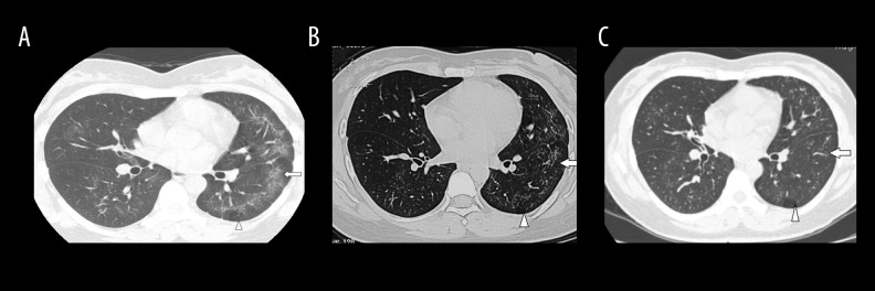
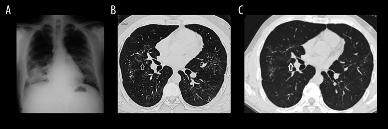
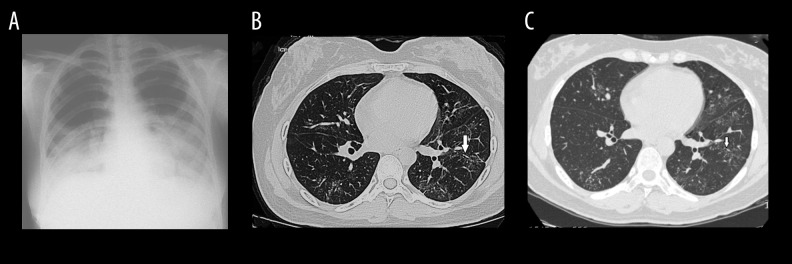
BACKGROUND SARS is not only an acute disease, but also leads to long-term impaired lung diffusing capacity in some survivors. However, there is a paucity of data regarding long-term CT findings in survivors after SARS. The aim of this study was to assess the changes in lung function and lung thin-section computed tomography (CT) features in patients recovering from severe acute respiratory syndrome (SARS), especially the dynamic changes in ground-glass opacity (GGO). MATERIAL AND METHODS Clinical and radiological data from 11 patients with SARS were collected. The serial follow-up thin-section CTs were evaluated at 3, 6, and 84 months after SARS presentation. The distribution and predominant thin-section CT findings of lesions were evaluated. RESULTS The extent of the lesions on the CT scans of the 11 patients decreased at 6 and 84 months compared to 3 months. The number of segments involved on 84-month follow-up CTs was less than those at 6 months (P<0.05). The predominant thin-section CT manifestation at 84 months (intralobular and interlobular septal thickening) was different than that at 6 months, at which GGO was predominant. CONCLUSIONS During convalescence after SARS, GGO and intralobular and interlobular septal thickening were the main thin-section CT manifestation. Intralobular and interlobular septal thickening predominated over GGO at 84 months.
Figures
Comment in
-
Post-Acute Sequelae of COVID-19 Pneumonia: Six-month Chest CT Follow-up.Radiology. 2021 Nov;301(2):E396-E405. doi: 10.1148/radiol.2021210834. Epub 2021 Jul 27. Radiology. 2021. PMID: 34313468 Free PMC article.
References
-
- World Health Organization. Savere acute respiratory syndrome (SARS) Weekly Emidepiol Rec. 2003;78:86.
-
- Gogna A, Tay KH, Tan BS. Severe acute respiratory syndrome: 11 years later – a radiology perspective. Am J Roentgenol. 2014;203:746–48. - PubMed
-
- Hui DS, Memish ZA, Zumla A. Severe acute respiratory syndrome vs. the Middle East respiratory syndrome. Curr Opin Pulm Med. 2014;20:233–41. - PubMed
-
- Wah TM, Moss HA, Robertson RJ, Barnard DL. Pulmonary complications following bone marrow transplantation. Br J Radiol. 2003;76:373–79. - PubMed
MeSH terms
LinkOut - more resources
Full Text Sources
Medical
Research Materials
Miscellaneous
