Review
. 2016 Aug;138 Suppl 1(Suppl Suppl 1):163-83.
doi: 10.1111/jnc.13668.

Prion-like propagation as a pathogenic principle in frontotemporal dementia

Affiliations
Review

Prion-like propagation as a pathogenic principle in frontotemporal dementia

Eva-Maria Hock et al. J Neurochem. 2016 Aug.

Abstract

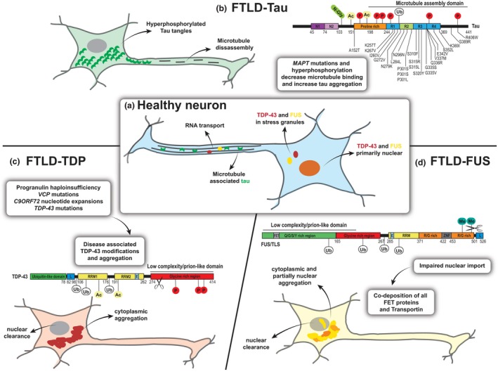
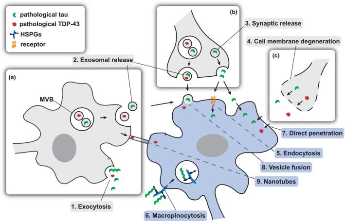
Frontotemporal dementia is a devastating neurodegenerative disease causing stark alterations in personality and language. Characterized by severe atrophy of the frontal and temporal brain lobes, frontotemporal dementia (FTD) shows extreme heterogeneity in clinical presentation, genetic causes, and pathological findings. Like most neurodegenerative diseases, the initial symptoms of FTD are subtle, but increase in severity over time, as the disease progresses. Clinical progression is paralleled by exacerbation of pathological findings and the involvement of broader brain regions, which currently lack mechanistic explanation. Yet, a flurry of studies indicate that protein aggregates accumulating in neurodegenerative diseases can act as propagating entities, amplifying their pathogenic conformation, in a way similar to infectious prions. In this prion-centric view, FTD can be divided into three subtypes, TDP-43 or FUS proteinopathy and tauopathy. Here, we review the current evidence that FTD-linked pathology propagates in a prion-like manner and discuss the implications of these findings for disease progression and heterogeneity. Frontotemporal dementia (FTD) is a progressive neurodegenerative disease causing severe personality dysfunctions, characterized by profound heterogeneity. Accumulation of tau, TDP-43 or FUS cytoplasmic aggregates characterize molecularly distinct and non-overlapping FTD subtypes. Here, we discuss the current evidence suggesting that prion-like propagation and cell-to-cell spread of each of these cytoplasmic aggregates may underlie disease progression and heterogeneity. This article is part of the Frontotemporal Dementia special issue.

Keywords: FTD; FUS; TDP-43; prion-like spread; tau.


Figures

Figure 1
Figure 1
The three main types of FTD pathology are characterized by the accumulation of distinct protein aggregates. (a) In healthy neurons, tau (green crescent) is mainly distributed in axons and regulates microtubule stability. TDP‐43 (red) and FUS (yellow) are primarily found in the nucleus, where they are involved in several steps of RNA metabolism. In the cytoplasm, both proteins participate in dynamic RNA granules, facilitating axonal transport or forming as part of the cell stress response. (b) In pathological conditions of FTLD‐tau, the protein is dissociated from microtubules and accumulates into pathogenic tangles in the cytoplasm. Mutations in the microtubule‐binding region of the microtubule‐associated protein tau (MAPT) gene are found FTLD‐17 MAPT and can lead to splicing changes and decreased microtubule‐binding affinity. Hyperphosphorylation (P) and other post‐translational modifications (N‐Glyc (N‑glycosylation), Ac (Acetylation), Ub (Ubiquitination) can further enhance the detachment, resulting in microtubule disassembly and the formation of tau aggregates in the cytoplasm (example modification sites are shown). (c) In FTLDTDP‐43 several underlying genetic causes (GRN, VCP, TDP‐43, C9ORF72 mutations) and sporadic cases are linked by a common TDP‐43 pathology. TDP‐43 undergoes several disease‐associated modifications including phosphorylation, ubiquitination and C‐terminal cleavage and forms pathogenic inclusions in the cytoplasm leading to a redistribution of the protein, associated with nuclear clearance. (d) Although called FTLDFUS, all FET proteins have now been found to co‐aggregate in ubiquitinated inclusions in severe cases of sporadic FTD. Factors impairing the nuclear import of these proteins might lead to cytoplasmic accumulation and following aggregation because of strongly aggregation‐prone low complexity domains. C9ORF72: chromosome 9 open reading frame 72, E: Nuclear export signal, FET: FUS, TAF‐15, EWS protein interacting domain, FTD: frontotemporal dementia, FTLD: frontotemporal lobar degeneration, FUS: fused in sarcoma, L: nuclear localization signal, N1, N2: near‐amino‐terminal inserts, Q/G/S/Y‐rich: glutamine‐glycine‐serine‐tyrosine rich region, R1, R2, R3, R4: carboxy‐terminal repeat domain, R/G‐rich: arginine/glycine rich region, RRM: RNA recognition motif, TDP‐43: TAR DNA‐binding protein 43, VCP: valosin‐containing protein, ZNF: zinc finger domain.
Figure 2
Figure 2
Proximity‐dependent and transsynaptic mechanisms of cell‐to‐cell spread. The transmission of pathological seeds (tau: green; TARDBP‐binding protein, 43 kD (TDP‐43): red) may happen between neighboring cells within the same brain region in a proximity‐dependent pattern through active release (a) or passive leakage (c) whereas spreading between distant anatomically connected brain regions may rely on synaptic connectivity (b). The protein seeds may be released from the donor cell (gray) via exocytosis (1), packaged into vesicles such as exosomes (released from multivesicular bodies (MVBs)) (2) or dispensed by synaptic vesicles (3), with all these mechanisms having been suggested for tau and TDP‐43. Furthermore, pre‐synaptic membrane leakage because of the degeneration of neurons can lead to extracellular protein seeds (4). Once released, free protein seeds may be internalized by receptor‐mediated endocytosis (5) or heparan sulfate proteoglycan (HSPG)‐dependent macropinocytosis (6) or directly penetrate the recipient cell (blue) (7). Membrane‐coated seeds can fuse with the receiving neuronal membrane (8). Direct transfer between two cells could also happen via tunneling nanotubes (9).

Similar articles

Cited by

References

    1. Agosta F., Scola E., Canu E. et al (2012) White matter damage in frontotemporal lobar degeneration spectrum. Cereb. Cortex 22, 2705–2714. - PubMed
    1. Agosta F., Weiler M. and Filippi M. (2015) Propagation of pathology through brain networks in neurodegenerative diseases: from molecules to clinical phenotypes. CNS Neurosci. Ther. 21, 754–767. - PMC - PubMed
    1. Aguzzi A. (2009) Cell biology: beyond the prion principle. Nature 459, 924–925. - PubMed
    1. Aguzzi A. and Polymenidou M. (2004) Mammalian prion biology: one century of evolving concepts. Cell 116, 313–327. - PubMed
    1. Aguzzi A. and Rajendran L. (2009) The transcellular spread of cytosolic amyloids, prions, and prionoids. Neuron 64, 783–790. - PubMed

Publication types

LinkOut - more resources