. 2016 Sep 30;119(8):909-20.
doi: 10.1161/CIRCRESAHA.116.309202. Epub 2016 Aug 8.

A Simplified, Langendorff-Free Method for Concomitant Isolation of Viable Cardiac Myocytes and Nonmyocytes From the Adult Mouse Heart

Affiliations

A Simplified, Langendorff-Free Method for Concomitant Isolation of Viable Cardiac Myocytes and Nonmyocytes From the Adult Mouse Heart

Matthew Ackers-Johnson et al. Circ Res. .

Abstract

Rationale: Cardiovascular disease represents a global pandemic. The advent of and recent advances in mouse genomics, epigenomics, and transgenics offer ever-greater potential for powerful avenues of research. However, progress is often constrained by unique complexities associated with the isolation of viable myocytes from the adult mouse heart. Current protocols rely on retrograde aortic perfusion using specialized Langendorff apparatus, which poses considerable logistical and technical barriers to researchers and demands extensive training investment.

Objective: To identify and optimize a convenient, alternative approach, allowing the robust isolation and culture of adult mouse cardiac myocytes using only common surgical and laboratory equipment.

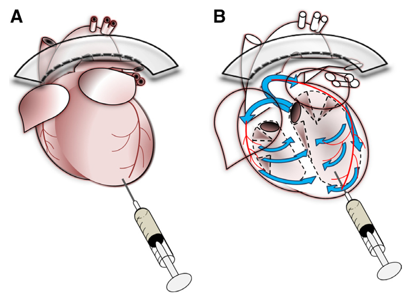
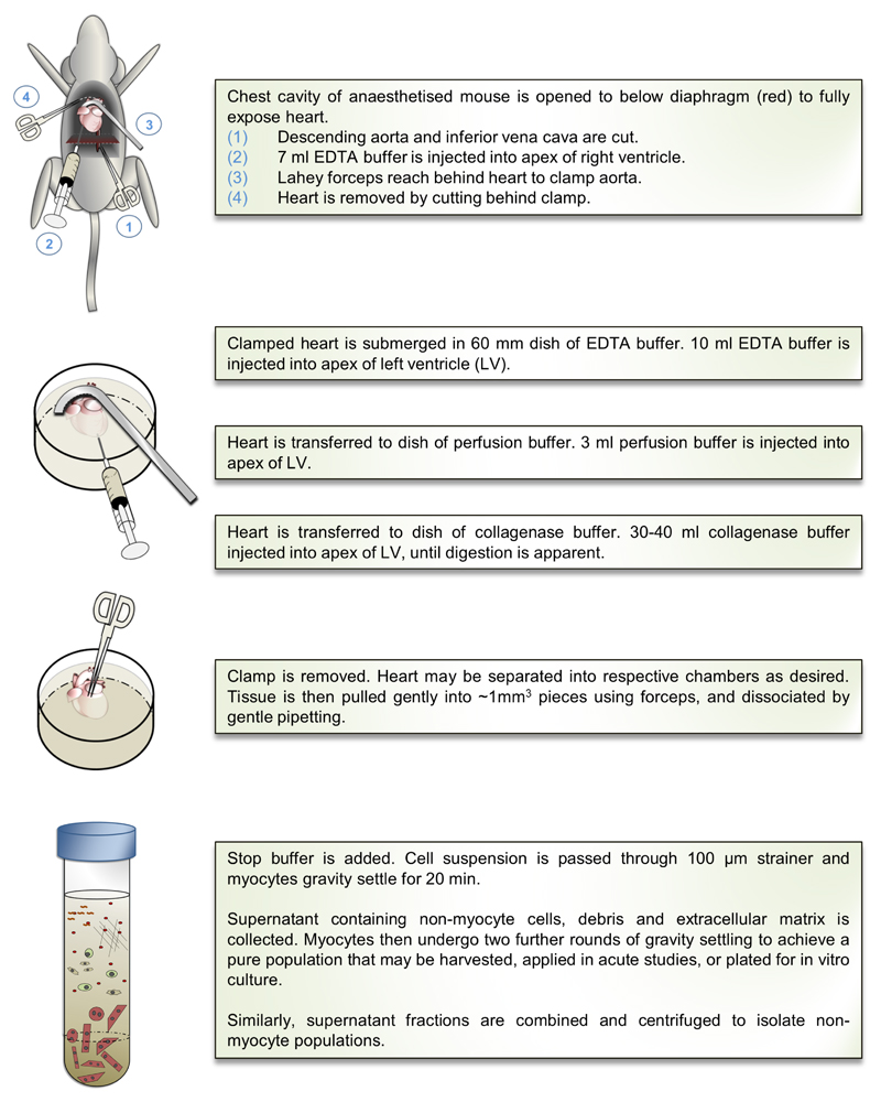
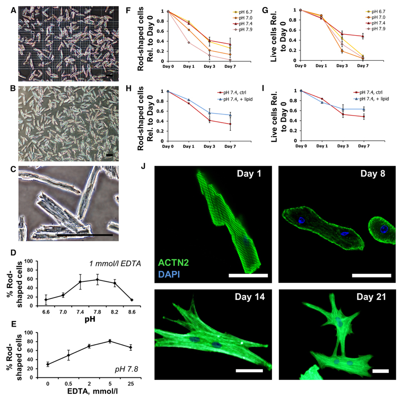
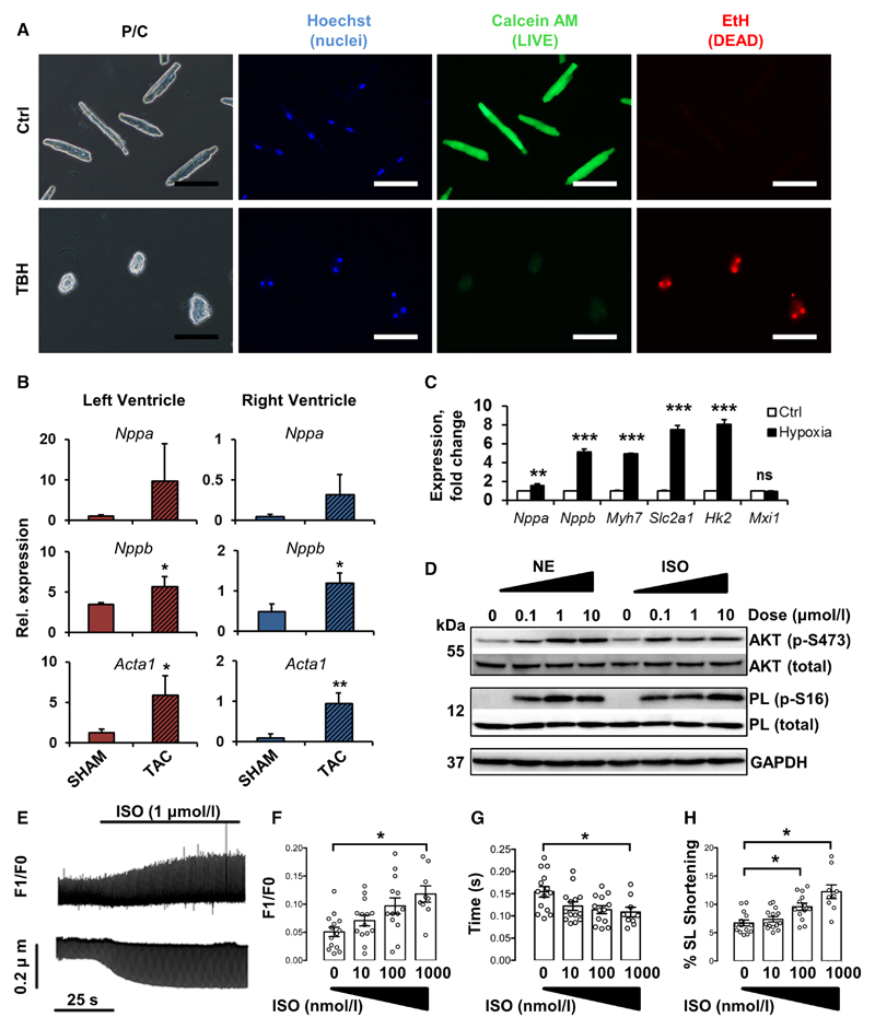
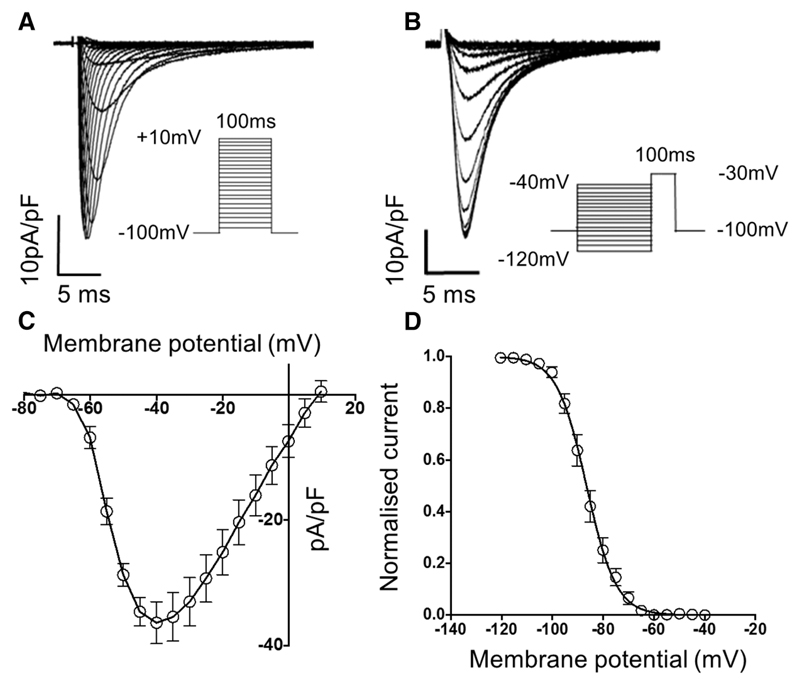
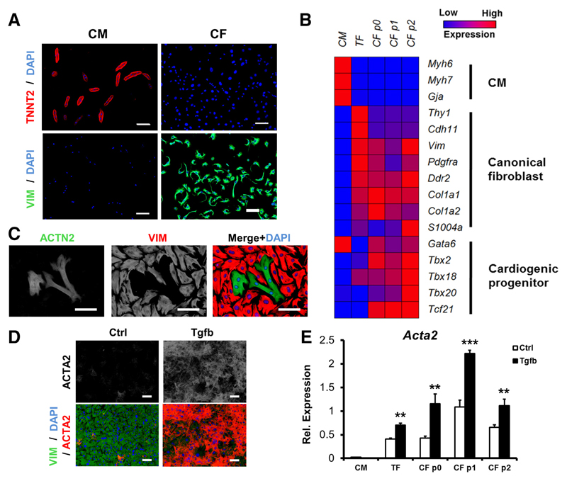
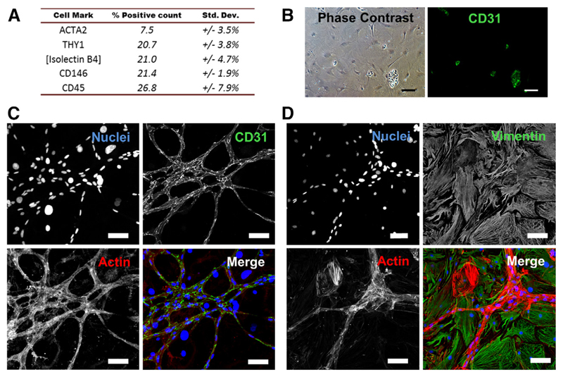
Methods and results: Cardiac myocytes were isolated with yields comparable to those in published Langendorff-based methods, using direct needle perfusion of the LV ex vivo and without requirement for heparin injection. Isolated myocytes can be cultured antibiotic free, with retained organized contractile and mitochondrial morphology, transcriptional signatures, calcium handling, responses to hypoxia, neurohormonal stimulation, and electric pacing, and are amenable to patch clamp and adenoviral gene transfer techniques. Furthermore, the methodology permits concurrent isolation, separation, and coculture of myocyte and nonmyocyte cardiac populations.

Conclusions: We present a novel, simplified method, demonstrating concomitant isolation of viable cardiac myocytes and nonmyocytes from the same adult mouse heart. We anticipate that this new approach will expand and accelerate innovative research in the field of cardiac biology.

Keywords: Langendorff-free; cardiac fibroblasts; cardiomyocytes; cardiovascular disease; coculture; mouse models; single-cell isolation.


Figures

Figure 1
Figure 1. A new method to isolate cardiac myocytes from the adult mouse heart.
Schematic diagrams illustrating the principle of the approach, in which traditional Langendorff-based retrograde aortic perfusion is replaced by simple injection of dissociation buffers into the left ventricle (A). Application of a hemostatic aortic clamp forces passage of buffers (blue arrows; B) through the coronary circulation (red), ensuring deep perfusion of the myocardium.
Figure 2
Figure 2. Summary of the cardiac myocyte isolation protocol.
A detailed, extended description of methodology, with images and video, is available in the Online Data Supplement. LV indicates left ventricle.
Figure 3
Figure 3. Protocol optimization and isolation of high yields of viable cardiac myocytes.
AC, Representative images of adult mouse left ventricular digestion products, before plating (A), and after 1-h culture (B and C), showing yields of 80% rod-shaped, viable myocytes, with organized sarcomeric striations. Scale bars=100 μm. D, Optimization of dissociation buffer pH. Highest viable yields were obtained at pH 7.8. EDTA concentration was 1 mmol/L. E, Optimization of EDTA concentration. Highest yields were obtained at 5 mmol/L EDTA. Buffer pH was 7.8. Data show mean±SD, n=3 independent experiments. FI, Quantification of myocyte rod shape morphology (F and H) and viability (exclusion of ethidium homodimer stain, G and I) for a time course of 7 d in culture at specified pH range (F and G) and with or without lipid supplementation at optimal pH 7.4 (H and I), as indicated. Data show mean±SD, n=2 independent experiments in biological triplicate. J, Immunologic staining and confocal imaging of myocytes with sarcomeric-α-actinin antibody (ACTN2; green) and DAPI (4’,6-diamidino-2-phenylindole), after increasing time in culture, as indicated. Loss of sarcomeric organization was observed after 8-d culture. 10% fetal bovine serum was included in cultures from day 8 onward, and 2,3-butanedione monoxime removed. Extended culture resulted in re-establishment of sarcomeric structures, formation of cell–cell contacts, and synchronized, spontaneous contractility. Scale bars=50 μm.
Figure 4
Figure 4. Cultured cardiac myocytes retain transcriptional and functional characteristics and are amenable to investigation.
A, Isolated myocytes are viable with intact plasma membranes. Freshly plated myocytes retain calcein (green) but exclude ethidium (EtH, red). Addition of 10 μmol/L butyl-peroxide (TBH) led to calcein escape and entry of EtH. Nuclear counterstain, Hoechst-33342. Scale bars=100 μm. B, Myocytes from both ventricles retain characteristic transcriptional signatures after isolation from a pressure-overload model of hypertrophy (TAC), compared with sham-operated controls. Expression was quantified by quantitative polymerase chain reaction, relative to 18S. Data show mean±SD, n=3 mice per group. *P<0.05, **P<0.01, Student t test. C, Hypoxia-regulated genes Nppa, Nppa, Myh7, Slc2a1, and Hk2, but not Mxi1, were significantly upregulated in cultured myocytes after 24-h hypoxic exposure. Data show mean±SD, n=3 independent experiments, expression relative to 18S. **P<0.01, ***P<0.001, Student t test. D–H, Myocytes are responsive to adrenergic stimulation in a dose-dependent manner. D, Western blot to demonstrate phosphorylation of AKT and phospholamban (PL), 20 min after addition of norepinephrine (NE) or isoproterenol (ISO), as indicated. E–H, Myocytes were loaded with fura2-AM and paced at 2 Hz in the presence of ISO as indicated. Calcium transients and sarcomere length shortening were measured using the integrated photometry/contractility system (Ionoptix). E, Representative raw traces of calcium transients (upper) and sarcomere length (lower) recorded from a single cell, ISO added as indicated. Calcium transient amplitude (F), calcium transient decay (G), and % sarcomere length (SL) shortening (H) were subsequently quantified in response to ISO addition as indicated. Data show mean±SE, n≥9 cells from 3 hearts, *P<0.05, 1-way ANOVA followed by Dunnett multiple comparisons test.
Figure 5
Figure 5. Isolated myocytes display normal sodium currents (INa).
INa were measured in freshly isolated left ventricular cardiac myocytes. A, Representative voltage-dependent INa raw traces recorded from a single ventricular cardiac myocyte. The voltage protocol is shown in the inset. B, Mean data for current–voltage relationship of INa current density (pA/pF; n=8 cells from 3 hearts). C, Representative raw traces showing voltage-dependent steady-state INa inactivation. The voltage protocol is shown in the inset. D, Mean data for INa inactivation curve (n=8 cells from 3 hearts).
Figure 6
Figure 6. Concomitant culture and study of cardiac myocytes and fibroblasts from the same mouse heart.
A, Immunologic staining of isolated cardiac myocytes (CM) and cardiac fibroblasts (CF) with cardiac troponin-T antibody (TNNT2, red), vimentin antibody (VIM, green), and DAPI (4’,6-diamidino-2-phenylindole), after 3-d culture. Specific staining demonstrates strong separation of myocyte and nonmyocyte fractions. B, Transcriptional analysis of cultured CM, tail fibroblasts (TF), and CF after 3-d culture, and CF after 1 (p1) or 2 (p2) passages in culture. Expression of selected cardiac myocyte–related (CM), canonical fibroblast–related, and cardiogenic-related genes was determined by quantitative polymerase chain reaction, relative to 18S, and presented in heat map format. Data represent mean expression, n=2 independent experiments in biological triplicate. C, Coculture of cardiac myocytes and fibroblasts from the same mouse heart. Cell fractions were isolated, recombined after 3-d separate culture, and maintained for 4 further days in the presence of 10% fetal bovine serum. Cells were fixed and costained with antibodies against sarcomeric-α-actinin (ACTN2, green, CM), vimentin (VIM, red, CF), and DAPI. D, Activation of isolated CFs after 24-h incubation with 10 ng/mL transforming growth factor beta (Tgfb) was detected by immunologic staining for smooth muscle α-actin (ACTA2) production. E, The potential for activation of isolated CFs with Tgfb persisted for at least 2 passages. Data show mean±SD, n=2 independent experiments in biological triplicate, Acta2 expression relative to 18S. **P<0.01, ***P<0.001, Student t test, compared with relevant unstimulated controls.
Figure 7
Figure 7. Isolated cardiac nonmyocytes represent a heterogeneous population.
A, Relative proportions of nonmyocyte fraction cells detected positive for putative identity markers: ACTA2 (smooth muscle), THY1 (cardiac fibroblast), CD146, GSL-Isolectin-B4 (endothelial), and CD45 (immunocyte), by flow cytometry. Data averaged from 3 independent experiments. B, Immunologic staining of CD31 (PECAM-1) in subconfluent nonmyocyte fraction cultures marked clusters of endothelial-like cells (green). C and D, In postconfluent cultures, these cells were observed to form CD31-positive (green), actin-rich (stained using fluorophore-conjugated phalloidin; red) networks (C) that stained negative for the fibroblast marker vimentin (VIM; green; D). All scale bars=100 μm.

Comment in

References

    1. Wong ND. Epidemiological studies of CHD and the evolution of preventive cardiology. Nat Rev Cardiol. 2014;11:276–289. doi: 10.1038/nrcardio.2014.26. - DOI - PubMed
    1. Cook C, Cole G, Asaria P, Jabbour R, Francis DP. The annual global economic burden of heart failure. Int J Cardiol. 2014;171:368–376. doi: 10.1016/j.ijcard.2013.12.028. - DOI - PubMed
    1. Braunwald E. The war against heart failure: the Lancet lecture. Lancet. 2015;385:812–824. doi: 10.1016/S0140-6736(14)61889-4. - DOI - PubMed
    1. Sambrano GR, Fraser I, Han H, Ni Y, O’Connell T, Yan Z, Stull JT. Navigating the signalling network in mouse cardiac myocytes. Nature. 2002;420:712–714. doi: 10.1038/nature01306. - DOI - PubMed
    1. Cervio E, Barile L, Moccetti T, Vassalli G. Exosomes for intramyocardial intercellular communication. Stem Cells Int. 2015;2015:482171. doi: 10.1155/2015/482171. - DOI - PMC - PubMed