. 2016 Aug 9:6:31226.
doi: 10.1038/srep31226.

A Nanoscale Interface Promoting Molecular and Functional Differentiation of Neural Cells

Affiliations

A Nanoscale Interface Promoting Molecular and Functional Differentiation of Neural Cells

Tamara Posati et al. Sci Rep. .

Abstract

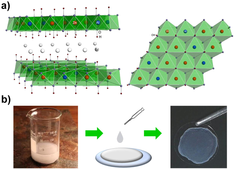
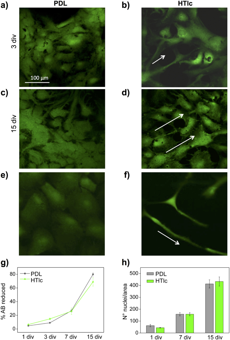
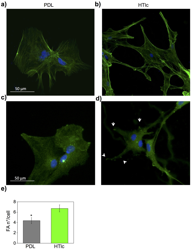
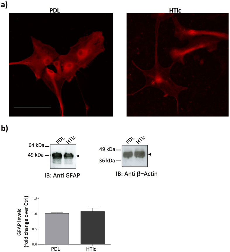
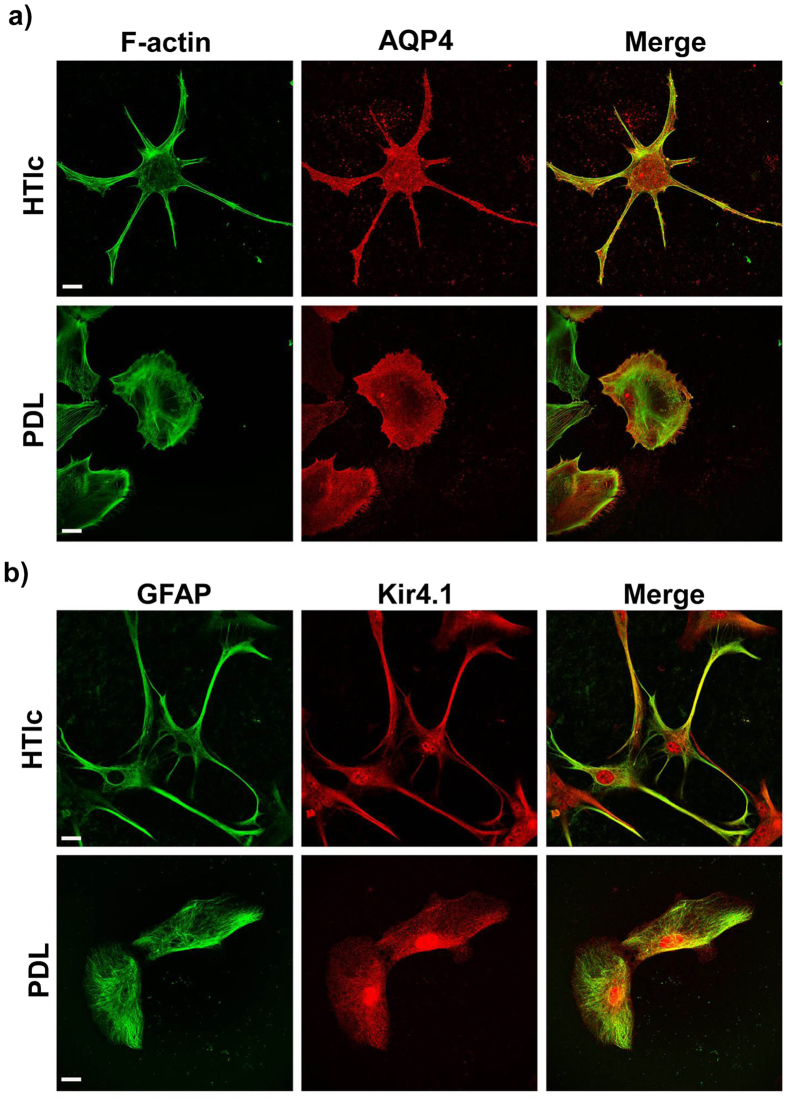
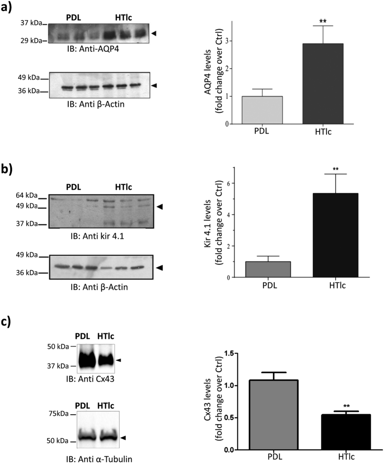
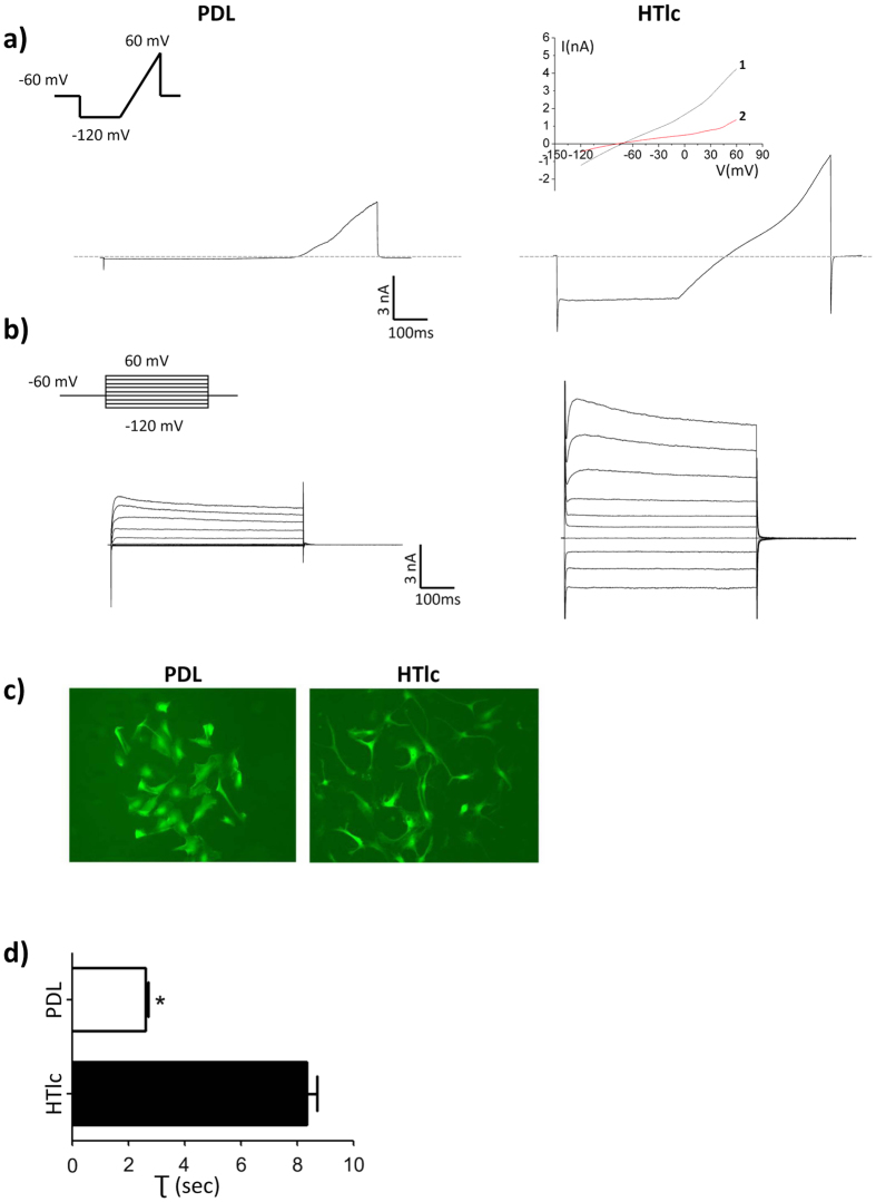
Potassium channels and aquaporins expressed by astrocytes are key players in the maintenance of cerebral homeostasis and in brain pathophysiologies. One major challenge in the study of astrocyte membrane channels in vitro, is that their expression pattern does not resemble the one observed in vivo. Nanostructured interfaces represent a significant resource to control the cellular behaviour and functionalities at micro and nanoscale as well as to generate novel and more reliable models to study astrocytes in vitro. However, the potential of nanotechnologies in the manipulation of astrocytes ion channels and aquaporins has never been previously reported. Hydrotalcite-like compounds (HTlc) are layered materials with increasing potential as biocompatible nanoscale interface. Here, we evaluate the effect of the interaction of HTlc nanoparticles films with primary rat neocortical astrocytes. We show that HTlc films are biocompatible and do not promote gliotic reaction, while favouring astrocytes differentiation by induction of F-actin fibre alignment and vinculin polarization. Western Blot, Immunofluorescence and patch-clamp revealed that differentiation was accompanied by molecular and functional up-regulation of both inward rectifying potassium channel Kir 4.1 and aquaporin 4, AQP4. The reported results pave the way to engineering novel in vitro models to study astrocytes in a in vivo like condition.


Figures

Figure 1
Figure 1. Structure and preparation of ZnAl-HTlc films.
(a) Schematic representation of the side view (left) and of the top view (right) of the ZnAl-HTlc structure. (b)Scheme showing ZnAl-HTlc film preparation.
Figure 2
Figure 2. Morphological characterization of HTlc films.
(a) SEM images at different magnifications of ZnAl-HTlc film (b) AFM topographical images of ZnAl-HTlc film (RMS~134 nm).
Figure 3
Figure 3. Viability and morphology of astrocytes on PDL and HTlc films.
(a–f) Single plane confocal images of astrocytes stained for FDA, representing viable cells plated on PDL (a,c) and on HTlc (b,d) captured after 3 div (a,b), and 15 div (c,d). (e,f) Low density plated astrocytes captured after 7 div; note differentiated astrocytes plated on HTlc (g) Time course of astrocyte viability on PDL and HTLc film, investigated by AB assay at different time points. Data are plotted as the averaged percentages of reduced AB ± Standard Error (SE) versus div. (h) Histogram plot reporting the averages of astrocyte nuclei number, counted on images of cells grown on PDL (gray bars) and on HTlc films (green bars). (p > 0.05, independent t test).
Figure 4
Figure 4
SEM imaging of cells plated on PDL (a) and HTlc (b–e): note the presence of elongated branches in astrocytes plated on HTlc that follow the rod like structure of HTlc. (ce) high magnification images of cells plated on HTlc films revealing lateral endfeet in astrocyte processes projecting to HTlc nanoplatelets.
Figure 5
Figure 5. F-actin cytoskeleton and Vinculin proteins expression in astrocytes plated on PDL and HTlc.
Micrographs representing astrocytes imunostained for actin (a,b) and vinculin (c,d) grown on PDL (a,c) and HTlc fims (b,d). (e) Hstogram plot reporting the number of Focal Adhesion (FA) contacts per astrocyte, which were counted in cells plated on PDL (gray bars) and on HTlc films (green bars). Data are expressed as means ± SE (n = 25; *p < 0.05).
Figure 6
Figure 6. GFAP protein expression in astrocytes plated on PDL and HTlc.
(a) Confocal imaging of astrocytes stained with GFAP. Scale bar is 50 μm. (b) immunoblotting and quantification of the expression level of GFAP. β-actin (right panel) is used as control for signal normalization. Lower panel: histogram plots evidencing protein fold of change over the control. Note that the expression level of GFAP was not significantly different between PDL and HTlc (n = 3). Cropped images of the entire blots are reported. However, no other specific signals were observed within the blot length.
Figure 7
Figure 7. AQP4 and Kir 4.1 protein expression in astrocytes plated on PDL and HTlc.
(a) Confocal imaging of astrocytes grown on HTLc NPs (upper panels) and PDL (lower panels) and stained for AQP4 (red), and Actin (green). (b) Confocal imaging of astrocytes grown on HTLc NPs (upper panels) and PDL (lower panels) and stained for Kir4.1 (red), and GFAP(green). A merged image of the channels is reported on the right side of the panel, revealing that AQP4 and Kir 4.1 are highly up-regulated in the endfeet of astrocytes grown on HTlc NPs.
Figure 8
Figure 8
Western blot analyses and quantification of protein expression of AQP4 (a) Kir4.1 (b) and Cx43 (c) on astrocytes plated on PDL and HTlc. β-actin (a,b, lower panels) and α-tubulin were re-blotted and used as controls for signal normalization. Histogram plots evidencing change in protein expression with respect to the control are reported in the right panels of the figures. Values are the mean ± SE. (n = 3; *p < 0.01). Cropped images of the entire blots are reported. However, no other specific signals were observed within the blot length.
Figure 9
Figure 9. Functional properties of HTlc-plated astrocytes.
(a,b) Typical current traces evoked by a holding potential (Vh) of −60 mV stimulating astrocytes with a voltage ramp or voltage step family (insets). Astrocytes (left panels) plated on PDL displayed only voltage-dependent outward rectifying K+ conductance. Astrocytes plated on HTlc coated coverslips (Right panels), display an inward conductance in response to a hyperpolarizing stimulus that is inhibited after extracellular superfusion with Ba2+ (a, inset, red trace 2). Note inhibition by Ba2+ of outward currents in HTLc-treated astrocytes, suggesting a weak rectification profile of the inward current typical of Kir4.1. (c) Micrograph of calcein-loaded astrocytes showing the different shape of astrocytes grown on PDL vs those grown on HTlc. Histogram summarizing the swelling rates (τ) for the astrocytes grown on PDL or on HTlc. Value is the mean ± SE. (n = 30; P < 0.05).

References

    1. Nedergaard M., Ransom B. & Goldman S. A. New roles for astrocytes: redefining the functional architecture of the brain. Trends Neurosci. 26, 523–530 (2003). - PubMed
    1. Benfenati V. & Ferroni S. Water transport between CNS compartments: functional and molecular interactions between aquaporins and ion channels. Neuroscience 168, 926–940 (2010). - PubMed
    1. Benfenati V. & Ferroni S. In Microdynamics of Water and Ion Homeostasis in the Brain: Role of Aquaporins and Ion Channels of Astroglial Cells; in Homeostatic control of brain function ; (eds Boison D. & Masino S. A.); First Edition, Oxford University Press (2015); “in press”.
    1. Kimelberg H. K. Astrocytic swelling in cerebral ischemia as a possible cause of injury and target for therapy. Glia 50, 389–397 (2005). - PubMed
    1. Steinhäuser C., Grunnet M. & Carmignoto G. Crucial role of astrocytes in temporal lobe epilepsy. Neuroscience, doi: 10.1016/j.neuroscience.2014.12.047 (2015). - DOI - PubMed

Publication types

MeSH terms

LinkOut - more resources