. 2016 Aug 8;4(1):80.
doi: 10.1186/s40478-016-0357-9.

Novel antibodies to phosphorylated α-synuclein serine 129 and NFL serine 473 demonstrate the close molecular homology of these epitopes

Affiliations

Novel antibodies to phosphorylated α-synuclein serine 129 and NFL serine 473 demonstrate the close molecular homology of these epitopes

Nicola J Rutherford et al. Acta Neuropathol Commun. .

Abstract

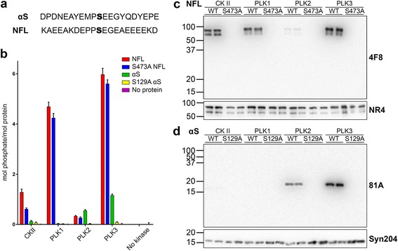
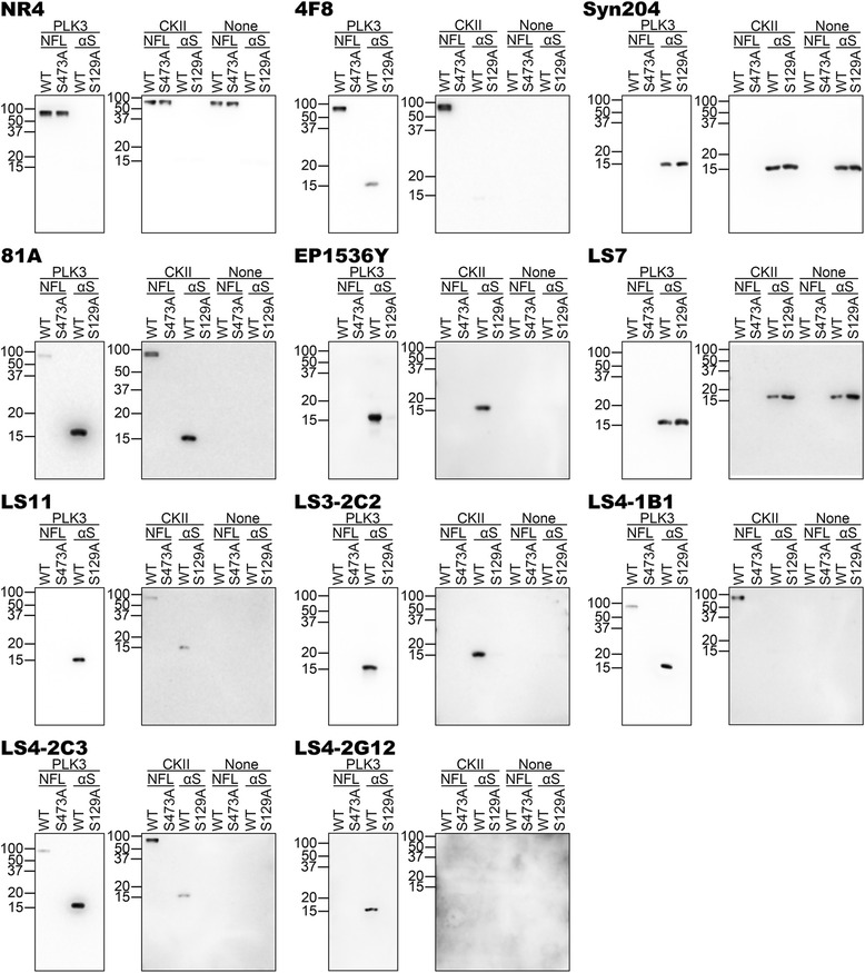
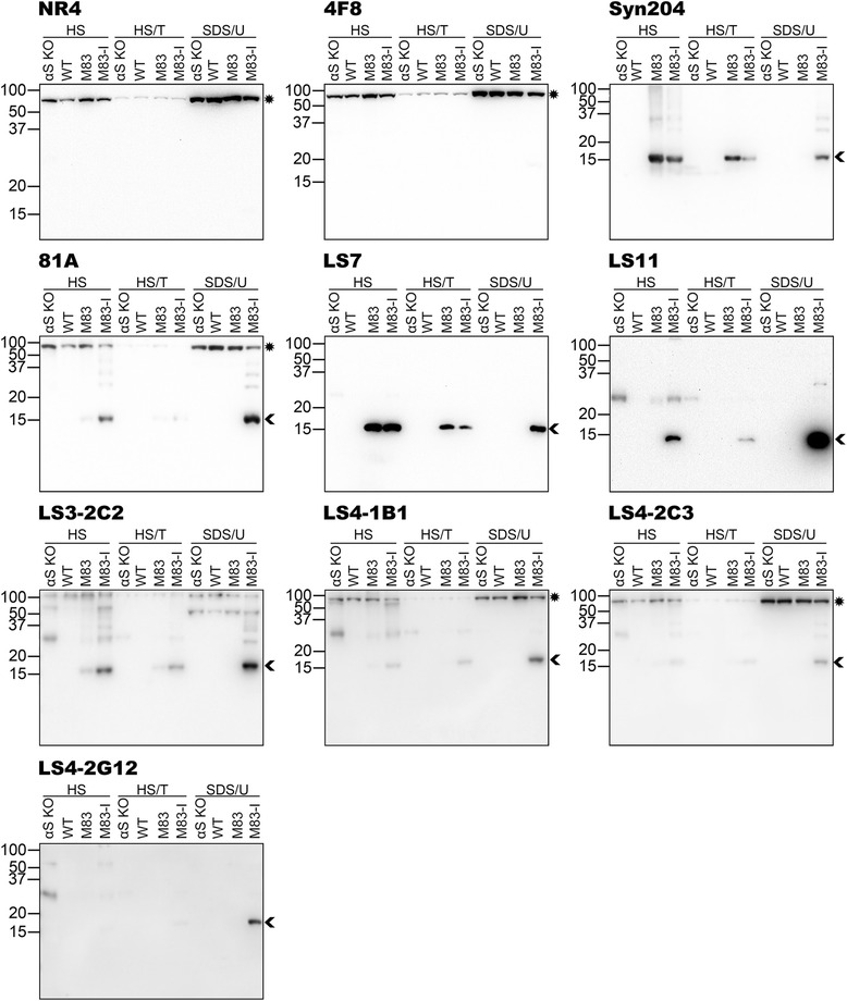
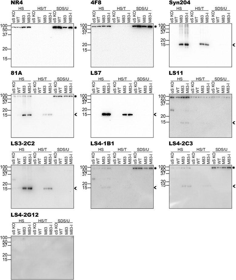
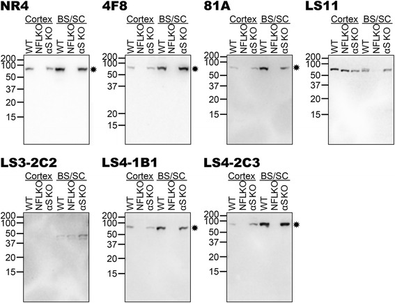
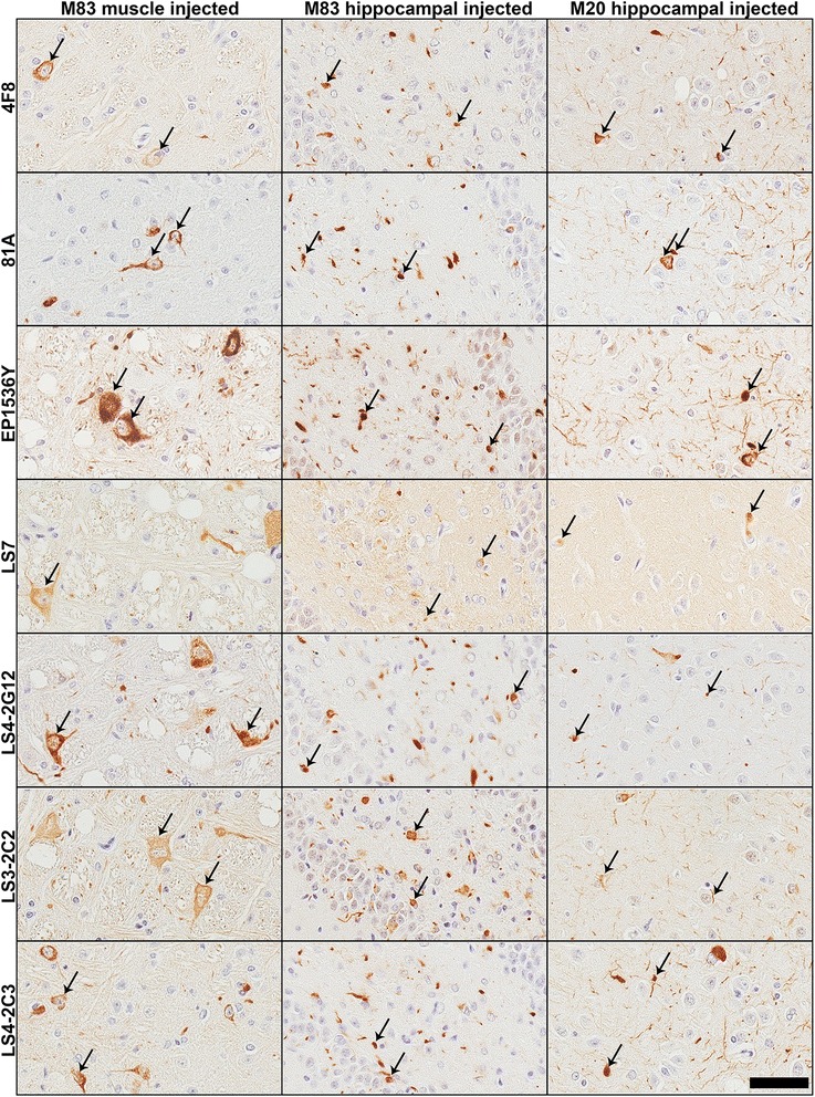
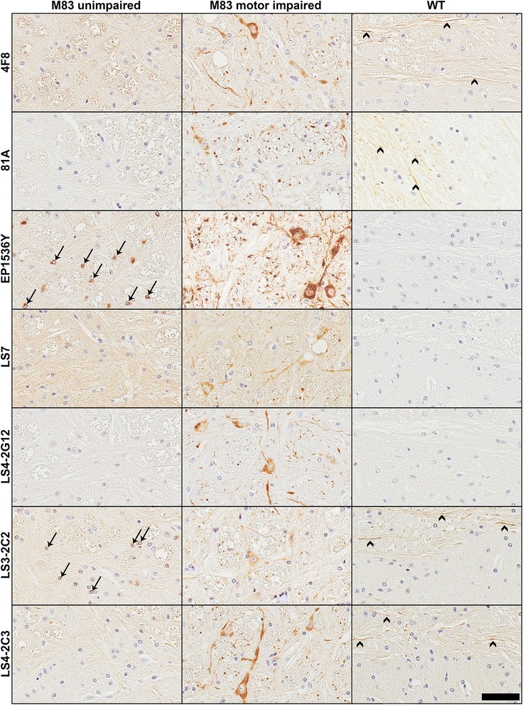
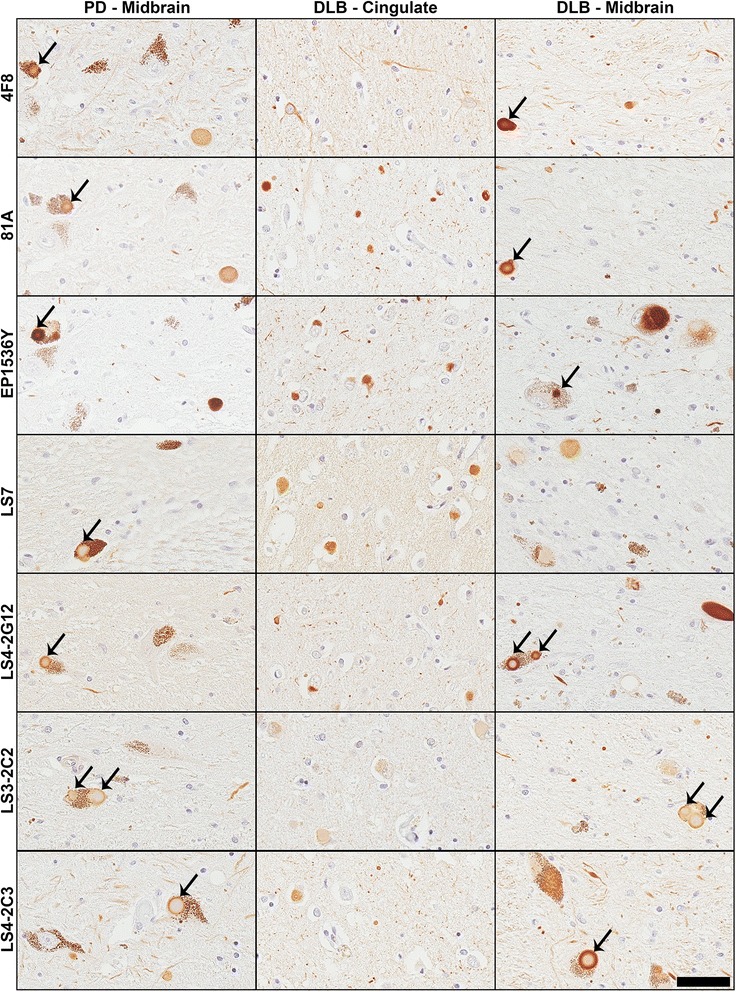
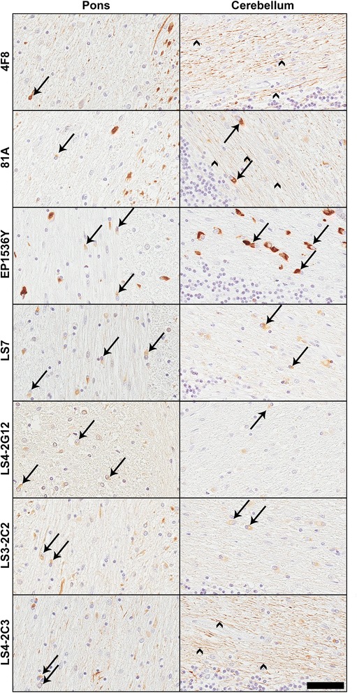
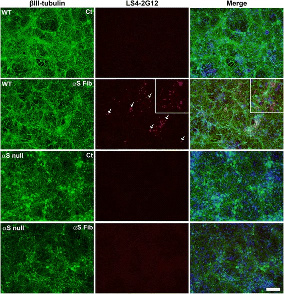
Pathological inclusions containing aggregated, highly phosphorylated (at serine129) α-synuclein (αS pSer129) are characteristic of a group of neurodegenerative diseases termed synucleinopathies. Antibodies to the pSer129 epitope can be highly sensitive in detecting αS inclusions in human tissue and experimental models of synucleinopathies. However, the generation of extensively specific pSer129 antibodies has been problematic, in some cases leading to the misinterpretation of αS inclusion pathology. One common issue is cross-reactivity to the low molecular mass neurofilament subunit (NFL) phosphorylated at Ser473. Here, we generated a series of monoclonal antibodies to the pSer129 αS and pSer473 NFL epitopes. We determined the relative abilities of the known αS kinases, polo-like kinases (PLK) 1, 2 and 3 and casein kinase (CK) II in phosphorylating NFL and αS, while using this information to characterize the specificity of the new antibodies. NFL can be phosphorylated by PLK1, 2 and 3 at Ser473; however CKII shows the highest phosphorylation efficiency and specificity for this site. Conversely, PLK3 is the most efficient kinase at phosphorylating αS at Ser129, but there is overlay in the ability of these kinases to phosphorylate both epitopes. Antibody 4F8, generated to the pSer473 NFL epitope, was relatively specific for phosphorylated NFL, however it could uniquely cross-react with pSer129 αS when highly phosphorylated, further showing the structural similarity between these phospho-epitopes. All of the new pSer129 antibodies detected pathological αS inclusions in human brains and mouse and cultured cell experimental models of induced synucleinopathies. Several of these pSer129 αS antibodies reacted with the pSer473 NFL epitope, but 2 clones (LS3-2C2 and LS4-2G12) did not. However, LS3-2C2 demonstrated cross-reactivity with other proteins. Our findings further demonstrate the difficulties in generating specific pSer129 αS antibodies, but highlights that the use of multiple antibodies, such as those generated here, can provide a sensitive and accurate assessment of αS pathology.

Keywords: Monoclonal antibodies; Neurofilament; Parkinson’s disease; Phosphorylation; α-synuclein.


Figures

Fig. 1
Fig. 1
In vitro phosphorylation of NFL and αS by CKII and PLK1, 2 and 3. a Amino acid sequences surrounding the target phosphorylation sites (bold) of Ser129 for αS, and Ser473 for NFL. b Quantitative analysis of the stoichiometric phosphorylation of recombinant αS, S129A αS, NFL and S473A NFL in in vitro kinase reactions as described in “Materials and methods” with CKII, PLK1, PLK2 or PLK3. All reactions were performed in triplicate. c Immunoblotting analyses of NFL and S473A NFL used in in vitro kinase reactions with CKII, PLK1, PLK2 or PLK3. Proteins were resolved onto 8 % polyacrylamide gel and probed with 4F8 (pSer473 NFL) and NR4 (anti-NFL) antibodies. d Immunoblotting analyses of αS and S129A αS used in in vitro kinase reactions with CKII, PLK1, PLK2 or PLK3. Proteins were resolved onto 13 % polyacrylamide gels and probed with 81A (pSer129 αS) and Syn 204 (anti-αS). The mobility of molecular mass markers are shown on the left
Fig. 2
Fig. 2
Specificity of novel pSer129 αS antibodies as determined by immunoblotting with NFL and αS proteins phosphorylated in vitro with PLK3 or CKII. NFL, S473A NFL, αS and S129A αS were incubated with PLK3 or CKII. The proteins were resolved onto 13 % polyacrylamide gels and analyzed by immunoblotting with novel αS antibodies (as indicated above each blot) LS7, LS11, LC2-2C2, LS4-1B1, LS4-2C3 and LS4-2G12 and novel NFL antibody 4F8, to demonstrate their specificity. NR4 (anti-NFL) and Syn 204 (anti-αS) were included to show each protein, respectively. Previously reported pSer129 αS antibodies 81A and EP1536Y were included for comparison. Reactions were performed in duplicate. Antibody 4F8, generated against the pSer473 NFL epitope, can cross react with αS phosphorylated at Ser129. Antibodies 81A, LS11, LS4-1B1 and LS4-2C3 generated against the pSer129 αS epitope can cross react with NFL phosphorylated at Ser473. Antibodies EP1536Y, LS3-2C2 and LS4-2G12 generated against the pSer129 αS epitope reacted only with phosphorylated WT αS. Antibody LS7 generated against the pSer129 αS epitope detected both phospho- and non-phospho-αS. The mobility of molecular mass markers are shown on the left
Fig. 3
Fig. 3
Characterization of the specificity of novel pSer129 αS antibodies by immunoblotting analyses using biochemically fractionated brainstem/spinal cord mouse tissues. Brainstem/spinal cord from an αS null (αS KO), a WT, a 2 month old non-symptomatic M83 (M83) and a 12 month old motor impaired M83 (M83-I) mouse were biochemically fractionated into high salt (HS), high salt/Triton X-100 (HS/T) and SDS/urea (SDS/U) fractions as described in “Materials and methods”. 10 μg of HS and HS/T, and 5 μg of SDS/U fractions were resolved onto 13 % polyacrylamide gels and analyzed by immunoblotting with each antibody indicated above. The protein band identified by an arrowhead is αS, while an asterisk depicts NFL. The mobility of molecular mass markers are shown on the left
Fig. 4
Fig. 4
Characterization of the specificity of novel pSer129 αS antibodies by immunoblotting analyses using biochemically fractionated cerebral cortex mouse tissues. Cerebral cortex from an αS null (αS KO), a WT, a 2 month old non-symptomatic M83 (M83) and a 12 month old motor impaired M83 (M83-I) mouse were biochemically fractionated into high salt (HS), high salt/Triton X-100 (HS/T) and SDS/urea (SDS/U) fractions as described in “Materials and methods”. 10 μg of HS and HS/T and 5 μg of SDS/U fractions were resolved onto 13 % polyacrylamide gels and analyzed by immunoblotting with each antibody indicated above. The protein band identified by an arrowhead is αS, while an asterisk depicts NFL. The mobility of molecular mass markers are shown on the left
Fig. 5
Fig. 5
Characterization of the specificity of novel pSer129 αS antibodies by immunoblotting analyses using total lysates of cortex and brainstem/spinal cord tissues from WT and NFL null mice. Cerebral cortex and brainstem/spinal cord (BS/SC) from a WT, an NFL null (NFL KO) and an αS null (αS KO) mouse were dissected and lysed in 2 % SDS/ 50 mM Tris pH 7.5 as described in “Materials and methods”. Equal amounts of proteins (5 μg) from each sample was resolved onto 10 % polyacrylamide gels and analyzed by immunoblotting with each antibody indicated above. The protein band identified by an asterisk depicts NFL. The mobility of molecular mass markers are shown on the left
Fig. 6
Fig. 6
Comparison of novel antibodies in detecting pathological inclusions in αS transgenic mice injected with αS fibrils in the periphery (intramuscular) or the brain (hippocampus). Representative images of IHC staining of tissue from M83 αS transgenic mice injected in the gastrocnemius muscle, and M83 and M20 αS transgenic mice injected in the hippocampus with recombinant preformed αS fibrils. Images were taken from the brainstem (muscle injection) and the hippocampus (hippocampal injection). Antibodies EP1536Y and 81A showed robust staining of induced inclusions (arrows). LS7 stained inclusions weakly with higher general labeling. Novel antibodies raised against the pSer129 αS epitope LS4-2G12, LS3-2C2 and LS4-2C3 or to the p473 NFL epitope all stained the induced inclusions in these models. Scale bar = 50 μm
Fig. 7
Fig. 7
Comparison of pSer129 antibody IHC staining using naïve αS transgenic and WT mice. Representative images of IHC staining of brainstem tissue from a 7 month old non-symptomatic homozygous M83 mouse (M83 unimpaired), a 12 month old motor impaired homozygous M83 mouse (M83 motor impaired) and a WT mouse. All antibodies stained perikaryal and neuritic inclusions in the M83 diseased mouse. LS7 showed weaker reactivity to αS pathology than the other antibodies and showed stronger diffused signal in the αS transgenic mice compared to the WT mouse. LS3-2C2 also showed weak staining of pathology. In addition to the pathology in motor impaired M83 mouse, antibodies 4F8, LS3-2C2, LS4-2C3 and 81A also labeled neuronal projections even in WT mice (arrowheads). EP1536Y exhibited some nuclear staining in some mouse sections (arrows), as did LS3-2C2, but much weaker than EP1536Y (eg. see arrows in the M83 unimpaired mouse). Scale bar = 50 μm
Fig. 8
Fig. 8
IHC analyses of brain sections of individuals with Parkinson’s disease and dementia with Lewy bodies. Representative IHC staining of brain tissue sections from PD and DLB patients. Images were taken from the midbrain (PD and DLB) or cingulate cortex (DLB only). All of the antibodies stained LBs within dopaminergic neurons in the midbrain sections for both PD and DLB. Arrows show LBs in some examples within cells containing neuromelanin. Scale bar = 50 μm
Fig. 9
Fig. 9
IHC analyses of brain sections of individuals with multiple system atrophy. Representative IHC staining of brain tissue sections from MSA patients. Images were taken from the pons or cerebellum. Arrows show GCIs. All of the antibodies stained inclusions, to some extent. EP1536Y showed the strongest staining. 4F8 stained only rare inclusions, but abundantly labeled axons (arrowheads). Antibodies 81A and LS4-2C3 also displayed strong reactivity to axons in the cerebellum. Antibodies LS4-2G12 and LS3-2C2 displayed weaker staining of inclusions in the cerebellum. Scale bar = 50 μm
Fig. 10
Fig. 10
Analysis of the induction of endogenous αS aggregation by treatment with exogenous αS mouse fibrils in primary neuronal-glial cultures using antibody LS4-2G12. Primary neuronal-glial cultures from WT mice or αS null mice were cultured for 6 days and either maintained without other treatment for 8 days (Ct) or treated with mouse αS fibrils (20 μg/ml; αS Fib) for 8 days. Double immunofluorescence analysis with antibodies LS4-2G12 (red) and specific neuronal marker βIII-tubulin (green) was performed. Cells were also counterstained with DAPI and merged images are shown. Higher magnification LS4-2G12 and merged images are shown on the far right. Arrows depict induced labeled αS aggregates. Scale bar = 100 μm and 250 μm for the higher magnification images on the right

Similar articles

Cited by

References

    1. Goedert M. Alpha-synuclein and neurodegenerative diseases. Nat Rev Neurosci. 2001;2:492–501. doi: 10.1038/35081564. - DOI - PubMed
    1. Goedert M, Spillantini MG, Del Tredici K, Braak H. 100 years of Lewy pathology. Nat Rev Neurol. 2013;9:13–24. doi: 10.1038/nrneurol.2012.242. - DOI - PubMed
    1. Spillantini MG, Schmidt ML, Lee VM, Trojanowski JQ, Jakes R, Goedert M. Alpha-synuclein in Lewy bodies. Nature. 1997;388:839–40. doi: 10.1038/42166. - DOI - PubMed
    1. Uchihara T, Giasson BI. Propagation of alpha-synuclein pathology: hypotheses, discoveries, and yet unresolved questions from experimental and human brain studies. Acta Neuropathol. 2016;131:49–73. doi: 10.1007/s00401-015-1485-1. - DOI - PMC - PubMed
    1. Spillantini MG, Crowther RA, Jakes R, Cairns NJ, Lantos PL, Goedert M. Filamentous alpha-synuclein inclusions link multiple system atrophy with Parkinson’s disease and dementia with Lewy bodies. Neurosci Lett. 1998;251:205–8. doi: 10.1016/S0304-3940(98)00504-7. - DOI - PubMed

MeSH terms