Review
. 2016 Oct;37(10):811-821.
doi: 10.1016/j.tips.2016.07.002. Epub 2016 Aug 5.

Tristetraprolin as a Therapeutic Target in Inflammatory Disease

Affiliations
Review

Tristetraprolin as a Therapeutic Target in Inflammatory Disease

Sonika Patial et al. Trends Pharmacol Sci. 2016 Oct.

Abstract

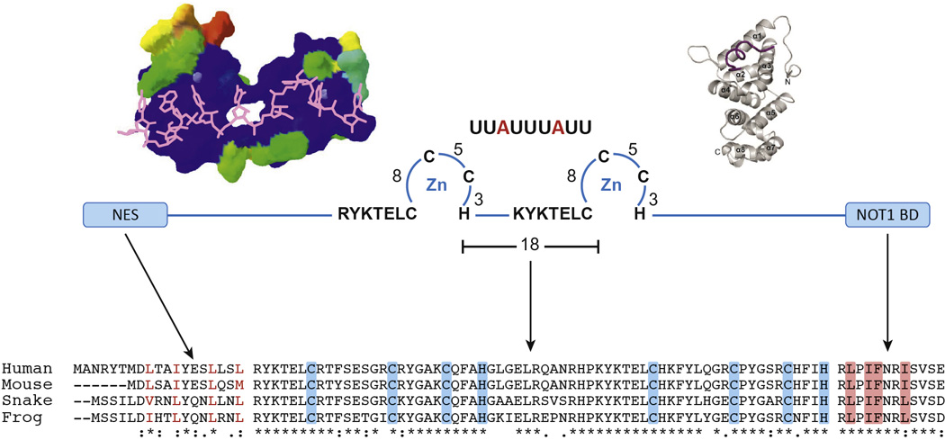
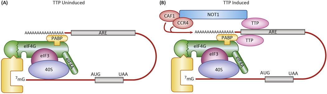
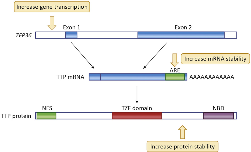
Members of the tristetraprolin (TTP) family of RNA-binding proteins are found in all major eukaryotic groups. TTP family members, from plants through humans, can bind adenosine-uridine rich elements in target mRNAs with high affinity. In mammalian cells, these proteins then promote deadenylation and decay of target transcripts. Four such proteins are found in mice, of which the best studied is TTP. When the gene encoding TTP is disrupted in mice, the animals develop a severe syndrome of arthritis, autoimmunity, cachexia, dermatitis, and myeloid hyperplasia. Conversely, recent overexpression studies have demonstrated protection against several experimental models of immune inflammatory disease. This endogenous anti-inflammatory protein could serve as the basis for novel approaches to therapy of similar conditions in humans.

Keywords: autoimmunity; deadenylation; inflammation; mRNA decay; tumor necrosis factor.


Figures

Fig. 1
Fig. 1. Critical domains of TTP
In the center of the figure is a schematic diagram of three critical domains of TTP: The N terminal nuclear export sequence (NES), the central tandem zinc finger domain (TZF domain), and the C terminal NOT1 binding domain (NOT1 BD). The key cysteines and histidines in each finger, as well as the conserved lead-in sequences, are indicated for the TZF domain, as is the ideal 9 base RNA binding site that is a component of many AU-rich regions in TTP target mRNAs. At the upper left is shown a structural model of the human TTP TZF domain bound to the same 9 base RNA sequence as shown at the top; RNA is in magenta in this figure. This structural model is taken from [46], with permission, and is based on the original structure of the ZFP36L2 (TIS11D) TZF domain [16]. At the upper right is a diagram of the crystal structure of the C-terminal NOT1 binding domain peptide of TTP (maroon), binding to the three internal helices from the human NOT1 protein (beige); this structure is taken from [12], with permission. At the bottom are shown parts of an amino acid sequence alignment done with Clustal Omega of human TTP with its orthologues from mouse, snake and frog. According to Clustal conventions, asterisks at the bottom indicate sequence identity at that site; a colon indicates a conserved residue; and a period indicates a less well conserved residue. Shown are the N termini of the proteins, with their conserved NES sequences; in this case, the branched chain amino acids are colored red. The TZF domain alignment is shown in the middle, with the critical cysteines and histidines shaded in blue. The NOT1 binding domains are shown to the right, with the sequences representing the extreme C termini of the proteins. Highlighted in orange are the amino acid residues that are inserted into the hydrophobic groove of the NOT1 protein central domain (upper right) [12]. The spaces in the sequence alignments represent gaps of various sizes. The sequences are from the following GenBank accession numbers: Human (Homo sapiens), NP_003398.2 ; mouse (Mus musculus), NP_035886.1 ; snake (Protobothrops mucrosquamatus (Taiwan habu)), XP_015676155.1; and frog (Xenopus tropicalis), NP_001106542.1.
Fig. 2
Fig. 2. Model of proposed TTP mechanism of action
The diagram shows a schematic representation of the “closed-loop” model [47] of a typical mRNA being translated in the absence of TTP (left), as in serum deprived fibroblasts or macrophages, and after the induction of TTP (right), as in serum-stimulated fibroblasts or LPS-stimulated macrophages. TTP binds to the AREs of its target mRNAs, often on multiple sites, and interacts with both polyA binding protein (PABP) as a possible means of localizing near the polyA tail, and NOT1, which can bring its associated deadenylases CAF1 and CCR4 into proximity with the 3’ end of the polyA tail. The net result of the TTP binding is destabilization and decreased translation of the mRNA. See the text for further details.
Fig. 3
Fig. 3. Sequences of mouse and human TTP mRNA 3’UTRs
Shown is a Clustal Omega alignment of the mouse and human TTP 3’UTRs, starting from the stop codons (underlined). The AU-rich segment that was deleted in the TTPΔARE mice is in red type, and the polyadenylation signals in both mouse and human mRNAs are shaded yellow. The asterisks indicate nucleotide identity at that site.
Fig. 4
Fig. 4. Effects of the homozygous TTPΔARE deletion on TTP protein expression in macrophages and liver
The top panel shows the responses of bone marrow derived macrophages from WT mice and their TTPΔARE counterparts to LPS stimulation. The time course of TTP protein expression is shown, as determined by western blotting. The different sizes of the protein bands are thought to represent variably phosphorylated forms of the protein. Immunoreactive tubulin is shown as a loading control. In the bottom panel is shown the expression of TTP protein in the livers of three TTPΔARE mice, along with livers from three WT mice and a single lane containing an equivalent amount of liver protein from a TTP KO mouse. Immunoreactive actin is shown as a loading control. Note the non-specific bands that appear in all samples. Taken from [8], with permission.
Fig. 5
Fig. 5. Schematic representation of steps in TTP accumulation that could be affected by proposed therapies
Shown schematically are steps in the biosynthesis and decay of TTP mRNA and protein that might be influenced by novel therapies designed to increase its overall accumulation. Small molecules might be identified that increase transcription of ZFP36, the human gene encoding TTP, that would have minimal effects on other genes that are often co-induced with TTP, such as pro-inflammatory cytokines. Other small molecules might be able to enhance splicing of the single ZFP36 intron. Changes in TTP mRNA stability might be influenced by “steric-blocking” oligonucleotides, which would inhibit the normally very rapid decay of the TTP mRNA. Small molecules might be identified that decrease the rate of decay of TTP protein and promote its overall accumulation in cells. Cell-based screening programs could be conducted without targeting specific steps in this biosynthetic pathway.

References

    1. Chastek B, et al. A Retrospective Cohort Study Comparing Utilization and Costs of Biologic Therapies and JAK Inhibitor Therapy Across Four Common Inflammatory Indications in Adult US Managed Care Patients. Advances in therapy. 2016;33:626–642. - PMC - PubMed
    1. Kalliolias GD, Ivashkiv LB. TNF biology, pathogenic mechanisms and emerging therapeutic strategies. Nat Rev Rheumatol. 2016;12:49–62. - PMC - PubMed
    1. Palladino MA, et al. Anti-TNF-alpha therapies: the next generation. Nature reviews. Drug discovery. 2003;2:736–746. - PubMed
    1. Ramiro S, et al. Safety of synthetic and biological DMARDs: a systematic literature review informing the 2013 update of the EULAR recommendations for management of rheumatoid arthritis. Annals of the rheumatic diseases. 2014;73:529–535. - PubMed
    1. Carballo E, et al. Bone marrow transplantation reproduces the tristetraprolin-deficiency syndrome in recombination activating gene-2 (−/−) mice. Evidence that monocyte/macrophage progenitors may be responsible for TNFalpha overproduction. The Journal of clinical investigation. 1997;100:986–995. - PMC - PubMed

Publication types