. 2016 Sep 28:238:272-280.
doi: 10.1016/j.jconrel.2016.08.005. Epub 2016 Aug 5.

Ultrasound-guided therapeutic modulation of hepatocellular carcinoma using complementary microRNAs

Affiliations

Ultrasound-guided therapeutic modulation of hepatocellular carcinoma using complementary microRNAs

Sayan Mullick Chowdhury et al. J Control Release. .

Abstract

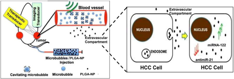
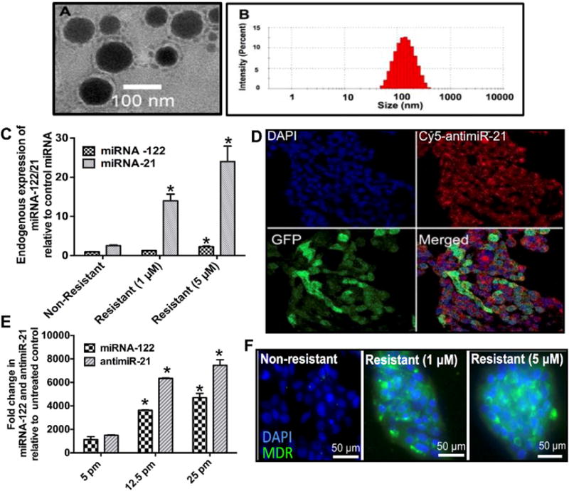
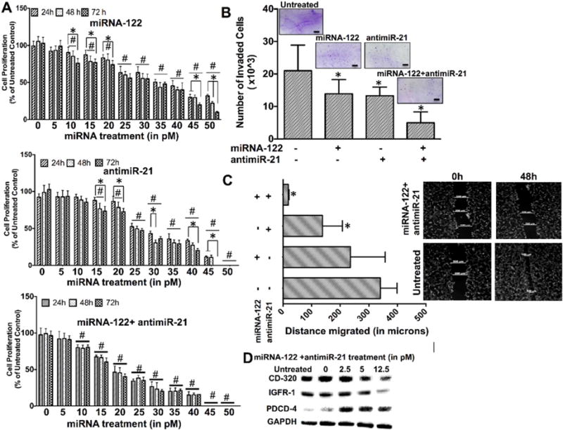
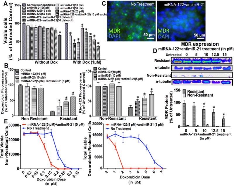
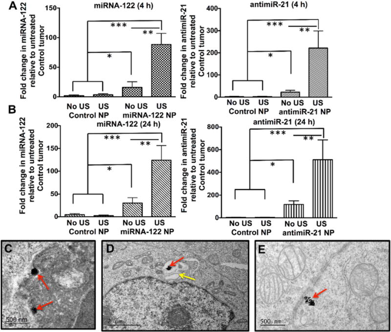
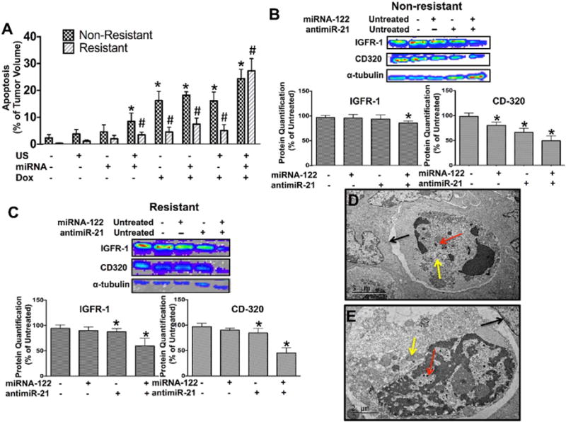
Treatment options for patients with hepatocellular carcinoma (HCC) are limited, in particular in advanced and drug resistant HCC. MicroRNAs (miRNA) are non-coding small RNAs that are emerging as novel drugs for the treatment of cancer. The aim of this study was to assess treatment effects of two complementary miRNAs (sense miRNA-122, and antisense antimiR-21) encapsulated in biodegradable poly (lactic-co-glycolic acid) nanoparticles (PLGA-NP), administered by an ultrasound-guided and microbubble-enhanced delivery approach in doxorubicin-resistant and non-resistant human HCC xenografts. Proliferation and invasiveness of human HCC cells after miRNA-122/antimiR-21 and doxorubicin treatment were assessed in vitro. Confocal microscopy and qRT-PCR were used to visualize and quantitate successful intracellular miRNA-loaded PLGA-NP delivery. Up and down-regulation of miRNA downstream targets and multidrug resistance proteins and extent of apoptosis were assessed in vivo in treated human HCC xenografts in mice. Compared to single miRNA therapy, combination therapy with the two complementary miRNAs resulted in significantly (P<0.05) stronger decrease in cell proliferation, invasion, and migration of HCC cells as well as higher resensitization to doxorubicin. Ultrasound-guided delivery significantly increased in vivo miRNA-loaded PLGA-NP delivery in human HCC xenografts compared to control conditions by 5-9 fold (P<0.001). miRNA-loaded PLGA-NP were internalized in HCC cells and anti-apoptotic proteins were down regulated with apoptosis in ~27% of the tumor volume of doxorubicin-resistant human HCC after a single treatment with complementary miRNAs and doxorubicin. Thus, ultrasound-guided delivery of complementary miRNAs is highly efficient in the treatment of doxorubicin- resistant and non-resistant HCC. Further development of this new treatment approach could aid in better treatment of patients with HCC.

Keywords: Cancer therapy; Complementary miRNA; Drug resistance; Hepatocellular carcinoma; Ultrasound.


Conflict of interest statement

of potential conflicts of interest There is no actual or potential conflicts of interest with regard to this paper.

Figures

Fig. 1
Fig. 1
Schematic shows experimental setting and transducer arrangement in mouse bearing two human HCC xenografts. One xenograft was used as non-insonated control tumor, one was treated with ultrasound and microbubble mediated sonoporation, causing leakage of miRNA-loaded PLGA-NP into the extravascular compartment. PLGA-NP are taken up by HCC cells via endocytosis and their miRNA cargo is released into the cytoplasm of HCC cells.
Fig. 2
Fig. 2
(A) Representative TEM images of PLGA-NP. (B) Distribution of the hydrodynamic diameter of PLGA-NP assessed by Dynamic Light Scattering (DLS). (C) Quantification of endogenous miRNA-122 and miRNA-21 expression levels in non-resistant and two doxorubicin-resistant HCC cells shows increased miRNA-21 levels with increased resistance while miRNA-122 levels remain relatively lower in all three cell lines. *, P < 0.05 compared to non-resistant cells; n = 6 each. (D) Confocal microscopy images confirm intracellular delivery of Cy5-labelled antimiR-21 in GFP expressing (green) HCC cells. (E) Dose-dependent intracellular uptake of miRNA-122- and antimiR-21-loaded PLGA-NP in HCC cells. *, P < 0.05 compared to untreated control cells; n = 6 each. (F) Confocal microscopy images show increasing Multi-drug Resistant (MDR) protein expression levels (green) in non-resistant and doxorubicin-resistant HCC cells.
Fig. 3
Fig. 3
(A) Cell proliferation assays of non-resistant HCC cells treated with miRNA-122 (upper row), antimiR-21 (middle row), and miRNA-122/antimiR-21 combination (lower row) for 24, 48, and 72 h show dose dependent decrease in proliferation with the miRNA combination treatment resulting in strongest anti-proliferation effects. *, P < 0.05 among time points for the same treatment concentration. #, P < 0.05 compared to untreated control; n = 6. (B) Cell invasion assays show strongest decrease of HCC cell invasion following the miRNA combination treatment. Inserts show crystal violet stained invaded HCC cells for the different treatments. *, P < 0.05 compared to untreated control cells; n = 3 each. (C) Cell migration assays show smallest migration distance when HCC cells were treated with the miRNA combination treatment. *, P < 0.05 compared to untreated control cells; n = 3. (D) Western blots shows strongest decrease of the two anti-apoptotic proteins CD-320 and IGFR-1 and strongest increase of the pro-apoptotic protein PDCD4 with the miRNA combination treatment in HCC cells. GAPDH was used as loading control.
Fig. 4
Fig. 4
(A) Cell proliferation assay of HCC cells with strong doxorubicin resistance (5 μM) treated with different doses of miRNA-122 and antimiR-21 (10, 25, 50 picomoles), either individually or in combination, shows that the miRNA combination therapy resulted in the strongest inhibition of cell proliferation. *, P < 0.05 compared to untreated control cells; n = 6 each. When HCC cells were treated with the miRNA combination treatment and doxorubicin, all cells died at a miRNA dose of 50 picomoles each. *, P < 0.05 compared to untreated control cells; n = 6 each. α tubulin was used as loading control. (B) Treatment with the miRNA combination therapy increased both doxorubicin and Rho-123 uptake in resistant HCC cells, suggesting decreased multi drug resistance (MDR) protein expression following miRNA treatment. *, P < 0.05 compared to untreated control cells; n = 12 each. This was confirmed by fluorescence imaging (C), and western blotting (D) showing decreased MDR protein expression in doxorubicin resistant HCC cells after treatment with the miRNA combination therapy. *, P < 0.05 compared to untreated control cells; n = 3 each. α tubulin was used as loading control. (E) Cell viability of non-resistant (left) and doxorubicin resistant (right) HCC cells treated with the miRNA combination therapy for 18 h followed by increasing doxorubicin doses shows increased therapeutic effects of doxorubicin in both cell types; n = 6 each.
Fig. 5
Fig. 5
Quantitative RT-PCR assessment of non-resistant human HCC xenografts in mice intravenously injected with either control PLGA-NP (no miRNA loading) or with miRNA-loaded PLGA-NP that were either treated or not treated with ultrasound (US). (A) shows fold increase of respective miRNA levels at 4 h, (B) shows them at 24 h following treatment. *, P = 0.005, **, P = 0.002; ***, P = 0.001, all compared to untreated control tumors; n = 5 each. Representative TEM image shows internalization of PLGA-NP (red arrows) into a HCC cell (C) through endocytosis. (D) PLGA-NP (red arrow) is shown within a vesicular structure (yellow arrow) in a HCC cell by TEM. (E) Degrading PLGA-NP (red arrow) aggregates in the cytoplasm of a HCC cell (red arrow).
Fig. 6
Fig. 6
(A) Bar graphs summarizing mean and standard deviations of % tumor volume apoptosis calculated from TUNEL stained non-resistant and doxorubicin-resistant HCC xenograft sections show highest extent of apoptosis in tumors treated with the miRNA combination therapy. * and #, P < 0.05 compared to untreated control tumors; n = 5 each. Western blotting and quantitative bar graph summary (mean ± standard deviations) of treatment effects on down-stream anti-apoptotic proteins, IGFR-1 and CD320, in non–resistant (B) and resistant (C) human HCC xenografts treated with miRNA-122 and antimiR-21, either isolated or in combination. *, P < 0.05 compared to control tumors; n = 3 each. α tubulin was used as loading control. (D and E) TEM images show HCC cells with multiple internalized PLGA-NP (red arrows), double layered vacuolar structures in the cytoplasm (yellow arrows) and evidence of detachment from surrounding HCC cells (black arrows), indicating apoptosis.

References

    1. Forner A, Llovet JM, Bruix J. Hepatocellular carcinoma. Lancet. 2012;379:1245–1255. - PubMed
    1. E.A.f.S.o. Liver. EASL-EORTC clinical practice guidelines: management of hepatocellular carcinoma. Eur J Cancer. 2012;48:599. - PubMed
    1. Lencioni R, Cioni D, Crocetti L, Bartolozzi C. Percutaneous ablation of hepatocellular carcinoma: state-of-the-art. Liver Transpl. 2004;10:S91–S97. - PubMed
    1. Yang T, Zhang J, Lu JH, Yang LQ, Yang GS, Wu MC, Yu WF. A new staging system for resectable hepatocellular carcinoma: comparison with six existing staging systems in a large Chinese cohort. J Cancer Res Clin Oncol. 2011;137:739–750. - PMC - PubMed
    1. Llovet JM, Fuster J, Bruix J. Intention-to-treat analysis of surgical treatment for early hepatocellular carcinoma: resection versus transplantation. Hepatology. 1999;30:1434–1440. - PubMed

MeSH terms