. 2017 May 9;26(5):773-787.
doi: 10.3727/096368916X692654. Epub 2016 Aug 5.

StemCell Keep™ Is Effective for Cryopreservation of Human Embryonic Stem Cells by Vitrification

StemCell Keep™ Is Effective for Cryopreservation of Human Embryonic Stem Cells by Vitrification

Akemi Ota et al. Cell Transplant. .

Abstract

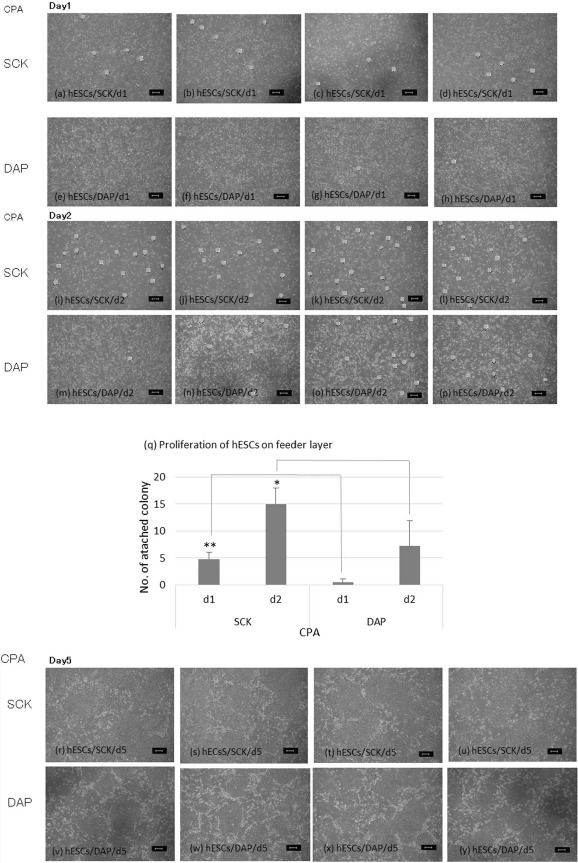
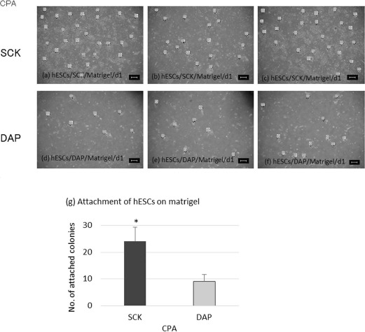
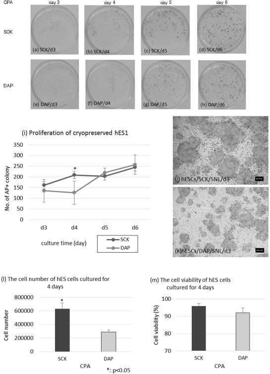
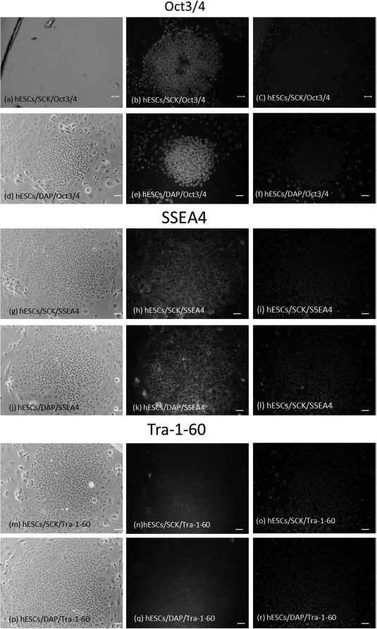
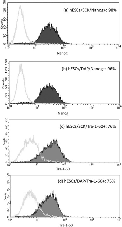
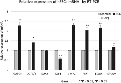
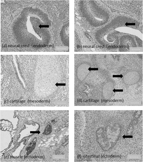
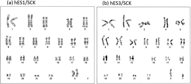
Safe and stable cryopreservation is critical for research involving human embryonic stem cells (hESCs). Dimethyl sulfoxide (DMSO) is a popular cryoprotective agent; however, its cytotoxicity cannot be ignored. Thus, there is a need for an alternate cryoprotectant. We reported previously that a novel cryopreservation reagent, StemCell Keep™ (SCK), was effective for cryopreserving human induced pluripotent stem cells (hiPSCs) by vitrification. Because hESCs and hiPSCs are not identical, the current study examined the use of SCK on hESCs. hESCs cryopreserved with SCK were thawed and cultured on SNL 76/7 cells, which were derived from a mouse fibroblast STO cell line transformed with neomycin resistance and murine LIF genes. After cryopreservation, cultured hESCs were assessed for their attachment ability and characterized by alkaline phosphatase (AP) and immunocytochemical (ICC) staining, fluorescence-activated cell sorting (FACS), reverse transcription polymerase chain reaction (RT-PCR), and karyotyping. The proliferation of SCK-cryopreserved hESCs cultured on SNL cells, or in feeder-free conditions, was higher than that of cells preserved in a solution of 2 M DMSO, 1 M acetamide, and 3 M propylene glycol (DAP). The cell number with SCK-cryopreserved hESCs was about twice that of hESCs cryopreserved in DAP. The pluripotency of SCK-cryopreserved hESCs was similar to that of DAP-cryopreserved hESCs based on AP staining. Data from ICC, FACS, and RT-PCR analyses showed that stem cell markers were continually expressed on SCK-cryopreserved hESCs. The teratoma assay showed that SCK-cryopreserved hESCs differentiated into three germ layers. Furthermore, SCK-cryopreserved hESCs had normal karyotypes. These data indicate that SCK was effective for cryopreservation of hESCs by vitrification.


Figures

Figure 1.
Figure 1.
Differential scanning calorimetry (DSC) thermograms of propylene glycol (DAP) and StemCell Keep™ (SCK) for cooling at 10°C/min and 30°C/min.
Figure 2.
Figure 2.
The attachment ability of human embryonic stem cells (hESCs) cryopreserved with SCK or DAP and cultured on a feeder cell layer. Freeze-thawed hESCs were cultured on a layer of SNL feeder cells, and their attachment to the bottom of a culture dish was observed under an inverted microscope. hESC colonies were photographed at 40x magnification. The number of colonies was counted by marking them with dots. hESC colonies after cryopreservation with SCK (a-d) or DAP (e-h) and culturing for 1 day (Day1). hESC colonies after cryopreservation with SCK (i-l) or DAP (m-p) and culturing for 2 days (Day2). (q) Comparison of the number of hESC colonies after cryopreservation with SCK or DAP and culturing for 1 or 2 days. Data are presented as means ± SD. Statistical evaluations were done by the Tukey-Kramer test. *p < 0.05, statistically significant difference. hESC colonies after cryopreservation with SCK (r-u) or DAP (v-y) and culturing for 5 days (Day5) (r-y). Both 5-day cultures had many colonies, and the numbers were not determined. Scale bars: 100 um. CPA, cryoprotective agent.
Figure 3.
Figure 3.
The ability of hESCs cryopreserved with SCK or a solution of 2 M dimethyl sulfoxide (DMSO), 1 M acetamide, and 3 M DAP to attach to the bottom of Matrigel-coated culture dishes. Freeze-thawed hESCs were cultured on a layer of feeder cells for several days, and then disaggregated and cultured on Matrigel-coated dishes for 1 day. Cells were then observed for their adherence under an inverted microscope. hESC colonies were photographed at 40x magnification. The number of colonies was counted by marking them with dots. hESC colonies had a star-shaped morphology. hESCs after 1 day (d1) of cryopreservation with (SCK) (a-c) or DAP (d-f). (g) Comparison of the number of hESC colonies following cryopreservation with SCK or DAP. Data are presented as means ± SD. Statistical evaluations were done by Student's t-test. *p < 0.05, statistically significant difference. Scale bars: 100 um. CPA, cryoprotective agent.
Figure 4.
Figure 4.
Proliferation profiles of human embryonic stem cells (hESCs) cryopreserved with SCK or a solution of 2 M dimethyl sulfoxide, 1 M acetamide, and 3 M DAP using AP staining. AP-stained hESCs cryopreserved with SCK (a-d) or DAP (e-h) after 3, 4, 5, and 6 days of culturing (day 3, day 4, day 5, and day 6). (i) Comparison of the number of AP+ hESC colonies after preservation with SCK or DAP and culturing for 3, 4, 5, and 6 days. AP+ colonies were counted in four fields at low magnification (10x). hESC colonies 3 days after cryopreservation in SCK (j) or DAP (k). AP staining was observed under an inverted microscopy at 40x magnification. The number (l) and viability (m) of hESCs cultured for 4 days after cryopreservation in SCK or DAP. All data are presented as means ± SD. Statistical evaluations were done by Student's t-test, *p < 0.05, statistically significant difference. Scale bars: 100 um. CPA, cryoprotective agent.
Figure 5.
Figure 5.
Immunocytochemical analyses of hESCs cryopreserved in SCK or DAP for the stem cell markers octamer-binding transcription factor 3/4 (Oct3/4), stage-specific embryonic antigen-4 (SSEA4), and Tra-1-60. Images of hESCs in bright-field, immunofluorescence, and 4′,6-diamidino-2-phenylindole (DAPI) staining were obtained on a microscope at a magnification of 40x. Scale bars: 100 um. hESCs were fixed with 4% PFA, permeabilized, and reacted with primary antibodies against Oct3/4, SSEA4, or Tra-1-60 at 4° C overnight, and further reacted with fluorescently tagged secondary antibodies in the dark. Mounting medium containing DAPI was added, and cells were observed on a fluorescence microscope. (a-c) hESC/SCK/Oct3/4. (d-f) hESC/DAP/Oct3/4. (g-i) hESC/SCK/ SSEA4. (j-l) hESC/DAP/SSEA4. (m-o) hESC/SCK/Tra-1-60. (p-r) hESC/DAP/Tra-1-60.
Figure 6.
Figure 6.
FACS analyses of the expression of Nanog and Tra-1-60 in human embryonic stem cells (hESCs) cryopreserved with SCK or a solution of 2 M DMSO, 1 M acetamide, and 3 M DAP. Freeze-thawed hESCs were cultured on a layer of feeder cells for 5 days, disaggregated by TrypLE Select, fixed with 4% PFA, and permeabilized. Cells were then reacted with primary antibodies against Nanog and Tra-1-60 for 1 h and further reacted with fluorescently tagged secondary antibodies in the dark. The control was hESCs without primary antibody treatment. hESCs were analyzed using the ABI FACSCalibur. Data were analyzed by Quest software and presented as the control (open histogram), and samples were treated with primary antibodies (closed histogram). (a) hESC/SCK/ Nanog. (b) hESC/DAP/Nanog. (c) hESC/SCK/Tra-1-60. (d) hESC/DAP/Tra-1-60.
Figure 7.
Figure 7.
Relative expression of eight genes in cryopreserved human embryonic stem cells (hESCs). RNA was extracted from hESCs cryopreserved with SCK or a solution of 2 M DMSO, 1 M acetamide, and 3 M DAP. cDNA was then synthesized, and the expression of eight Homo sapiens genes was assessed: GAPDH, SOX2, OCT3/4, KLF4, c-MYC, REX2, ECAD, and EPCAM, determined by RT-PCR. The 2-Ct SCK/2-Ct DAP ratio was calculated for each gene. SOX2 was used as the control gene. Data for the comparative Ct method are shown as mean fold changes ± SD of the expression of each gene. **p < 0.01, *p < 0.05, statistically significant difference versus DAP.
Figure 8.
Figure 8.
Pluripotency of hESCs cryopreserved with SCK. hESCs developed into a teratoma when transplanted subcutaneously into SCID mice. (a, b) Hematoxylin and eosin (H&E)-stained sections of neural crests, (c, d) cartilage, (e) muscle-like tissue, and (f) intestinal-like tissue. Scale bars: 100 μm.
Figure 9.
Figure 9.
Karyograms of hESCs cryopreserved with SCK. Karyotypes of (a) hESC1 and (b) hESC3 after vitrification with SCK. The karyograms were created by taking a photo of a G-banded metaphase obtained from hESCs. Both karyograms show normal profiles of KhES1 and KhES3.

References

    1. Adler S.; Pellizzer C.; Paparella M.; Hartung T.; Bremer S. The effects of solvents on embryonic stem cell differentiation. Toxicol. In Vitro 20: 265–271; 2006. - PubMed
    1. Baharvand H.; Salekdeh G. H.; Taei A.; Mollamohammadi S. An efficient and easy-to-use cryopreservation protocol for human ES and iPS cells. Nat. Protoc. 5: 588–594; 2010. - PubMed
    1. Beier A. F.; Schulz J. C.; Zimmermann H. Cryopreservation with a twist—Towards a sterile, serum-free surface-based vitrification of hESCs. Cryobiology 66: 8–16; 2013. - PubMed
    1. Chen H. H.; Chuang C. Y.; Lee W. C.; Huang H. P.; Wu H. C.; Ho H. N.; Chen Y. J.; Kuoet H. C. Surface marker epithelial cell adhesion molecule and e-cadherin facilitate the identification and selection of induced pluripotent stem cells. Stem Cell Rev. Rep. 7: 722–735; 2011. - PubMed
    1. Fujioka T.; Yasuchika K.; Nakamura Y.; Nakatsuji N.; Suemori F. A simple and efficient cryopreservation method for primate embryonic stem cells. Int. J. Dev. Biol. 48: 1149–1154; 2004. - PubMed

Publication types

MeSH terms

LinkOut - more resources