. 2016 Aug 30;113(35):E5182-91.
doi: 10.1073/pnas.1600311113. Epub 2016 Aug 8.

Sickle cell anemia mice develop a unique cardiomyopathy with restrictive physiology

Affiliations

Sickle cell anemia mice develop a unique cardiomyopathy with restrictive physiology

Nihal Bakeer et al. Proc Natl Acad Sci U S A. .

Abstract

Cardiopulmonary complications are the leading cause of mortality in sickle cell anemia (SCA). Elevated tricuspid regurgitant jet velocity, pulmonary hypertension, diastolic, and autonomic dysfunction have all been described, but a unifying pathophysiology and mechanism explaining the poor prognosis and propensity to sudden death has been elusive. Herein, SCA mice underwent a longitudinal comprehensive cardiac analysis, combining state-of-the-art cardiac imaging with electrocardiography, histopathology, and molecular analysis to determine the basis of cardiac dysfunction. We show that in SCA mice, anemia-induced hyperdynamic physiology was gradually superimposed with restrictive physiology, characterized by progressive left atrial enlargement and diastolic dysfunction with preserved systolic function. This phenomenon was absent in WT mice with experimentally induced chronic anemia of similar degree and duration. Restrictive physiology was associated with microscopic cardiomyocyte loss and secondary fibrosis detectable as increased extracellular volume by cardiac-MRI. Ultrastructural mitochondrial changes were consistent with severe chronic hypoxia/ischemia and sarcomere diastolic-length was shortened. Transcriptome analysis revealed up-regulation of genes involving angiogenesis, extracellular-matrix, circadian-rhythm, oxidative stress, and hypoxia, whereas ion-channel transport and cardiac conduction were down-regulated. Indeed, progressive corrected QT prolongation, arrhythmias, and ischemic changes were noted in SCA mice before sudden death. Sudden cardiac death is common in humans with restrictive cardiomyopathies and long QT syndromes. Our findings may thus provide a unifying cardiac pathophysiology that explains the reported cardiac abnormalities and sudden death seen in humans with SCA.

Keywords: arrhythmias; cardiomyopathy; restrictive physiology; sickle cell anemia; sudden death.


Conflict of interest statement

The authors declare no conflict of interest.

Figures

Fig. 1.
Fig. 1.
Sickle mice exhibit evidence of diastolic dysfunction by tissue Doppler imaging (TDI). Six- to 9-mo-old age- and gender-matched sickle (Berk-SS) and WT mice underwent cardiac echocardiography with TDI. Select panels are shown that depict (A) mitral valve early filling (MV E), (B) mitral valve late filling (MV A), and (C) ratio of transmitral E and e′ (MV IVS E/e′). Data shown as mean ± SEM, n = 8–14 mice in WT and 5–10 mice in Berk-SS; Mann–Whitney U test; *P ≤ 0.05; **P ≤ 0.01.
Fig. 2.
Fig. 2.
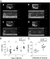
Sickle mice exhibit progressive LAE compared with their age- and gender-matched WT controls, which is unique to SCA and does not develop in the IDA mice. (A) Representative images of parasternal long-axis M-mode imaging through the aortic root and LA demonstrating cross-sectional atrial dilation in a 1- and 8-mo-old sickle cell and WT mouse. The smaller upper image in each set shows the 2D picture of this region of the heart in the parasternal long axis, and the lower image records the movement of each pixel during the cardiac cycle, allowing quantification of structure sizes. Under normal conditions, the LA and aortic root are of approximately equal size. The subpanels are: (a) 1-mo-old WT control mouse; (b) 8-mo-old WT control mouse; (c) 1-mo-old sickle cell mouse (Berk-SS); (d) 8-mo-old Berk-SS. Ao, aortic root; LA, left atrium. (B) A temporal analysis of LAD by M-mode imaging of WT and Berk-SS mice starting soon after weaning to 8 mo of age. (C) Assessment of LAD by transthoracic echocardiography in experimentally induced IDA to achieve hemoglobin levels similar to sickle mice for 3 mo compared with LAD in sickle mice with a similar 3-mo duration of anemia. Data shown as mean ± SEM, n = 4 mice in WT, 6–11 mice in Berk-SS, and 10 mice in IDA; Mann–Whitney U test; ns, P > 0.05; *P ≤ 0.05; **P ≤ 0.01; ***P ≤ 0.001.
Fig. 3.
Fig. 3.
Sickle mice exhibit LV hypertrophy and dilation with preserved systolic function but increased myocardial fibrosis by CMR. Six- to 9-mo-old age- and gender-matched sickle (Berk-SS) and WT mice underwent CMR. Panels depict (A) left and right end-diastolic volumes (LVEDV and RVEDV, respectively); (B) LV ejection fraction (LVEF) and stroke volume (SV); (C) LVM; and (D) myocardial extracellular volume (ECV%) measured by contrast-enhanced T1 mapping. Data shown as mean ± SEM n= 3–7 mice in WT and 7–9 mice in Berk-SS; Mann–Whitney U test; ns, P > 0.05; **P ≤ 0.01; ***P ≤ 0.001.
Fig. 4.
Fig. 4.
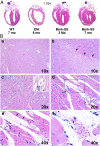
Sickle mice exhibit biventricular hypertrophy and microscopic multifocal ischemic changes and fibrosis. (A) Representative H&E-stained four-chamber views of WT, IDA, and sickle (Berk-SS) mouse hearts. Compared with an age- and gender-matched WT control, the 8-mo-old IDA mouse heart exhibits ventricular dilation but not hypertrophy after 3 mo of anemia. Biventricular hypertrophy and LA dilation are noted as early as 3 mo of age in the Berk-SS mouse. These findings worsen with age as evident in the 7-mo-old Berk-SS mouse. (B) H&E-stained mouse myocardium. Compared with an age- and gender-matched WT control (a), an 8-mo-old male Berk-SS mouse shows early ischemic changes characterized by loss of cross striations and decreased cardiomyocyte cytoplasmic eosinophilia (black arrows) (b). Late and more-severe ischemic changes, such as vanishing dead cardiomyocytes, are also noted in the Berk-SS mouse (d and e), and an occluded/congested microvessel with sickled RBC is noted adjacent to these ischemic changes (f, Inset). Microvessels are patent in the WT mouse (c, Inset).
Fig. 5.
Fig. 5.
Patchy microscopic fibrosis is seen in sickle mice hearts. Sirius red (A and B) and Masson’s trichrome (C and D) staining of the myocardium of 8-mo-old sickle (Berk-SS) and WT mice. Patchy microscopic fibrosis can be seen in the Berk-SS mouse (B and D) but not the age- and gender-matched WT control mice (A and C).
Fig. 6.
Fig. 6.
Sickle cardiomyocytes exhibit hypoxia/ischemia-related mitochondrial ultrastructural changes, contracted sarcomeres, and chaotic remodeling. Representative EM images of WT and sickle mouse LV posterior wall myocardium. Compared with WT control (A and C), a subset of cardiomyocytes in an 8-mo-old sickle mouse show ultrastructural abnormalities (B, D, and E). Electron lucent areas between mitochondria and sarcomeres, and membranes of the SR and sarcomeres are noted (B, arrows). Mitochondria are hypertrophied, more abundant, more rounded, with less densely packed and often disrupted cristae (D, arrow). Sarcomeres in some of the sickle cells were contracted and hence shorter in diastole (D, double-headed arrow) compared with WT (C, double-headed arrow). Chaotic cardiomyocyte remodeling is noted in a 2-mo-old Berk-SS mouse (E). Sarcomere lengths of two, 8-mo-old sickle (Berk-SS) and age- and gender-matched WT controls are shown in F. Data shown as mean ± SEM, n = 187 sarcomere in WT and 150 sarcomere in Berk-SS, two mice per group, Mann–Whitney U test, ****P ≤ 0.0001.
Fig. 7.
Fig. 7.
(A) Gene Ontology pathways that are differentially regulated between WT and Berk-SS mouse myocardium (n = 3 WT and 3 in Berk-SS), following a sickle myocardial transcriptome (with associated P values) compared with WT myocardial transcriptome; arrows indicate up- or down-regulated pathways in the sickle myocardium. (B) Immunohistochemistry staining of CTGF in mouse myocardium. Compared with age- and gender-matched WT myocardium (a, c, e, g), an 8-mo-old Berk-SS mouse shows increased overall background expression (b, d, f, h) with several areas of pronounced focal staining (perivascular regions and areas of myocyte loss).
Fig. 8.
Fig. 8.
Biological network analysis of the SS down-regulated genes in the heart showed dramatic loss of expression of an extensive set of genes associated with maintenance of the electrophysiological and structural integrity of the heart. RNAseq showed that 31 ion channel genes and genes associated with electrophysiologic function were down-regulated in Berk-SS mouse myocardium compared with WT (n = 3 WT and 3 in Berk-SS). The seven key down-regulated genes (yellow: CPT1A, CPT2, SCN4B, AKAP9, SCN4A, KCNJ2, CACNA1S) are associated with arrhythmias and sudden cardiac death. The cardiac networks they affect and how they interact with the rest of the down-regulated genes associated with rhythm dysfunction, including prolonged QTc, are shown.
Fig. 9.
Fig. 9.
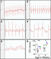
QTc prolongation, cardiac ischemic events, and fatal arrhythmias are evident in antemortem EKG tracings of sickle mice. Compared with the normal sinus rhythm in a 2-mo-old WT control (1), antemortem EKG tracings of an age-matched Berk-SS mouse shows ST depression (2). Another 2-mo-old Berk-SS mouse’s antemortem EKG tracings show slower heart rate and P-wave irregularity (3), which progressed into outright atrial flutter with 4–5:1 AV conduction (4), and eventual ventricular fibrillation (5). Sudden and significant QTc prolongation of three 6-wk-old Berk-SS male mice few days before their death (filled triangle) compared with their age- and gender-matched WT (open triangles) controls (6). Data shown as mean ± SEM n = 8 mice in WT and 3 mice in Berk-SS; Mann-Whitney U test; ns, P > 0.05; *P ≤ 0.05.

Comment in

References

    1. Quinn CT, Rogers ZR, Buchanan GR. Survival of children with sickle cell disease. Blood. 2004;103(11):4023–4027. - PMC - PubMed
    1. Lanzkron S, Carroll CP, Haywood C., Jr Mortality rates and age at death from sickle cell disease: U.S., 1979–2005. Public Health Rep. 2013;128(2):110–116. - PMC - PubMed
    1. Fitzhugh CD, et al. Cardiopulmonary complications leading to premature deaths in adult patients with sickle cell disease. Am J Hematol. 2010;85(1):36–40. - PMC - PubMed
    1. Darbari DS, et al. Circumstances of death in adult sickle cell disease patients. Am J Hematol. 2006;81(11):858–863. - PubMed
    1. Gladwin MT, et al. Pulmonary hypertension as a risk factor for death in patients with sickle cell disease. N Engl J Med. 2004;350(9):886–895. - PubMed

Publication types

MeSH terms