. 2016 Aug 23;113(34):9539-44.
doi: 10.1073/pnas.1600702113. Epub 2016 Aug 8.

Differential lipid binding of vinculin isoforms promotes quasi-equivalent dimerization

Affiliations

Differential lipid binding of vinculin isoforms promotes quasi-equivalent dimerization

Krishna Chinthalapudi et al. Proc Natl Acad Sci U S A. .

Abstract

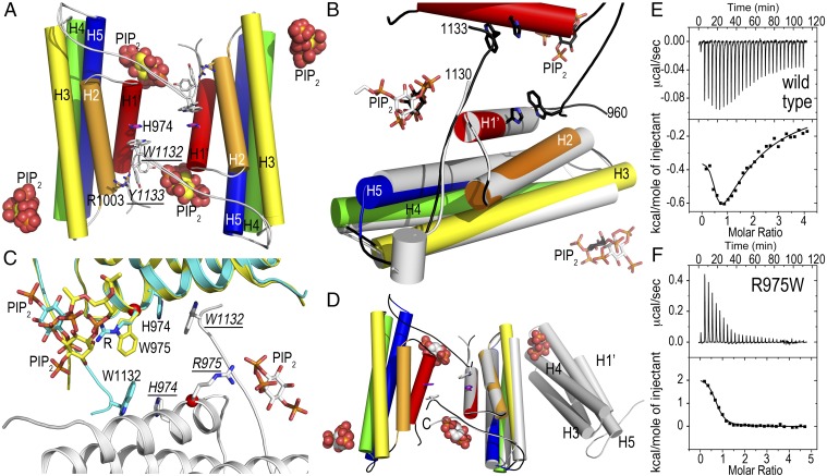
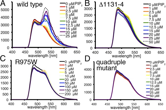
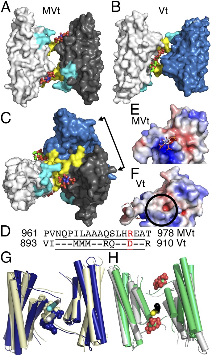
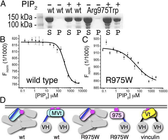
The main cause of death globally remains debilitating heart conditions, such as dilated cardiomyopathy (DCM) and hypertrophic cardiomyopathy (HCM), which are often due to mutations of specific components of adhesion complexes. Vinculin regulates these complexes and plays essential roles in intercalated discs that are necessary for muscle cell function and coordinated movement and in the development and function of the heart. Humans bearing familial or sporadic mutations in vinculin suffer from chronic, progressively debilitating DCM that ultimately leads to cardiac failure and death, whereas autosomal dominant mutations in vinculin can also provoke HCM, causing acute cardiac failure. The DCM/HCM-associated mutants of vinculin occur in the 68-residue insert unique to the muscle-specific, alternatively spliced isoform of vinculin, termed metavinculin (MV). Contrary to studies that suggested that phosphoinositol-4,5-bisphosphate (PIP2) only induces vinculin homodimers, which are asymmetric, we show that phospholipid binding results in a domain-swapped symmetric MV dimer via a quasi-equivalent interface compared with vinculin involving R975. Although one of the two PIP2 binding sites is preserved, the symmetric MV dimer that bridges two PIP2 molecules differs from the asymmetric vinculin dimer that bridges only one PIP2 Unlike vinculin, wild-type MV and the DCM/HCM-associated R975W mutant bind PIP2 in their inactive conformations, and R975W MV fails to dimerize. Mutating selective vinculin residues to their corresponding MV residues, or vice versa, switches the isoform's dimeric constellation and lipid binding site. Collectively, our data suggest that MV homodimerization modulates microfilament attachment at muscular adhesion sites and furthers our understanding of MV-mediated cardiac remodeling.

Keywords: cardiomyopathy; cell adhesion; cytoskeleton; metavinculin; vinculin.


Conflict of interest statement

The authors declare no conflict of interest.

Figures

Fig. 1.
Fig. 1.
Structural and thermodynamic analyses of PIP2-induced MV dimerization. (A) Cartoon drawing of the 3.1-Å crystal structure of MVt (residues 959–1134) in complex with PIP2. There are four five-helix bundle MVt molecules in the asymmetric unit (for clarity, only the dimer is shown) and the four polypeptide chains can be superimposed with rmsd ranging from 0.2 Å to 0.29 Å for over 851 atoms. There are five PIP2 molecules in the asymmetric unit and the four PIP2 binding sites near the C terminus (proximal to R1128) are occupied, indicating that this site is important for dimerization, and the second site (K1012 and R1013) is occupied in one molecule in the asymmetric unit. W1132 from one MVt molecule stacks against H974 from a twofold-related MVt molecule in a domain swap-like arrangement. MVt α-helices are labeled H1′ and H2–H5 and are colored spectrally (red, residues 964–979, H1′; orange, 986–1006, H2; yellow, 1009–1039, H3; green, 1042–1074, H4; and blue, 1081–1116, H5). PIP2 molecules are shown as spheres. (B) Close-up view of the superposition (rmsd of 0.399 Å for 910 atoms) of the MVt/PIP2 structure onto the 2.3-Å truncated (Δ1131–1134) PIP2-bound MVt crystal structure that does not dimerize (gray). Although the two PIP2 binding sites are conserved (black sticks for PIP2 in the MVt Δ1131–1134 /PIP2 structure), the truncated C terminus does not allow for the lipid-induced dimerization. MVt α-helices are labeled H1′ and H2–H5 and are colored spectrally; MVt Δ1131–1134 is shown in gray. (C) Close-up view of the superposition (rmsd of 0.393 Å for 885 atoms) of the MVt/PIP2 structure onto the 2.9-Å PIP2-bound DCM/HCM-associated MVt R975W crystal structure that does not dimerize. The two polypeptide chains of the dimeric wild-type MVt are colored in cyan and gray, respectively, and the DCM/HCM-associated MVt R975W in yellow. (D) Superposition (rmsd of 0.496 Å for 949 atoms) of wild-type MVt/PIP2 (α-helices are colored spectrally; PIP2 is shown as spheres) onto the R975Q–K979Q–R1107Q–R1128Q mutant MVt/PIP2 structure (shown in gray). The quadruple mutant binds PIP2 and does not dimerize, and a symmetry-related molecule occupies the second PIP2 binding site. In the mutant MVt structure, the loop after the last α-helix H5 (residues 1116–1123) adopts a conformation in between the one found for the wild-type MVt/PIP2 and the full-length MV structures (not shown for clarity). Otherwise, the mutant and wild-type MVt/PIP2 structures are very similar for residues 959–1129. (E) Isothermal titration calorimetry binding traces for calorimetric titrations of PIP2 to MVt. (Upper) Sequence of peaks corresponding to each injection where the monitored signal is the additional thermal power needed to be supplied or removed to keep a constant temperature relative to the reference cell. (Lower) Integrated heat plot of the area of each peak per mole versus the molar ratio. The solid line corresponds to theoretical curves with Kd = 0.7 μM, ΔH = −0.335 ± 0.008 kcal/mol and n = 0.483 ± 0.054 for the high-affinity binding site and Kd = 0.59 μM, ΔH = −2.3 ± 0.222 kcal/mol and n = 0.98 ± 0.81 for the lower affinity binding site. MVt protein samples were in the cell at a concentration of 25–30 μM and PIP2 was in the syringe at a concentration of 600–700 μM at 1:20 or 1:30 molar ratio at 25 °C. (F) ITC binding traces for calorimetric titrations of PIP2 to MVt R975W. Kd = 2.3 μM, ΔH = 2.1 ± 0.064 kcal/mol, and n = 0.71 ± 0.015.
Fig. 2.
Fig. 2.
PIP2-induced MV dimerization in solution. (A–D) PIP2 micelles induce dimerization of wild-type but not mutant MV. Emission spectra of CFP-MVt and YPF-MVt FRET pairs. (A) Wild type, (B) Δ1131–1134, (C) R975W, and (D) R975Q, K979Q, R1107Q, R1128Q mutant in the absence (black trace) and presence of increasing concentrations of PIP2 (colored spectrally; black dotted trace, highest concentration) upon excitation at 414 nm. The ordinate shows the relative fluorescence.
Fig. 3.
Fig. 3.
The PIP2-induced Vt dimer differs from PIP2-induced MVt dimer despite almost identical polypeptide chains via quasi-equivalent intermolecular contacts. (A) Surface representation of the MVt/PIP2 dimer. Individual subunits colored in white and gray, respectively. Each MV molecule binds one PIP2, resulting in a symmetric homodimer with PIP2 (shown as spheres; oxygen, red; carbon, blue) binding near the N terminus (residues 959–965, cyan) and C terminus (residues 1122–1134, yellow). (B) Surface representation of the Vt/PIP2 dimer. Individual subunits are colored in white and blue, respectively. One PIP2 molecule (carbon atoms, green) is sandwiched between two vinculin molecules, resulting in an asymmetric dimer. PIP2 interacts with the termini of both protomers. (C) Superposition of the MVt/PIP2 and Vt/PIP2 dimers rotated 90° down (or 90° up) with respect to the view shown in A (or B, respectively). The double arrow shows the relative (Vt versus MVt) movement of about 75° of the second protomer. (D) Structure-based sequence alignment of the region that differs in the two isoform tail domains. Residues residing on α-helix H1′ or H1 in MV and vinculin, respectively, are underlined. R975, which is mutated to a tryptophan in HCM/DCM and binds to PIP2 in MV, is highlighted in red and corresponds to D907 in vinculin, which is not in contact with PIP2. (E) Electrostatic surface potential representation of the PIP2-bound MVt protomer. The electrostatic potential gradient is from −5 to +5 kBT (red, negative; blue, positive), where kB is Boltzmann constant and T is the temperature. The positive electrostatic surface potential is mainly contributed by R1128, R975, R1107, and K979. (F) Electrostatic surface potential representation of the PIP2-bound Vt protomer. Aspartate 907 replaces arginine 975 of MVt, thereby generating a negative electrostatic potential at this site (circle). The PIP2 binding site in Vt is mainly contributed by K1061, K915, and K924. (G) Mutating Q971, R975, and T978 in MVt (light yellow) to the corresponding Vt residues (RDR) converts the lipid-induced symmetric MVt dimer into an asymmetric Vt-like dimer. The Vt/PIP2 dimer is superimposed and shown in blue, whereby one mutant MVt subunit (Left, light yellow) is superimposed onto one Vt subunit (Left, blue) highlighting the ∼160°/200° rotation axes for the mutated second MVt (white) or Vt (cyan), resulting in a relative movement of the second Vt (Right, blue) of 3° with respect to the second mutated MVt (Right, light yellow) via quasi-equivalent intermolecular interactions. (H) Mutating R903, D907, and R910 in Vt (gray) to the corresponding MVt residues (QRT) converts the lipid-induced asymmetric Vt dimer into a symmetric MVt-like dimer. One mutant Vt subunit (Left, gray) is superimposed onto one MVt subunit (Left, green) highlighting the 180° axes of the mutated Vt or MVt dyads (black and yellow, respectively), resulting in a relative movement of the second mutated Vt (Right, gray) of ∼20° with respect to the second MVt subunit (Right, green) via quasi-equivalent intermolecular interactions.
Fig. 4.
Fig. 4.
Membrane attachment differs in the vinculin isoforms. (A) Lipid cosedimentation assays of human full-length wild type (first four lanes) and DCH/HCM-associated mutant R975W MV (last four lanes). P, pellet; S, supernatant; wt, wild type. (B and C) Microscale thermophoresis analyses show that PIP2 binds (B) wild-type MV and (C) the DCM/HCM-associated mutant MV with affinities of 313.8 ± 8.8 μM (SD n = 3) and 53.7 ± 3.4 μM (SD n = 3), respectively. (D) Full-length wild-type (wt) MV (individual VH domains, gray; MVt extended coil, blue; and five-helix MVt bundle, cyan) binds PIP2 (magenta) in its closed conformation. Upon activation, PIP2 induces MV dimerization by releasing the extended coil (VH, gray sphere). The DCM/HCM-associated MV R975W mutant (five-helix MVt bundle, pink) also binds PIP2 in its closed conformation but does not dimerize at the cell membrane. Finally, only activated vinculin (five-helix Vt bundle, yellow) binds PIP2, which induces dimerization.

References

    1. Xu W, Baribault H, Adamson ED. Vinculin knockout results in heart and brain defects during embryonic development. Development. 1998;125(2):327–337. - PubMed
    1. Rodríguez Fernández JL, Geiger B, Salomon D, Ben-Ze’ev A. Overexpression of vinculin suppresses cell motility in BALB/c 3T3 cells. Cell Motil Cytoskeleton. 1992;22(2):127–134. - PubMed
    1. Coll JL, et al. Targeted disruption of vinculin genes in F9 and embryonic stem cells changes cell morphology, adhesion, and locomotion. Proc Natl Acad Sci USA. 1995;92(20):9161–9165. - PMC - PubMed
    1. Belkin AM, Ornatsky OI, Glukhova MA, Koteliansky VE. Immunolocalization of meta-vinculin in human smooth and cardiac muscles. J Cell Biol. 1988;107(2):545–553. - PMC - PubMed
    1. Belkin AM, Ornatsky OI, Kabakov AE, Glukhova MA, Koteliansky VE. Diversity of vinculin/meta-vinculin in human tissues and cultivated cells. Expression of muscle specific variants of vinculin in human aorta smooth muscle cells. J Biol Chem. 1988;263(14):6631–6635. - PubMed

Publication types

MeSH terms

LinkOut - more resources