Intranodal Palisaded Myofibroblastoma: Radiological and Cytological Overview
- PMID: 27504146
- PMCID: PMC4959455
- DOI: 10.12659/PJR.895743
Intranodal Palisaded Myofibroblastoma: Radiological and Cytological Overview
Abstract
Background: Intranodal palisaded myofibroblastoma is a benign and very rare mesenchymal neoplasm of the lymph nodes originating from differentiated smooth muscle cells and myofibroblasts.
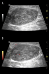
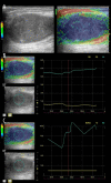
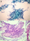
Case report: We report a case of intranodal palisaded myofibroblastoma in an 84-year-old woman with Parkinson's disease that presented as a left inguinal mass. The diagnosis was made using ultrasound-guided fine needle aspiration biopsy and consequent cytopathological examination that included immunohistochemical analysis. Herein, we discuss the presentation of a rare intranodal palisaded myofibroblastoma with emphasis on its ultrasonographic and cytopathologic features.
Conclusions: Intranodal palisaded myofibroblastoma should be considered in the differential diagnosis of inguinal lymphadenopathy and the diagnosis is possible with cytopathologic exam and immunohistochemical analysis using ultrasound-guided FNA biopsy, guiding the clinician to nodal excision rather than aggressive measures.
Keywords: Biopsy, Fine-Needle; Histocytological Preparation Techniques; Lymphatic Abnormalities; Ultrasonography.
Figures


Similar articles
-
Parotid's Intranodal Palisaded Myofibroblastoma: A Very Rare Tumour.Indian J Otolaryngol Head Neck Surg. 2023 Sep;75(3):2702-2706. doi: 10.1007/s12070-023-03771-9. Epub 2023 Apr 29. Indian J Otolaryngol Head Neck Surg. 2023. PMID: 37636792 Free PMC article.
-
Intranodal palisaded myofibroblastoma: a review of the literature.Int J Surg Pathol. 2013 Aug;21(4):337-41. doi: 10.1177/1066896913489348. Epub 2013 May 27. Int J Surg Pathol. 2013. PMID: 23714684 Review.
-
Cytopathological findings of intranodal palisaded myofibroblastoma: Case report and review of the literature.Diagn Cytopathol. 2023 Aug;51(8):E248-E254. doi: 10.1002/dc.25172. Epub 2023 May 27. Diagn Cytopathol. 2023. PMID: 37243568 Review.
-
Axillary intranodal palisaded myofibroblastoma: report of a case associated with chronic mastitis.BMJ Case Rep. 2014 Oct 16;2014:bcr2014205877. doi: 10.1136/bcr-2014-205877. BMJ Case Rep. 2014. PMID: 25323283 Free PMC article.
-
Intranodal palisaded myofibroblastoma originating from retroperitoneum: an unusual origin.BMC Clin Pathol. 2011 Jun 30;11:7. doi: 10.1186/1472-6890-11-7. BMC Clin Pathol. 2011. PMID: 21718465 Free PMC article.
Cited by
-
Parotid's Intranodal Palisaded Myofibroblastoma: A Very Rare Tumour.Indian J Otolaryngol Head Neck Surg. 2023 Sep;75(3):2702-2706. doi: 10.1007/s12070-023-03771-9. Epub 2023 Apr 29. Indian J Otolaryngol Head Neck Surg. 2023. PMID: 37636792 Free PMC article.
-
Intranodal palisaded myofibroblastoma in the submandibular gland region: a case report.Front Oncol. 2024 Jul 31;14:1362090. doi: 10.3389/fonc.2024.1362090. eCollection 2024. Front Oncol. 2024. PMID: 39148907 Free PMC article.
-
Sonoelastographic evaluation for benign neck lymph nodes and parathyroid lesions.J Ultrason. 2018;18(75):284-289. doi: 10.15557/JoU.2018.0041. J Ultrason. 2018. PMID: 30763011 Free PMC article.
-
Intranodal Palisaded Myofibroblastoma: A Diagnostic Differential for Inguinal Lymphadenopathy.Am J Case Rep. 2021 Dec 18;22:e934752. doi: 10.12659/AJCR.934752. Am J Case Rep. 2021. PMID: 34921129 Free PMC article.
-
Intranodal palisaded myofibroblastoma expressing DOG1: focusing on the potential diagnostic pitfalls.Rom J Morphol Embryol. 2021 Jul-Sep;62(3):849-853. doi: 10.47162/RJME.62.3.25. Rom J Morphol Embryol. 2021. PMID: 35263416 Free PMC article.
References
-
- Sarma NH, Arora KS, Varalaxmi KP. Intranodal palisaded myofibroblastoma: A case report and an update on etiopathogenesis and differential diagnosis. J Can Res Ther. 2013;9:295–98. - PubMed
-
- Bhullar JS, Varshney N, Dubay L. Intranodal palisaded myofibroblastoma: A review of the literature. Int J Surg Pathol. 2013;21:337–41. - PubMed
-
- Kleist B, Poetsch M, Schmoll J. Intranodal palisaded myofibroblastoma with overexpression of cyclin D1. Arch Pathol Lab Med. 2003;127:1040–43. - PubMed
Publication types
LinkOut - more resources
Full Text Sources
Other Literature Sources
