

Effect of Gestational Diabetes on Purkinje and Granule Cells Distribution of the Rat Cerebellum in 21 and 28 days of Postnatal Life
- PMID: 27504151
- PMCID: PMC4741272
Effect of Gestational Diabetes on Purkinje and Granule Cells Distribution of the Rat Cerebellum in 21 and 28 days of Postnatal Life
Abstract
Introduction: Diabetes mellitus is associated with nervous system alterations in both human and animal models. This study was done to determine the effect of gestational diabetes on the Purkinje and granular cells in the cerebellum of rat offspring.
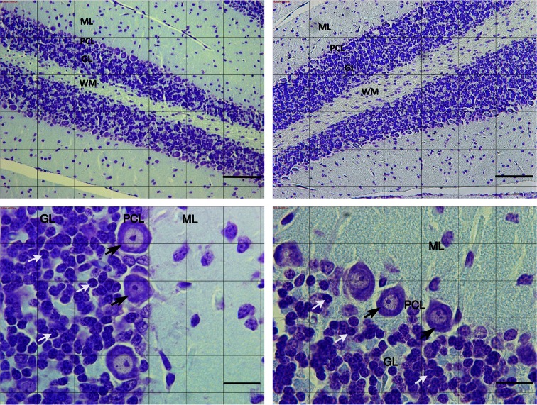
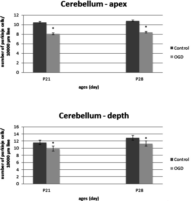
Methods: 10 Wistar rats Dams were randomly allocated in control and diabetic group. The experimental group received 40 mg/kg/body weight of streptozotocin (STZ) at the first day of gestation and control groups received saline injection intraperitoneally (IP). Six male offsprings of gestational diabetic mothers and control dams, at the 21, 28 postnatal days were randomly scarified and coronal sections of cerebellum (6 micrometer) serially collected. The neurons were stained with cresyl violet.
Results: The Purkinje cells density in the apex and depth of cerebellum in P21, in the experimental group was reduced 23% and 15% in comparison with the control group (P<0.001). The granular cells density in the experimental group was reduced 19.58% and 18.3% in comparison with the controls (P<0.001). The Purkinje cells density of cerebellum in P28, in the diabetic group reduced to 22.12% and 12.62% in comparison with the control group (P<0.001). The granular cells density in the diabetic group reduced 17.14% and 16.12% in comparison with the control group (P<0.001).
Discussion: The Purkinje and granular cells significantly reduced in gestational diabetes rat offspring.
Keywords: Cerebellum; Gestational diabetes; Granular cell; Purkinje cell; Rat.
Figures
Similar articles
-
Gestational diabetes induced neuronal loss in CA1 and CA3 subfields of rat hippocampus in early postnatal life.Folia Morphol (Warsz). 2012 May;71(2):71-7. Folia Morphol (Warsz). 2012. PMID: 22648583
-
Purkinje cells loss in off spring due to maternal morphine sulfate exposure: a morphometric study.Anat Cell Biol. 2012 Jun;45(2):121-7. doi: 10.5115/acb.2012.45.2.121. Epub 2012 Jun 30. Anat Cell Biol. 2012. PMID: 22822467 Free PMC article.
-
Effect of intrauterine morphine sulfate exposure on cerebellar histomorphological changes in neonatal mice.Folia Neuropathol. 2011;49(4):328-34. Folia Neuropathol. 2011. PMID: 22212923
-
Gestational diabetes influences retinal Muller cells in rat's offspring.Iran J Basic Med Sci. 2017 Feb;20(2):216-221. doi: 10.22038/ijbms.2017.8251. Iran J Basic Med Sci. 2017. PMID: 28293400 Free PMC article.
-
Preliminary morphological and morphometric study of rat cerebellum following sodium arsenite exposure during rapid brain growth (RBG) period.Toxicology. 2007 May 5;234(1-2):10-20. doi: 10.1016/j.tox.2007.01.024. Epub 2007 Feb 11. Toxicology. 2007. PMID: 17374429
Cited by
-
Inflammatory Consequences of Maternal Diabetes on the Offspring Brain: a Hippocampal Organotypic Culture Study.Neurotox Res. 2019 Aug;36(2):357-375. doi: 10.1007/s12640-019-00070-6. Epub 2019 Jun 13. Neurotox Res. 2019. PMID: 31197747 Free PMC article.
-
Restorative effects of Momordica charantia extract on cerebellar GFAP and NGF expression in pregnant diabetic rats and their offspring.PLoS One. 2025 Apr 4;20(4):e0321022. doi: 10.1371/journal.pone.0321022. eCollection 2025. PLoS One. 2025. PMID: 40184394 Free PMC article.
-
Cerebral Effects of Neonatal Dysglycemia.Clin Perinatol. 2022 Jun;49(2):405-426. doi: 10.1016/j.clp.2022.02.008. Epub 2022 Apr 21. Clin Perinatol. 2022. PMID: 35659094 Free PMC article. Review.
-
Gestational Diabetes Mellitus and its Effects on the Developing Cerebellum: A Narrative Review on Experimental Studies.Iran J Child Neurol. 2024 Spring;18(2):9-22. doi: 10.22037/IJCN.V18I2.36632. Epub 2024 Mar 12. Iran J Child Neurol. 2024. PMID: 38617398 Free PMC article. Review.
-
Dietary Marine Oils Selectively Decrease Obesogenic Diet-Derived Carbonylation in Proteins Involved in ATP Homeostasis and Glutamate Metabolism in the Rat Cerebellum.Antioxidants (Basel). 2024 Jan 15;13(1):103. doi: 10.3390/antiox13010103. Antioxidants (Basel). 2024. PMID: 38247527 Free PMC article.
References
-
- Ahmadpour S. H., Haghir H. (2011). Diabetes mellitus type 1 induces dark neuron formation in the dentate gyrus: a study by Gallyas’ method and transmission electron microscopy. Romanian Journal of Morphology and Embryology, 52 (2), 575– 579. - PubMed
-
- Allen D. A., Yaqoob M. M., Harwood S. M. (2005). Mechanisms of high glucose induced apoptosis and its relationship to diabetic complications. Journal of Nutritional Biochemistry, 16 (12), 705– 713. - PubMed
-
- Beauquis J., Roig P., Homo-Delarche F., De Nicola A., Saravia F. (2006). Reduced hippocampal neurogenesis and number of hilarneurones in streptozotocin-induced diabetic mice: reversion by antidepressant treatment. European Journal of Neuroscience, 23 (6), 1539– 1546. - PubMed
-
- Biessels G. J., Heide L. P. V., Kamal A., Bleys R. L. A. W., Gispen W. H. (2002). Ageing and diabetes: implications for brain function. European Journal of Pharmacology, 441, 1– 14. - PubMed
-
- Chen G., Goeddel D. V. (2002). TNF-R1 signaling: a beautiful pathway. Science, 296 (5573), 1634– 1635. - PubMed
LinkOut - more resources
Full Text Sources
Miscellaneous