

doi: 10.1155/2016/1498135.
Epub 2016 Jul 18.
Spontaneous Minced Cartilage Procedure for Unexpectedly Large Femoral Condyle Surface Defect
Affiliations
- PMID: 27504207
- PMCID: PMC4967685
- DOI: 10.1155/2016/1498135
Item in Clipboard
Spontaneous Minced Cartilage Procedure for Unexpectedly Large Femoral Condyle Surface Defect
Case Rep Orthop.
2016.
Abstract
Articular cartilage defects at the knee joint are being identified and treated with increasing frequency. Chondrocytes may have strongest potential to generate high-quality repair tissue within the defective region, in particular when large diameter defects are present. Autologous chondrocyte implantation is not available in every country. We present a case where we spontaneously covered an acute cartilage defect, which was significantly larger than expected and loose during initial arthroscopic inspection after reading preoperative MRI, by mincing the separated fragment and directly implanting the autologous cartilage chips into the defective region.
Figures
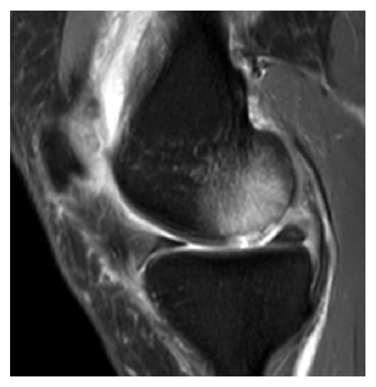
Sagittal T2-weighted MRI of left knee joint depicting cartilage lesion and large underlying BME at dorsomedial femoral condyle.
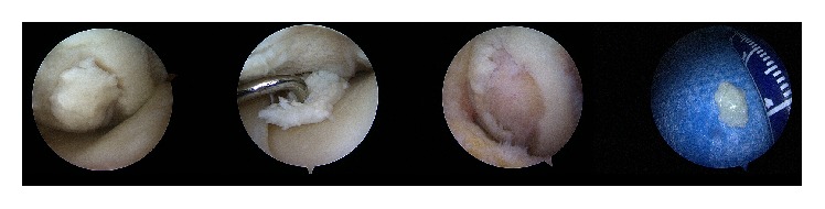
Intraoperative arthroscopic images in display of large separated fragment in situ, unstable under probing with remaining large cartilage lesion at dorsomedial femoral condyle after removal.
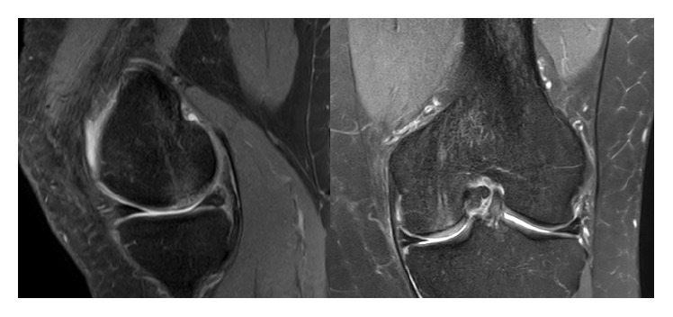
Sagittal and coronal T2-weighted MRI of left knee joint illustrating well repaired previous defective area with almost isointense, firmly integrated neocartilage formation at similar height to the surrounding cartilage without significant BME and healthy appearing subchondral bone at dorsomedial femoral condyle (6 months postoperatively).
References
-
- Marlovits S., Singer P., Zeller P., Mandl I., Haller J., Trattnig S. Magnetic resonance observation of cartilage repair tissue (MOCART) for the evaluation of autologous chondrocyte transplantation: determination of interobserver variability and correlation to clinical outcome after 2 years. European Journal of Radiology. 2006;57(1):16–23. doi: 10.1016/j.ejrad.2005.08.007. - DOI - PubMed
LinkOut - more resources
Full Text Sources
Other Literature Sources
