

doi: 10.3390/jdb4030022.
Hedgehog Signalling in the Embryonic Mouse Thymus
Affiliations
- PMID: 27504268
- PMCID: PMC4975939
- DOI: 10.3390/jdb4030022
Item in Clipboard
Hedgehog Signalling in the Embryonic Mouse Thymus
J Dev Biol.
.
Abstract
T cells develop in the thymus, which provides an essential environment for T cell fate specification, and for the differentiation of multipotent progenitor cells into major histocompatibility complex (MHC)-restricted, non-autoreactive T cells. Here we review the role of the Hedgehog signalling pathway in T cell development, thymic epithelial cell (TEC) development, and thymocyte-TEC cross-talk in the embryonic mouse thymus during the last week of gestation.
Keywords: Gli1; Gli2; Gli3; Hedgehog; Ihh; Shh; T cell development; thymic epithelial cell (TEC); thymus.
Conflict of interest statement
The authors declare no conflict of interest.
Figures
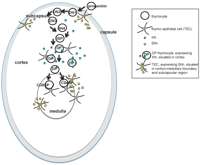
Hedgehog (Hh) expression in different microenvironments as thymocytes migrate through the embryonic thymus. The cartoon illustrates the stages of T cell development in the embryonic thymus, as developing thymocytes move through different thymus microenvironments and receive different amounts of Hh signal. Molecules of Indian hedgehog (Ihh, expressed by double positive (DP) thymocytes) are shown in green, and molecules of Sonic hedgehog (Shh, expressed by TEC in the sucbcapsular region and medulla) are shown in yellow. Progenitor cells first enter the embryonic thymus through the capsule on ~E12.5, and migrate towards the centre of the thymus as they differentiate. DP cells first appear on E16.5 and are located in the cortex. Mature single positive (SP) T cells and mature medullary TEC (mTEC) and cortical TEC (cTEC) populations are present by birth.
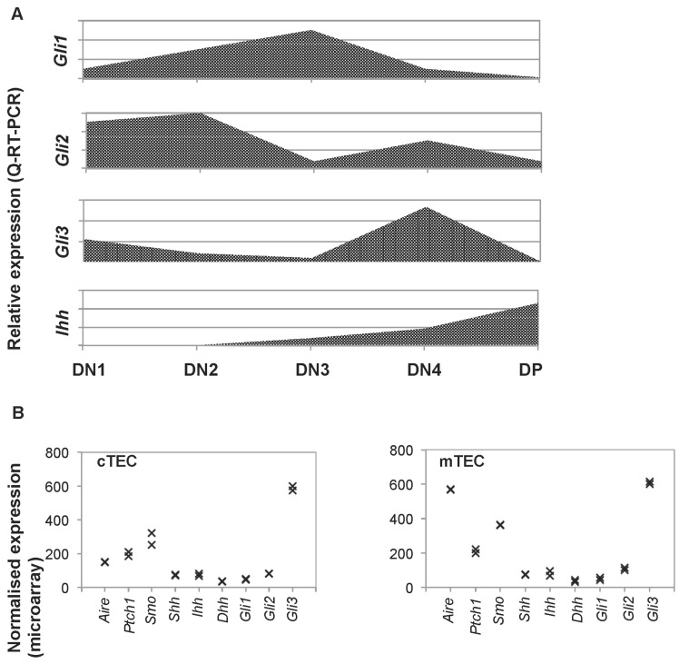
Expression of components of the Hh signalling pathway in embryonic thymocytes and TEC. (A) Graphs illustrate the relative expression of Gli1, Gli2, Gli3, and Ihh in sorted DN1, DN2, DN3, DN4, and DP thymocytes from E16.5 thymus, determined by quantitative reverse transcription polymerase chain reaction (QRT-PCR); (B) Plots show the normalised expression of Aire, Ptch1, Smo, Shh, Ihh, Dhh, Gli1, Gli2, and Gli3 by microarray analysis (GSE81433) from fluorescence activated cell sorting (Facs)-sorted cTEC (CD45−EpCam1+Ly51+UEA-1−, left-hand plot), and mTEC (CD45−EpCam1+Ly51−UEA-1+, right-hand plot); populations prepared from E14.5 WT (C57BL/6) foetal thymus organ culture (FTOC) after 7 days in culture. TEC were isolated as described [29].
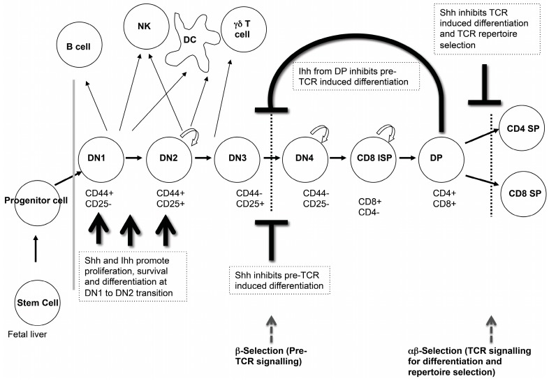
Hh signalling regulates multiple stages of thymocyte development. The cartoon illustrates the different stages of thymocyte differentiation that are regulated by Hh signalling in the embryonic thymus. (TCR: T cell receptor.)
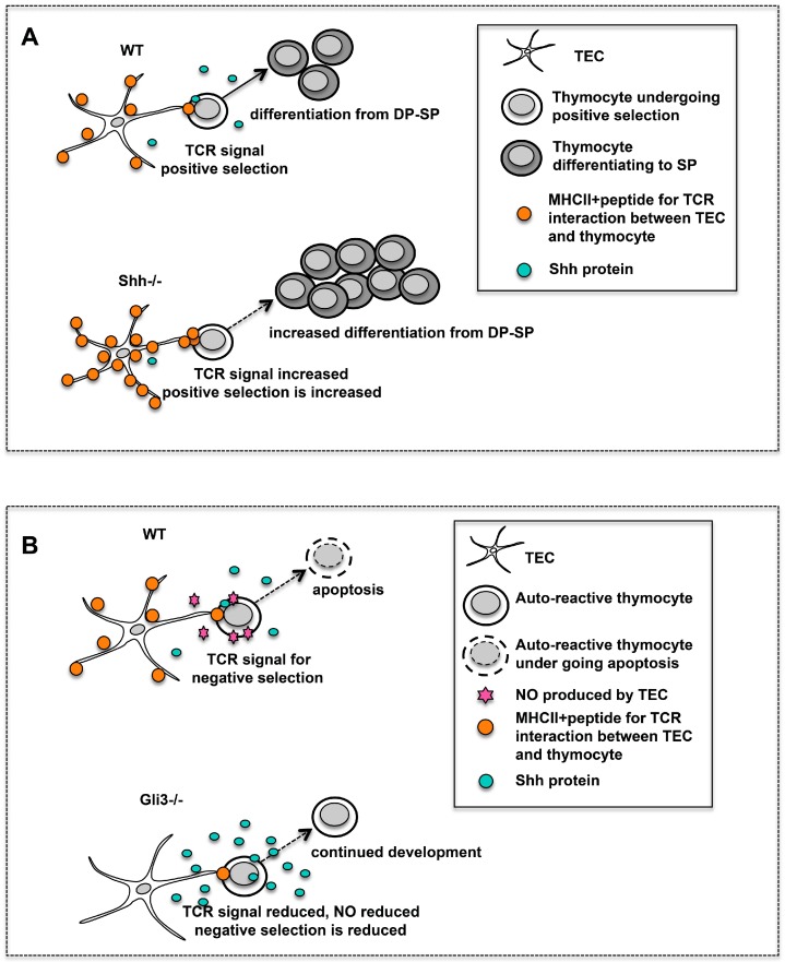
Hh signalling at the transition from DP to SP thymocyte. (A) The cartoon illustrates the influence of Shh on positive selection. Positive selection is increased in the Shh−/− thymus and the proportion of CD4SP cells is increased. Cell surface expression of major histocompatibility complex class II (MHCII) is increased on Shh−/− TEC compared to wild type (WT) TEC. (B) The influence of Gli3 on negative selection. In the Gli3−/− thymus MHCII expression is decreased in TEC and nitric oxide (NO) activity is decreased. This may allow SP thymocytes to escape from negative selection.
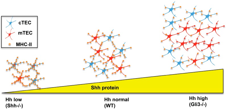
Impact of Shh-deficiency and Gli3-deficiency on TEC differentiation and function. In the Shh−/− embryonic thymus, Hh protein levels are low, there are fewer TEC, and the ratio of mTEC (shown in red) to cTEC (shown in blue) is decreased, but expression of MHCII is increased on individual TEC, compared to WT. In the Gli3−/− embryonic thymus—which has increased levels of Hh pathway activation—the opposite phenotype is seen. TEC numbers are increased, but MHCII expression on individual TEC is reduced compared to WT.
References
Grants and funding
LinkOut - more resources
Full Text Sources
Other Literature Sources
Molecular Biology Databases
Research Materials