. 2016 Jul 20:3:162-71.
doi: 10.1016/j.ejro.2016.05.007. eCollection 2016.

Role of dual energy spectral computed tomography in characterization of hepatocellular carcinoma: Initial experience from a tertiary liver care institute

Affiliations

Role of dual energy spectral computed tomography in characterization of hepatocellular carcinoma: Initial experience from a tertiary liver care institute

S T Laroia et al. Eur J Radiol Open. .

Abstract

Objective: To investigate dual-energy spectral CT in characterization of hepatocellular Carcinoma (HCC) in patients with chronic liver disease.

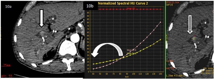
Methods: Dynamic computed tomography (CT) was performed in 3600 patients (2879 males; 721 females, mean age 50.9 ± 11.9 years) with working clinical diagnosis of liver cirrhosis for hepatocellular carcinoma screening and other clinical indications. The study was conducted over a period of 3 years. During dynamic CT scanning, spectral (monochromatic) and routine (polychromatic) CT acquisitions were obtained on a single tube, dual energy, 64 slice multi-detector CT scanner. Imaging findings were studied on routine CT. On the basis of routine CT findings, indeterminate lesions (lesions not showing characteristic hypervascularity followed by washout on dynamic routine CT scan) that were referred for biopsy or surgery were segregated. A retrospective blinded review of the lesions, acquired by the spectral CT acquisitions was done with the help of gem stone imaging (GSI) software to characterize these lesions. All the above lesions were analyzed qualitatively in the arterial phase for lesion conspicuity as well as quantitatively using the monochromatic data sets and nodule Iodine concentration on material density maps, respectively. This data was studied with respect to predictability of HCC using the spectral CT technique. Iodine density of the lesion, surrounding liver parenchyma, and lesion to liver parenchyma ratio (LLR) were derived and statistically analyzed. Histopathology of the lesion in question was treated as gold standard for analysis.

Results: It was observed via statistical analysis that the value of iodine density of the lesion on material density sets of ≥29.5 mg/dl, enabled a discriminatory power of 86.5%, sensitivity of 90.5% with 95% confidence Interval (CI) (69.2-98.8%) and specificity of 81.2% with 95% Confidence Interval (54.4-95.9%) in predicting HCC. Qualitative assessment also showed higher lesion conspicuity with spectral CT image sets as compared to routine CT data.

Conclusion: This study reveals that spectral imaging is an excellent qualitative as well as a quantitative tool for assessing and predicting hepatocellular carcinoma in cirrhotic patients.

Keywords: CI, confidence interval; CT, computed tomography; DECT, dual energy computed tomography; Dual energy computed tomography; Functional imaging; GSI, gem stone imaging; HCC, hepatocellular carcinoma; Hepatocellular carcinoma; Iodine quantification; LLR, lesion to liver parenchyma ratio; MMD, monochromatic material density; Material density images; Spectral computed tomography.


Figures

Fig. 1
Fig. 1
55 year old male with hepatitis B related chronic liver disease on follow up CT scan with three sets of image datasets for analysis. a. Polychromatic routine dynamic arterial phase image to depict faintly enhancing lesion in segment IV of liver (bold arrow). b. Monochromatic image viewed at 55 keV from the spectrum of images derived between 40 to 140 kVp with lesion conspicuity more than previous routine image (bold arrow). c. Material decomposition image showing Iodine containing structures with rest of the components appearing suppressed, the focal lesion (bold arrow) shows iodine enhancement on the Iodine map.
Fig. 2
Fig. 2
55 year old male with hepatitis B related chronic liver disease on follow up CT scan showing axial section of the superior aspect of the liver in the arterial phase with spectral HU curve of enhancing lesion. a. Axial section of the superior aspect of liver showing ROI’s in the enhancing portion of the liver lesion labeled as (1), the surrounding liver parenchyma as (3) and within the aortic lumen as (2). b Spectral HU curve of the same lesion denoting the normalized aorta on the top of the graph (bold vertical arrow), the graph of the liver lesion in comparison to the normalized aorta (horizontal arrow without fill) and the normal liver curve (vertical arrow without fill).
Fig. 3
Fig. 3
45 year old male patient with a cirrhotic liver showing hypervascular lesions. Axial material decomposition images showing iodine density values and scatter plot of the lesions. a. Axial iodine density map of the material decomposition image dataset depicting iodine quantification done within the liver lesion using an ROI labeled (1), the surrounding liver parenchyma labeled as (2) and the Aorta as (3). b The values of the ROI’s measuring Iodine density in mg/ml of the liver lesion (horizontal arrow without fill) and the surrounding liver curve (vertical arrow without fill) has been plotted on the scatter plot with values normalized against the density in the Aorta ((bold vertical arrow).
Fig. 4
Fig. 4
Bland and Altmann graph depicting 2 observer agreement analysis on routine CT observations of qualitative lesion conspicuity.
Fig. 5
Fig. 5
Bland and Altmann graph depicting 2 observer agreement analysis on spectral CT observations of qualitative lesion conspicuity.
Fig. 6
Fig. 6
ROC curve for GSI lesion in HCC group.
Fig. 7
Fig. 7
55 year old male with hepatitis B related chronic liver disease on follow up CT scan. Comparison of an enhancing heterogeneous lesion in segment VIII of liver on routine versus low keV spectral CT. a. Enhancing component (open arrow) on routine CT. b. The enhancing lesion (bold arrow) is better visualized on low (55) keV spectral CT monochromatic image. c. The low attenuation (central ROI within it) showing fluid attenuation of 5HU on the routine CT.
Fig. 8
Fig. 8
55 year old male with hepatitis B related chronic liver disease on follow up CT scan. Spectral HU curve plot and GSI scatter plots to depict the attenuation and iodine density values of the low density portion of the nodule in segment VIII, seen in Fig. 7c. a. Spectral HU curve depicting the negative attenuation (green tracing on the graph) suggestive of fat component with the arrow showing the approximate values compared to the lesion (royal blue color trace) and the aorta enhancement (light blue trace). The fatty attenuation of the area marked (*). b. GSI scatter plot showing the iodine density of the same three lesions as depicted in Fig. 8a.
Fig. 9
Fig. 9
Gross specimen post hepatectomy showing the nodule (dotted circle) − steatohepatic variety of HCC) containing fat (*) within it.
Fig. 10
Fig. 10
63 year old cirrhotic male patient presented with suspicious blush at the porta, evaluated on a dynamic CT followed by analysis on spectral CT. a. Low keV (55kev) CT shows a definite focal (arrow, black outline) area of blush. b. Spectral HU curve shows that lesion (open white arrow in iodine density map) is visualized (white curved arrow pointing at attenuation difference) only at low keV as analyzed (yellow line tracing of the lesion).
Fig. 11
Fig. 11
Gross specimen of the mass (white arrow with black outlines) (moderately differentiated Grade 2 HCC on histopathology) with bile duct tumor thrombus, small vessels invasion, embolization and the bile duct, seen separate from mass.

References

    1. Bruix J., Sherman M. Management of hepatocellular carcinoma: an update AASLD, American Association for the Study of Liver Diseases. Hepatology. 2011;53(March (3)):1020–1022. PMID: 21374666. - PMC - PubMed
    1. European Association for the Study of the Liver, European Organisation for Research and Treatment of Cancer EASL-EORTC clinical practice guidelines: management of hepatocellular carcinoma. J. Hepatol. 2012;56:908–943. - PubMed
    1. Omata M., Lesmana L.A., Tateishi R., Chen P.J., Lin S.M., Yoshida H. Asian Pacific Association for the Study of the Liver consensus recommendations on hepatocellular carcinoma. Hepatol. Int. 2010;4:439–474. - PMC - PubMed
    1. Kudo M., Izumi N., Kokudo N., Matsui O., Sakamoto M., Nakashima O. HCC expert panel of Japan society of hepatology: management of hepatocellular carcinoma in japan: consensus-based clinical practice guidelines proposed by the Japan society of hepatology (JSH) 2010 updated version. Dig. Dis. 2011;29:339–364. - PubMed
    1. Forner A., Vilana R., Ayuso C., Bianchi L., Solé M., Ayuso J.R., Boix L., Sala M., Varela M., Llovet J.M., Brú C., Bruix J. Diagnosis of hepatic nodules 20 mm or smaller in cirrhosis: prospective validation of the noninvasive diagnostic criteria for hepatocellular carcinoma. Hepatology. 2008;47(January (1)):97–104. - PubMed

LinkOut - more resources