. 2016 Aug 8;38(3):227-33.
doi: 10.1016/j.devcel.2016.07.022.

Keratins Are Going Nuclear

Affiliations

Keratins Are Going Nuclear

Ryan P Hobbs et al. Dev Cell. .

Abstract

Previously thought to reside exclusively in the cytoplasm, the cytoskeletal protein keratin 17 (K17) has been recently identified inside the nucleus of tumor epithelial cells with a direct impact on cell proliferation and gene expression. We comment on fundamental questions raised by this new finding and the associated significance.


Figures

Figure 1
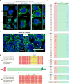
Figure 1. Nuclear Localization and Nuclear Export and Import Sequences for K17 and All IF Proteins
(A and B) Indirect immunostaining for K17 (green) in representative single-plane confocal micrographs of (A) cultured human epithelial tumor cells derived from three tissue types (breast, BT-20 [left]; cervix, HeLa [middle]; vulva, A431 [right]), treated with 40 nM LMB (bottom row) or vehicle (Veh, 70% methanol) (top row), and (B) human biopsy of basal cell carcinoma in situ (image at right is a zoomed view of the dotted box in image at left). Nuclei are stained with DAPI (blue). All scale bars for the xy plane are 10 mm. Scale bars for the z planes are 5 mm. BT-20 insets in (A) highlight diffuse pattern for nuclear K17. Arrows denote nuclear punctae containing K17. For detailed methods on cell culture, tissue acquisition, immunostaining, and imaging, see Hobbs et al. (2015). (C) Schematic of K17 protein (head in red, rod in green, tail in blue) highlighting the conservation of NLS (above) and NES (below) motifs across orthologs. Conserved residues are highlighted (red-to-yellow color gradient reflects most to least conserved). Numbers refer to the human K17 primary amino acid sequence (NCBI reference sequence NP_000413.1). Bolded residues are experimentally validated functional elements. # denotes NES residues predicted with ValidNESs (Fu et al., 2013). * denotes NES residues predicted with NetNES1.1 (la Cour et al., 2004). Alignment of K17 orthologs to the human K17 sequence was done using the Molecular Evolutionary Genetics Analysis (MEGA) software tool. (D) Schematic of an IF protein (head in red, rod in green, tail in blue) highlighting the domain location for predicted monopartite (M) or bipartite (B) NLS sequences across the entire IF superfamily. ‘‘B’’ located on a dotted line indicates an NLS spanning the boundary between rod and tail domains. Classical importin-α-dependent NLS sequences were predicted using the cNLS Mapper online software (Kosugi et al., 2009).
Figure 2
Figure 2. Model for Keratin Localization and Function in the Nucleus
(1) Newly synthesized keratin proteins in the cytoplasm are (2) assembled into oligomeric subunits, which become either (3) incorporated into mature fully polymerized keratin filaments or (4) specified for nuclear targeting, likely by a post-translational modification (PTM). Appropriately specified keratin oligomers can then be imported into the nucleus by either (5) classical importin-a-mediated NLS recognition or (6) non-classical nuclear localization mechanisms. Once inside the nucleus, keratins may localize (7) in a diffuse pattern and/or (8) in small rounded punctae adjacent to PML bodies and in chromatin-sparse regions (Hobbs et al., 2015). The two known functions for nuclear K17 include roles in (9) cell-cycle regulation (via p27KIP1 association [Escobar-Hoyos et al., 2015]) and (10) transcription (via Aire and p65 association [Hobbs et al., 2015]). To be exported, (11) nuclear keratin oligomers are recognized by the exportin machinery and shuttled to the cytoplasm through the nuclear pore complex. Newly exported keratin oligomers may then be ‘‘re-specified’’ and (12) targeted for degradation, (13) become incorporated into mature fully polymerized keratin filaments, or (14) be targeted for another round of nuclear import.

References

    1. Aebi U, Cohn J, Buhle L, Gerace L. The nuclear lamina is a meshwork of intermediate-type filaments. Nature. 1986;323:560–564. - PubMed
    1. Akoumianaki T, Kardassis D, Polioudaki H, Georgatos SD, Theodoropoulos PA. Nucleocytoplasmic shuttling of soluble tubulin in mammalian cells. J. Cell Sci. 2009;122:1111–1118. - PubMed
    1. Bader BL, Magin TM, Freudenmann M, Stumpp S, Franke WW. Intermediate filaments formed de novo from tail-less cytokeratins in the cytoplasm and in the nucleus. J. Cell Biol. 1991;115:1293–1307. - PMC - PubMed
    1. Badock V, Steinhusen U, Bommert K, Wittmann-Liebold B, Otto A. Apoptosis-induced cleavage of keratin 15 and keratin 17 in a human breast epithelial cell line. Cell Death Differ. 2001;8:308–315. - PubMed
    1. Bastos R, Engel P, Pujades C, Falchetto R, Aligué R, Bachs O. Increase of cytokeratin D during liver regeneration: association with the nuclear matrix. Hepatology. 1992;16:1434–1446. - PubMed

Publication types