. 2016 Aug 10:6:31342.
doi: 10.1038/srep31342.

Interaction of Staphylococcus aureus persister cells with the host when in a persister state and following awakening

Affiliations

Interaction of Staphylococcus aureus persister cells with the host when in a persister state and following awakening

Elin G Mina et al. Sci Rep. .

Abstract

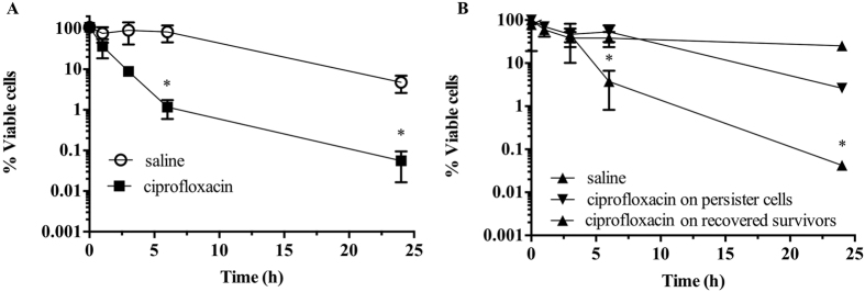
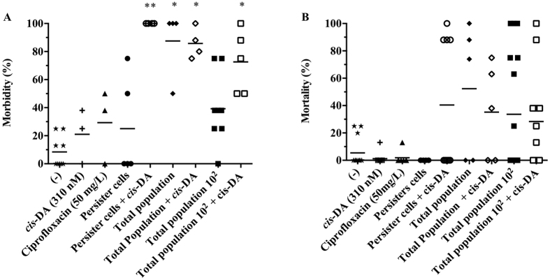
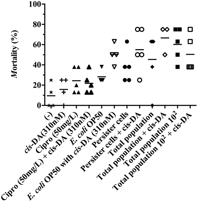
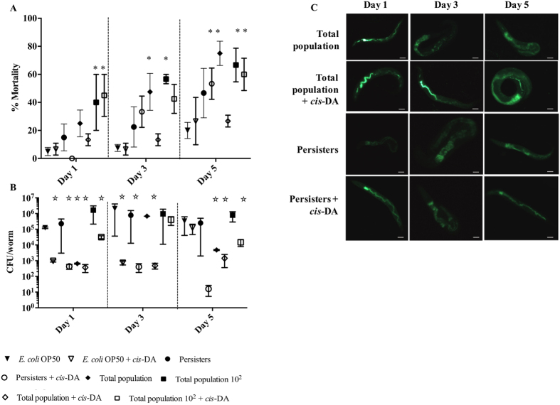
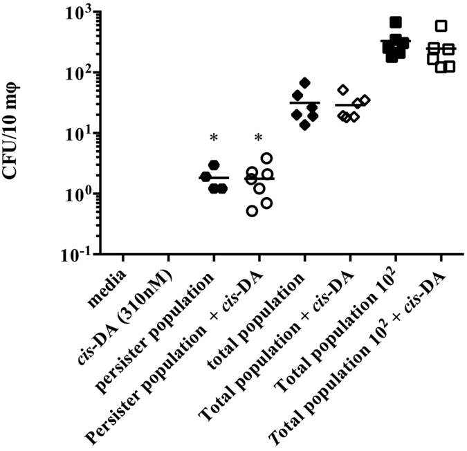
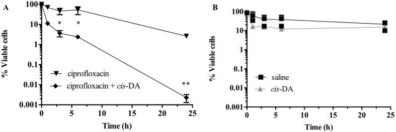
Persister cells, a tolerant cell sub-population, are commonly associated with chronic and recurrent infections. However, little is known about their ability to actually initiate or establish an infection, become virulent and cause pathogenicity within a host. Here we investigated whether Staphylococcus aureus persister cells initiate an infection and are recognized by macrophages, while in a persister cell status, and upon awakening due to exposure to cis-2-decenoic acid (cis-DA). Our results show that S. aureus persister cells are not able to initiate infections in A. thaliana and present significantly reduced virulence towards C. elegans compared to total populations. In contrast, awakened S. aureus persister cells are able to initiate infections in A. thaliana and in C. elegans albeit, with lower mortality than total population. Furthermore, exposure of S. aureus persister cells to cis-DA led to a loss of tolerance to ciprofloxacin, and an increase of the bacterial fluorescence to levels found in total population. In addition, macrophage engulfment of persister cells was significantly lower than engulfment of total population, both before and following awakening. Overall our findings indicate that upon awakening of a persister population the cells regain their ability to infect hosts despite the absence of an increased immune response.


Figures

Figure 1
Figure 1. Isolation of persister cells from S. aureus planktonic populations.
Stationary-phase planktonic cultures were exposed to saline or ciprofloxacin in saline for a period of 24 h (A). Cell viability was determined at 0, 1, 3, 6 and 24 h. To confirm that only persister cells were present, cultures were washed and isolated persister cells were exposed to saline or ciprofloxacin in saline for 24 h (B). Persister cells were also re-grown in LB medium and recovered survivors were re-challenged with ciprofloxacin for a period of 24 h (B). The averages of data from 3 experiments with 2 replicates per experiment are shown. Error bars indicate standard deviations (*P < 0.001- significantly different from cells exposed to saline alone, as indicated by one-way ANOVA).
Figure 2
Figure 2. Effect of persister cells on A. thaliana morbidity and mortality.
A. thaliana plants were infected with S. aureus persister cells, total population (at identical concentration as persister cells), and total population 102, in the presence and absence of cis-DA (310 nM). Controls consisted of ½ MS salts (−), cis-DA (310 nM) alone and ciprofloxacin (50 mg/L). A. thaliana morbidity 5 days post infection (A). A. thaliana mortality 7 days post infection (B). Results shown consist of a minimum of 5 experiments, using 8 plants per experiment. Lines indicate average. *Significant infection compared to (−), and cis-DA (P < 0.001); **Significant infection compared to: (−), cis-DA, and equivalent treatment without cis-DA (P < 0.01); as indicated by one-way ANOVA followed by Tukey’s multicomparison test.
Figure 3
Figure 3. Effect of persister cells on C. elegans mortality when using the slow killing assay.
C. elegans nematodes were infected with S. aureus persister cells, total population (at identical concentration as persister cells), and total population 102, in the presence and absence of cis-DA. C. elegans mortality was assessed daily for a period of 15 d. Overall mortality was assessed at 15 d. Results shown consist of a minimum of 4 experiments, using 8 nematodes per experiment.
Figure 4
Figure 4. Effect of persister cells on C. elegans mortality when using the fast killing assay.
C. elegans nematodes were infected with S. aureus persister cells, total population (at identical concentration as persister cells), and total population 102, in the presence and absence of cis-DA (310 nM). C. elegans infection was followed for a period of 5 days. C. elegans mortality was scored at 1, 3 and 5 d (A). S. aureus CFU per worm were determined at 1, 3 and 5 d (B). Bacterial fluorescence and distribution within the nematode was also assessed (C). Results shown consist of a minimum of 4 experiments, using 10 nematodes per experiment. Bar corresponds to 100 μm. Error bars indicate standard error of the mean. *Significant death compared to E. coli (P < 0.001); Significant lower CFU/worm compared to E. coli (P < 0.001); as indicated by one-way ANOVA followed by Tukey’s multicomparison test.
Figure 5
Figure 5. Macrophage uptake of S. aureus populations.
THP-1 monocytes (5 × 105) were allowed to differentiate for a period of 72 h, after which, they were exposed to S. aureus persister cells, total population (at identical concentration as persister cells), and total population 102, in the presence and absence of cis-DA. Following 1.5 h, intracellular S. aureus CFUs were evaluated. Results shown consist of a minimum of 4 experiments. Lines indicate average. (*P < 0.001- significant lower engulfment compared to total population and total population 102, as indicated by one-way ANOVA).
Figure 6
Figure 6. Tolerance of persister cells to ciprofloxacin is reduced in the presence of cis-DA.
Persister cell sub-populations isolated from S. aureus were exposed to ciprofloxacin and ciprofloxacin with cis-DA to assess whether they would lose their tolerance upon bacterial awakening (A). Persister cells were also exposed to saline and cis-DA (310 nM) in saline for a period of 24 h (B). The averages of data from 3 experiments with 2 replicates per experiment are shown in A, while individual data are shown in B. Error bars indicate standard deviations (*P < 0.01; **P < 0.001- significantly different from cells exposed to saline alone, as indicated by one-way ANOVA).

References

    1. Bigger J. The bactericidal action of penicillin on Staphylococcus pyogenes. Irish J. Med. Sci. 19, 553–568 (1944).
    1. Keren I., Kaldalu N., Spoering A., Wang Y. & Lewis K. Persister cells and tolerance to antimicrobials. FEMS Microbiol. Lett. 230, 13–18 (2004). - PubMed
    1. Shah D. et al.. Persisters: a distinct physiological state of E. coli. BMC Microbiol 6, 53 (2006). - PMC - PubMed
    1. Korch S. B., Henderson T. A. & Hill T. M. Characterization of the hipA7 allele of Escherichia coli and evidence that high persistence is governed by (p)ppGpp synthesis. Mol. Microbiol. 50, 1199–1213 (2003). - PubMed
    1. Fauvart M., De Groote V. N. & Michiels J. Role of persister cells in chronic infections: clinical relevance and perspectives on anti-persister therapies. J. Med. Microbiol. 60, 699–709 (2011). - PubMed

Publication types

MeSH terms

LinkOut - more resources