. 2016 Nov;24(11):2012-2020.
doi: 10.1038/mt.2016.161. Epub 2016 Aug 10.

Type I Interferons Interfere with the Capacity of mRNA Lipoplex Vaccines to Elicit Cytolytic T Cell Responses

Affiliations

Type I Interferons Interfere with the Capacity of mRNA Lipoplex Vaccines to Elicit Cytolytic T Cell Responses

Ans De Beuckelaer et al. Mol Ther. 2016 Nov.

Abstract

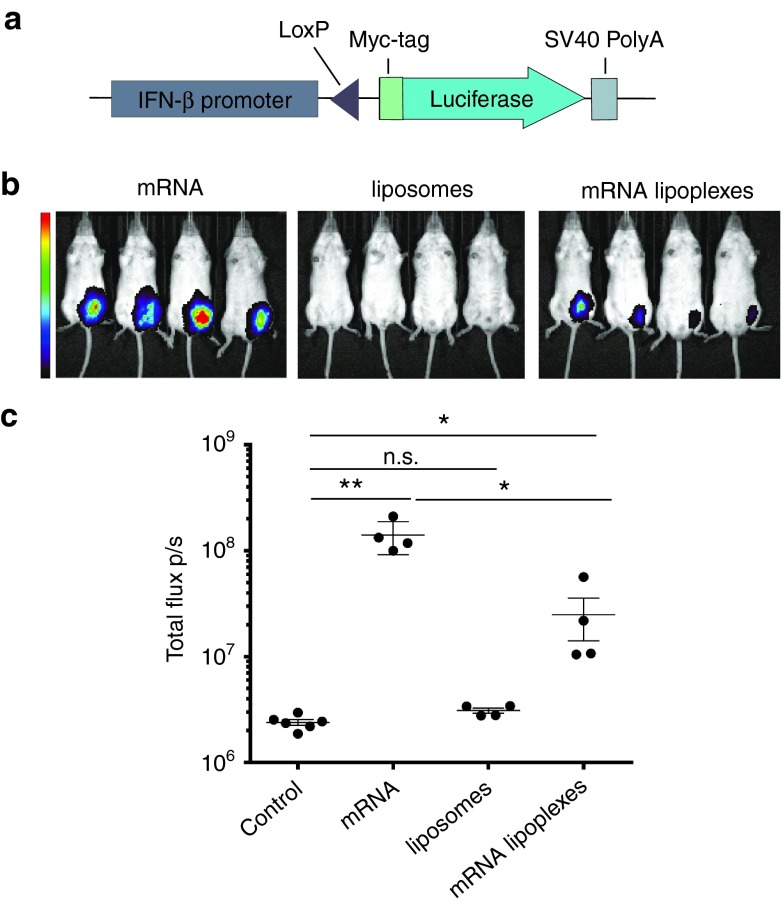
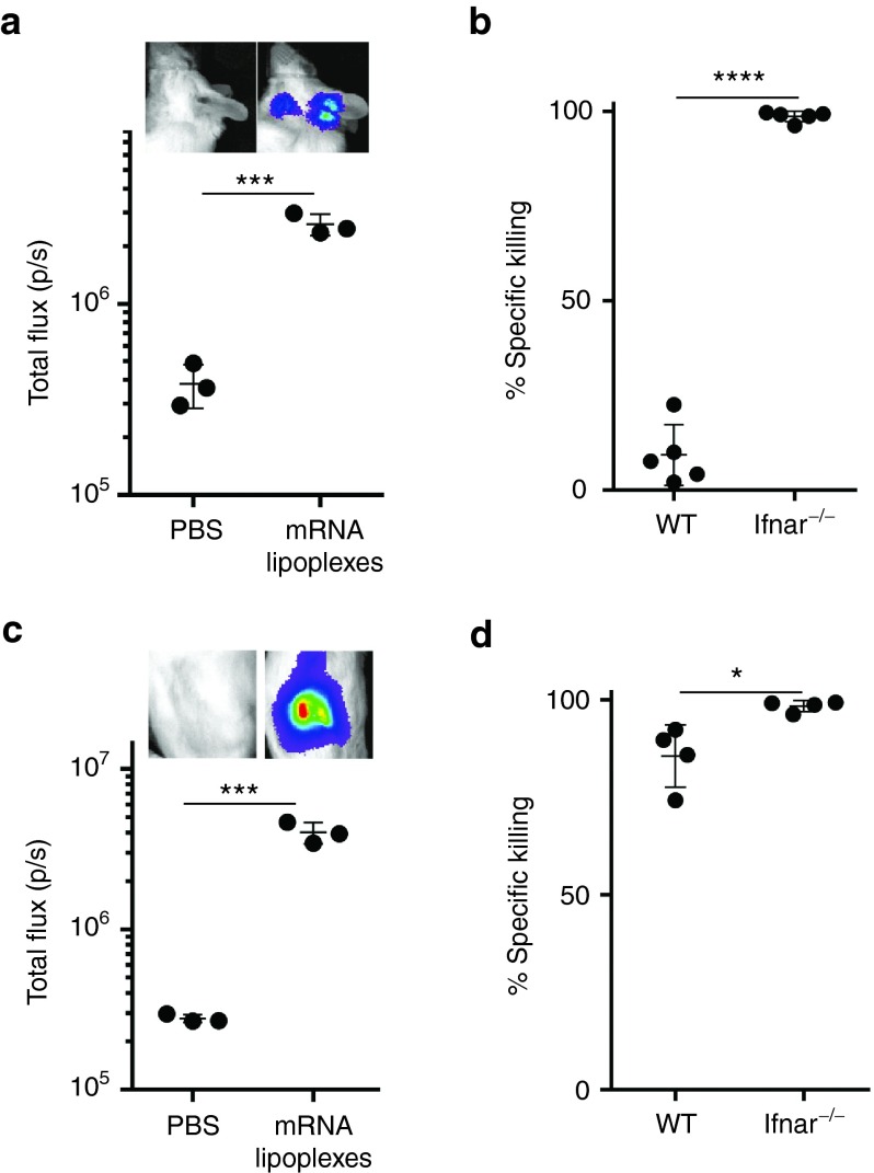
Given their high potential to evoke cytolytic T cell responses, tumor antigen-encoding messenger RNA (mRNA) vaccines are now being intensively explored as therapeutic cancer vaccines. mRNA vaccines clearly benefit from wrapping the mRNA into nano-sized carriers such as lipoplexes that protect the mRNA from degradation and increase its uptake by dendritic cells in vivo. Nevertheless, the early innate host factors that regulate the induction of cytolytic T cells to mRNA lipoplex vaccines have remained unresolved. Here, we demonstrate that mRNA lipoplexes induce a potent type I interferon (IFN) response upon subcutaneous, intradermal and intranodal injection. Regardless of the route of immunization applied, these type I IFNs interfered with the generation of potent cytolytic T cell responses. Most importantly, blocking type I IFN signaling at the site of immunization through the use of an IFNAR blocking antibody greatly enhanced the prophylactic and therapeutic antitumor efficacy of mRNA lipoplexes in the highly aggressive B16 melanoma model. As type I IFN induction appears to be inherent to the mRNA itself rather than to unique properties of the mRNA lipoplex formulation, preventing type I IFN induction and/or IFNAR signaling at the site of immunization might constitute a widely applicable strategy to improve the potency of mRNA vaccination.


Figures

Figure 1
Figure 1
mRNA lipoplexes induce a potent type I IFN response in vivo. (a) Graphical scheme of the IFN-β reporter construct. The myc-tagged luciferase gene is brought under the control of the IFN-β promoter by the Cre-Lox system. (b,c) IFN-β+/Δβ-luc mice were subcutaneous (s.c.) injected with 10 µg of OVA mRNA, mRNA lipoplexes and liposomes. Luminescence was measured 6 hours postinjection. Data are shown as mean ± SD of four mice. **P < 0.001. *P < 0.05 (Mann–Whitney test). Control = 5% glucose water; liposomes = DOTAP/DOPE lipids; mRNA lipoplexes = messenger RNA complexed to liposomes. mRNA, messenger RNA; IFN, interferon, OVA, ovalbumin; SD, standard deviation.
Figure 2
Figure 2
Type I IFNs impact the magnitude and functional characteristics of the vaccine elicited CD8+ T cell response. (a) Gating strategy used for OVA- specific CD8+ T cell counting and proliferation. Cells are gated based on FSC and SCC, before single cells are gated based on SSC-area and height. Living cells are selected and gated for CD3+CD19- T cells. Within CD8+ T cells, OVA-specificity is gated by labeling with MHC-I SIINFEKL–PE dextramer. Proliferation of CFSE positive OVA-specific CD8+ T cells is shown. (b) Two days prior to immunization CFSE-labeled OT-I cells were adoptively transferred to wild type (WT) and Ifnar−/− mice. Subcutaneous (s.c.) immunization was performed at tail base with 10 µg OVA mRNA lipoplexes, naked mRNA or liposomes alone. Four days after immunization inguinal lymph nodes were isolated and CD8+ T cell proliferation was analyzed by flow cytometry. Data are shown as mean of 2–3 mice. ***P < 0.001 (Chi-square test). (c) Wild type (WT) and Ifnar−/− mice were s.c. injected with 20 µg OVA mRNA lipoplexes or naked OVA mRNA as a control. Blood was isolated 5 days later and the percentage OVA-specific CD8+ T cells was determined by dextramer staining followed by flow cytometry. Data are shown as mean of four mice per group. ***P < 0.001 (Chi-square test). (d) Wild type (WT) and Ifnar−/− mice were immunized s.c. with 20 µg OVA mRNA lipoplexes or naked mRNA as a control. After 2 weeks, mice were boosted with the same formulation. Spleens were isolated 2 weeks after the boost immunization, and the number of OVA-specific interferon-γ spot-forming CD8+ and CD4+ T cells (SFC) was determined by enzyme-linked immunosorbent spot (ELISPOT). Data are shown as mean of 2–4 mice per group. ***P < 0.001 (Chi-square test). (e,f) Wild type (WT) and Ifnar−/− mice were immunized with a two-week interval with naked OVA mRNA or OVA mRNA lipolexes. Two weeks after the boost immunization, a mixture of CFSE-labeled cells pulsed with control (CFSElow) or OVA peptide (CFSEhigh) were adoptively transferred. Specific killing was measured 2 days later by flow cytometry. Data are presented as means of 100 −100x ((CFSEhigh / CFSElow)immunized mice / (CFSEhigh / CFSElow)mock-mice) of 3–4 mice per group. **P < 0.01 (Chi-square test). mRNA = OVA-coding messenger RNA; mRNA lipoplexes = messenger RNA complexed to DOTAP/DOPE liposomes. CFSE, carboxyfluorescein diacetate succinimedyl ester; mRNA, messenger RNA; IFN, interferon, OVA, ovalbumin; SD, standard deviation.
Figure 3
Figure 3
Impact of type I IFNs on the efficacy of antitumor immunity elicited by mRNA lipoplex vaccination. (a) Prophylactic vaccination scheme. Wild type (WT) mice (b) and Ifnar−/− mice (c) were either mock s.c. immunized (i.e., injected with PBS only) or immunized with 20 μg of mRNA lipoplexes. After 2 weeks, mice were boosted with the same formulation. At week 4, mice were inoculated with 100,000 OVA-expressing B16 melanoma cells. (n = 12–16 mice/group). (d) Therapeutic vaccination scheme. Wild type (WT) mice (e) and Ifnar−/− mice (f) were inoculated with 75,000 B16.OVA melanoma cells. After 4 and 6 days, immunization was performed with similar preparations as in the prophylactic setting. (n = 5–6 mice/group). mRNA lipoplexes = OVA-coding messenger mRNA complexed to DOTAP/DOPE liposomes. **P < 0.01; ***P < 0.001; ****P < 0.0001 (Mantel-Cox log-rank test). PBS, phosphate-buffered saline; mRNA, messenger RNA; IFN, interferon, OVA, ovalbumin; SD, standard deviation.
Figure 4
Figure 4
Antibody-mediated blocking of IFNAR improves the efficacy of the mRNA vaccine evoked antitumor immune response. (a,b) Two days prior to immunization CFSE-labeled OT-I cells were adoptively transferred to wild type (WT) mice. Immunization was performed in the footpad with 10 µg mRNA lipoplexes in the absence or presence of 20 µg IFNAR blocking antibody or isotype control. Four days after immunization inguinal lymph nodes were isolated and CD8+ T cell proliferation was analyzed by flow cytometry. Data are shown as mean of 3–6 mice per group. ***P < 0.001 (Chi-square test). (a) A representative sample out of 3–6 mice each group is presented. (c) Prophylactic vaccination scheme. (d) Wild type (WT) mice were immunized s.c with 20 μg of mRNA lipoplexes in absence or presence of 20 µg of the IFNAR blocking antibody or isotype control. After 2 weeks, mice were boosted with the same formulation. At week 4, mice were inoculated with 100,000 B16.OVA melanoma cells (n = 6–8 mice/group). *P < 0.05 (Mantel-Cox log-rank test). (e) Therapeutic vaccination scheme. (f) Wild type (WT) mice were inoculated with 75,000 B16.OVA melanoma cells. After 4 and 9 days, immunization was performed using 20 μg of mRNA lipoplexes in absence or presence of the IFNAR blocking antibody or isotype control (20 µg) (n = 6–8 mice/group). *P < 0.05 (Mantel-Cox log-rank test). mRNA lipoplexes = OVA- coding messenger mRNA complexed to DOTAP/DOPE liposomes. CFSE, carboxyfluorescein diacetate succinimedyl ester; mRNA, messenger RNA; IFN, interferon, OVA, ovalbumin; SD, standard deviation.
Figure 5
Figure 5
Type I IFNs inhibit the induction of cytolytic T cells regardless of the route of immunization. (a) IFN-β+/Δβ-luc mice were intradermally injected with 10 µg of OVA mRNA lipoplexes complexed or PBS. In vivo bioluminescence was measured 6 hours postinjection. Data are shown as mean ± SD of three mice. ***P < 0,001 (t-test). (b) Wild type (WT) and Ifnar−/− mice were immunized with a two-week interval with 10 µg of mRNA lipoplexes. Two weeks after boost immunization, a mixture of CFSE-labeled cells pulsed with control (CFSElow) or OVA peptide (CFSEhigh) were adoptively transferred. Specific killing was measured after 2 days by flow cytometry. Killing percentages were calculated with the following formula: 100 − 100x ((CFSEhigh/CFSElow)immunized mice/(CFSEhigh/CFSElow)mock-mice) of five mice per group. ****P < 0.0001 (t-test). (c) IFN-β+/Δβ-luc mice were intranodally injected with 10 µg of OVA mRNA lipoplexes or mock treated. In vivo bioluminescence was measured 6 hours postinjection. Data are shown as mean ± SD of three mice. ***P < 0.001 (t-test). (d) Wild type (WT) and IFNAR−/− mice were immunized with a two-week interval with 10 µg of OVA mRNA lipoplexes and killing was performed as previously described. *P < 0.05 (t-test). mRNA lipoplexes = OVA- coding messenger mRNA complexed to DOTAP/DOPE liposomes. CFSE, carboxyfluorescein diacetate succinimedyl ester; mRNA, messenger RNA; IFN, interferon, OVA, ovalbumin; PBS, phosphate-buffered saline; SD, standard deviation.

References

    1. Appay, V, Douek, DC and Price, DA (2008). CD8+ T cell efficacy in vaccination and disease. Nat Med 14: 623–628. - PubMed
    1. Sahin, U, Karikó, K and Türeci, Ö (2014). mRNA-based therapeutics–developing a new class of drugs. Nat Rev Drug Discov 13: 759–780. - PubMed
    1. Palucka, K and Banchereau, J (2013). Dendritic-cell-based therapeutic cancer vaccines. Immunity 39: 38–48. - PMC - PubMed
    1. Holtkamp, S, Kreiter, S, Selmi, A, Simon, P, Koslowski, M, Huber, C et al. (2006). Modification of antigen-encoding RNA increases stability, translational efficacy, and T-cell stimulatory capacity of dendritic cells. Blood 108: 4009–4017. - PubMed
    1. Anderson, BR, Muramatsu, H, Jha, BK, Silverman, RH, Weissman, D and Karikó, K (2011). Nucleoside modifications in RNA limit activation of 2′-5′-oligoadenylate synthetase and increase resistance to cleavage by RNase L. Nucleic Acids Res 39: 9329–9338. - PMC - PubMed

MeSH terms