. 2016 Nov;59(11):2448-2458.
doi: 10.1007/s00125-016-4067-4. Epub 2016 Aug 9.

Islet cell hyperexpression of HLA class I antigens: a defining feature in type 1 diabetes

Affiliations

Islet cell hyperexpression of HLA class I antigens: a defining feature in type 1 diabetes

Sarah J Richardson et al. Diabetologia. 2016 Nov.

Abstract

Aims/hypothesis: Human pancreatic beta cells may be complicit in their own demise in type 1 diabetes, but how this occurs remains unclear. One potentially contributing factor is hyperexpression of HLA class I antigens. This was first described approximately 30 years ago, but has never been fully characterised and was recently challenged as artefactual. Therefore, we investigated HLA class I expression at the protein and RNA levels in pancreases from three cohorts of patients with type 1 diabetes. The principal aims were to consider whether HLA class I hyperexpression is artefactual and, if not, to determine the factors driving it.

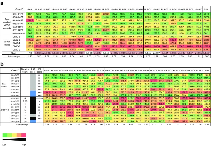
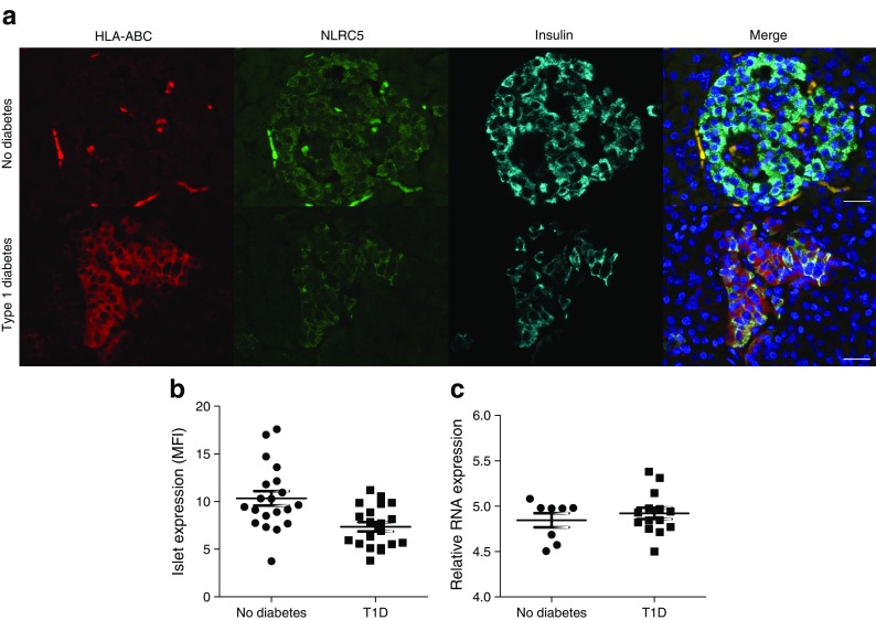
Methods: Pancreas samples from type 1 diabetes patients with residual insulin-containing islets (n = 26) from the Network for Pancreatic Organ donors with Diabetes (nPOD), Diabetes Virus Detection study (DiViD) and UK recent-onset type 1 diabetes collections were immunostained for HLA class I isoforms, signal transducer and activator of transcription 1 (STAT1), NLR family CARD domain containing 5 (NLRC5) and islet hormones. RNA was extracted from islets isolated by laser-capture microdissection from nPOD and DiViD samples and analysed using gene-expression arrays.

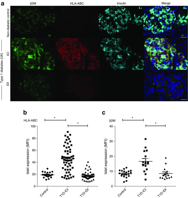
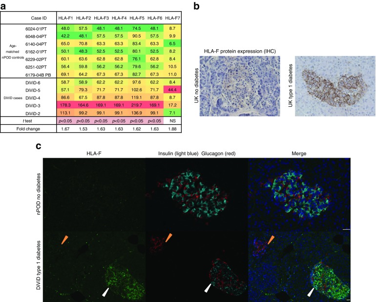
Results: Hyperexpression of HLA class I was observed in the insulin-containing islets of type 1 diabetes patients from all three tissue collections, and was confirmed at both the RNA and protein levels. The expression of β2-microglobulin (a second component required for the generation of functional HLA class I complexes) was also elevated. Both 'classical' HLA class I isoforms (i.e. HLA-ABC) as well as a 'non-classical' HLA molecule, HLA-F, were hyperexpressed in insulin-containing islets. This hyperexpression did not correlate with detectable upregulation of the transcriptional regulator NLRC5. However, it was strongly associated with increased STAT1 expression in all three cohorts. Islet hyperexpression of HLA class I molecules occurred in the insulin-containing islets of patients with recent-onset type 1 diabetes and was also detectable in many patients with disease duration of up to 11 years, declining thereafter.

Conclusions/interpretation: Islet cell HLA class I hyperexpression is not an artefact, but is a hallmark in the immunopathogenesis of type 1 diabetes. The response is closely associated with elevated expression of STAT1 and, together, these occur uniquely in patients with type 1 diabetes, thereby contributing to their selective susceptibility to autoimmune-mediated destruction.

Keywords: DiViD; HLA class I; HLA-F; Islet cell; Pancreas; STAT1; Type 1 diabetes; nPOD.


Conflict of interest statement

Funding

We are pleased to acknowledge financial support from the European Union’s Seventh Framework Programme PEVNET (FP7/2007-2013, grant agreement number 261441). The participants of the PEVNET consortium are described at www.uta.fi/med/pevnet/publications.html. Additional support was provided by a Diabetes Research Wellness Foundation Non-Clinical Research Fellowship and, since 2014, a JDRF Career Development Award (5-CDA-2014-221-A-N) to SJR. KD-J has received grants from the South-Eastern Norway Regional Health Authority, the Novo Nordisk Foundation and through the PEVNET Consortium. The research was also performed with the support of nPOD, a collaborative type 1 diabetes research project sponsored by JDRF International (JDRF 25-2013-268 to MAA, including a subcontract to JSK), and of JDRF research grants awarded to the nPOD-V Consortium (JDRF 25-2012-516 to AP and JDRF 25-2012-770 to MAA). Additional funding was provided by JDRF to CEM (JDRF 47-2013-520) and from the National Institutes of Health (UC4 DK104155) to ICG and MvH (R01 AI092453).

Duality of interest

The authors declare that there is no duality of interest associated with this manuscript.

Contribution statement

SJR and NGM designed the study, performed data analysis and interpretation, and drafted, revised and approved the manuscript. TR-C, ICG, MZ, MAR and PL performed data collection, analysis and interpretation, and revised and approved the manuscript. LK and KD-J collected patient material, and revised and approved the manuscript. ICG and CEM designed the Affymetrix array component of the study, and revised and approved the manuscript. JSK provided statistical expertise, and revised and approved the manuscript. MvH, AP and MAA provided critical interpretation of the data, and revised and approved the manuscript.

SJR and NGM are the guarantors of this work and, as such, had full access to all the data in the study and take responsibility for the integrity of the data and the accuracy of the data analysis.

Figures

Fig. 1
Fig. 1
Immunocytochemical analysis of the expression HLA-ABC in pancreas tissue. (a) Pancreas sections from two individuals with recent-onset type 1 diabetes from the DiViD cohort showing insulin and HLA-ABC immunostaining on serial sections. ICIs are indicated with red asterisks (magnification ×40 for the whole tissue section and ×100 for the islet). (b) Immunofluorescence analysis of HLA-ABC expression in frozen pancreas from a patient with recent-onset type 1 diabetes from the nPOD cohort. Hyperexpression of HLA-ABC (red) was predominantly seen only in ICIs (green; inset) (magnification ×40 for the whole tissue section and ×400 for the islet)
Fig. 2
Fig. 2
Correlation between HLA-ABC, β2M and insulin expression in controls and individuals with type 1 diabetes. (a) Analysis of β2M (green), HLA-ABC (red) and insulin (light blue) in an ICI from a non-diabetic control individual and a type 1 diabetes patient, and an IDI from the same individual. Scale bar, 25 μm. (b) The MFI of islet HLA-ABC expression was measured in 5–14 islets among non-diabetic control individuals (n = 4) and in the ICI and IDIs of nine individuals with type 1 diabetes (T1D) (five from the DiViD cohort, three from the UK cohort and one from the nPOD cohort). (c) The MFI of islet β2M expression was measured in 15 ICIs and 15 IDIs from each of three type 1 diabetes patients (two from the UK cohort and one from the nPOD cohort). This was compared with the expression in 20 islets from each of four non-diabetic control individuals (three from the UK cohort and one from the nPOD cohort). *p < 0.001
Fig. 3
Fig. 3
Heat map illustrating the relative expression of HLA-ABC and B2M genes in control individuals and those with type 1 diabetes (T1D). The expression of each probe set is displayed separately in islets of: (a) seven nPOD non-diabetic controls age-matched to five DiViD patients; and (b) eight nPOD non-diabetic controls and nine nPOD type 1 diabetic donors. Expression values are shown in arbitrary units and the heat map illustrates relative expression ranging from low (green) to high (red). In (b), a comparison with the level of expression scored after immunohistochemical analysis of islets present in nearby pancreatic blocks from the same patients is provided (black, hyperexpression; blue, elevated expression; grey, normal expression), together with an indication of the extent of insulin immunopositivity
Fig. 4
Fig. 4
Expression of HLA-F in control individuals and type 1 diabetes patients. (a) Heat map illustrating the relative expression of the HLA-F probe sets in seven nPOD non-diabetic controls age-matched to five DiViD patients. (b) Representative immunostaining of islets from an individual without diabetes and a patient with type 1 diabetes with anti-HLA-F. (c) Immunofluorescence staining of HLA-F (green), insulin (light blue) and glucagon (red) in an ICI (white arrowheads) and an IDI (orange arrowheads) of a DiViD type 1 diabetes patient, and an islet from an nPOD control donor. Scale bar, 25 μm
Fig. 5
Fig. 5
Expression of NLRC5 in the islets of control individuals and those with type 1 diabetes. (a) Representative islets from an individual without diabetes and a patient with type 1 diabetes are shown (red, HLA-ABC; green, NLRC5; light blue, insulin; dark blue, DAPI). Scale bar, 25 μm. (b) MFI values for NLRC5 protein expression were quantified after immunostaining in five islets per section from four control and four type 1 diabetes (three from the UK, one from nPOD) samples (p = 0.0704). (c) Expression of NLRC5 was compared in RNA isolated from islets of individuals with and without type 1 diabetes (p = 0.4504)
Fig. 6
Fig. 6
Expression of STAT1 and HLA-ABC in islets from control individuals and those with type 1 diabetes. (a) Representative islets from a control individual and from a type 1 diabetes patient were immunostained for STAT1 (green), HLA-ABC (red), insulin (light blue) and DAPI (dark blue). The localisation of STAT1 is shown in beta cells (white arrowheads) and non-beta cells (orange arrowheads). Scale bar, 25 μm. (b) MFI values for STAT1 and HLA-ABC expression were quantified and correlated from a minimum of seven ICIs in seven patients with type 1 diabetes (T1D) among the DiViD and UK (E560) cohorts (Spearman’s rank coefficient =0.5454, p < 0.0001). (c) Correlation between the expression of STAT1 mRNA and disease duration in nPOD type 1 diabetes patients with residual ICIs (p < 0.05). (d) Analysis of STAT1 expression in RNA isolated from islets from DiViD patients and age-matched control donors (nPOD) (p = 0.0263)

References

    1. Atkinson MA. The pathogenesis and natural history of type 1 diabetes. Cold Spring Harb Perspect Med. 2012;2:2:a007641. doi: 10.1101/cshperspect.a007641. - DOI - PMC - PubMed
    1. Atkinson MA, Eisenbarth GS, Michels AW. Type 1 diabetes. Lancet. 2014;383:69–82. doi: 10.1016/S0140-6736(13)60591-7. - DOI - PMC - PubMed
    1. Patterson CC, Dahlquist GG, Gyurus E, Green A, Soltesz G, Group ES Incidence trends for childhood type 1 diabetes in Europe during 1989-2003 and predicted new cases 2005-20: a multicentre prospective registration study. Lancet. 2009;373:2027–2033. doi: 10.1016/S0140-6736(09)60568-7. - DOI - PubMed
    1. Foulis AK, Liddle CN, Farquharson MA, Richmond JA, Weir RS. The histopathology of the pancreas in type 1 (insulin-dependent) diabetes mellitus: a 25-year review of deaths in patients under 20 years of age in the United Kingdom. Diabetologia. 1986;29:267–274. doi: 10.1007/BF00452061. - DOI - PubMed
    1. Campbell-Thompson M. What can organ donor specimens tell us about type 1 diabetes? Pediatr Diabetes. 2015;16:320–330. doi: 10.1111/pedi.12286. - DOI - PMC - PubMed

Publication types

Substances