

Comment
doi: 10.1073/pnas.1610806113.
Epub 2016 Aug 9.
Implications for Alzheimer's disease of an atomic resolution structure of amyloid-β(1-42) fibrils
Affiliations
- PMID: 27506787
- PMCID: PMC5003263
- DOI: 10.1073/pnas.1610806113
Item in Clipboard
Comment
Implications for Alzheimer's disease of an atomic resolution structure of amyloid-β(1-42) fibrils
Proc Natl Acad Sci U S A.
.
No abstract available
Conflict of interest statement
The authors declare no conflict of interest.
Figures
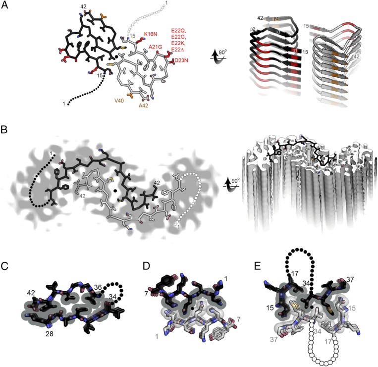
Comparison of two molecular models for amyloid-β(1–42) fibrils. (A) The near atomic resolution model determined by solid-state NMR by Wälti et al. (1). Two double-horseshoe–shaped molecules of amyloid-β(1–42) are shown (black and gray) related by a twofold axis (marked by a circle), which runs down the center of the fibril. The N-terminal 14 residues are disordered; one possible conformation is shown here by dotted lines. Many of the known familial mutations are carried by residues located on the outer surface (red). The surface hydrophobic patch formed by residues V40 and A42 (orange) may explain the greater rate of secondary nucleation by the 1–42 species compared with 1–40 (3). (B). The lower resolution (5–7 Å) model determined by Schmidt et al. (7) by cryo-EM is another polymorph of amyloid-β(1–42), also with two molecules per layer, related by a twofold axis, and also with a poorly ordered N terminus. The gray color represents a slice through the cryo-EM map. The two molecules appear to be related by a homo-steric zipper type of bonding. In both A and B the models are viewed down the fibril axis on the left and nearly perpendicular to the fibril axis on the right. (C) A hetero-zipper in amyloid-β(1–42) fibrils from Wälti et al. (1). (D) A homo-zipper from GNNQQNY (4). (E) A noncontiguous homo-zipper from Wälti et al. (1). Dotted lines represent intervening residues.
Comment on
-
Atomic-resolution structure of a disease-relevant Aβ(1-42) amyloid fibril.Proc Natl Acad Sci U S A. 2016 Aug 23;113(34):E4976-84. doi: 10.1073/pnas.1600749113. Epub 2016 Jul 28. Proc Natl Acad Sci U S A. 2016. PMID: 27469165 Free PMC article.
References
-
- Sawaya MR, et al. Atomic structures of amyloid cross-beta spines reveal varied steric zippers. Nature. 2007;447(7143):453–457. - PubMed
Publication types
MeSH terms
Substances
Grants and funding
LinkOut - more resources
Full Text Sources
Other Literature Sources
Medical
