. 2016 Aug 23;113(34):E4966-75.
doi: 10.1073/pnas.1603244113. Epub 2016 Aug 9.

Peroxidation of polyunsaturated fatty acids by lipoxygenases drives ferroptosis

Affiliations

Peroxidation of polyunsaturated fatty acids by lipoxygenases drives ferroptosis

Wan Seok Yang et al. Proc Natl Acad Sci U S A. .

Abstract

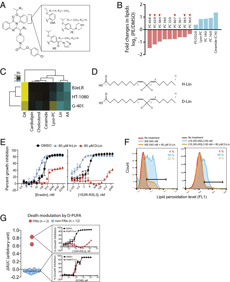
Ferroptosis is form of regulated nonapoptotic cell death that is involved in diverse disease contexts. Small molecules that inhibit glutathione peroxidase 4 (GPX4), a phospholipid peroxidase, cause lethal accumulation of lipid peroxides and induce ferroptotic cell death. Although ferroptosis has been suggested to involve accumulation of reactive oxygen species (ROS) in lipid environments, the mediators and substrates of ROS generation and the pharmacological mechanism of GPX4 inhibition that generates ROS in lipid environments are unknown. We report here the mechanism of lipid peroxidation during ferroptosis, which involves phosphorylase kinase G2 (PHKG2) regulation of iron availability to lipoxygenase enzymes, which in turn drive ferroptosis through peroxidation of polyunsaturated fatty acids (PUFAs) at the bis-allylic position; indeed, pretreating cells with PUFAs containing the heavy hydrogen isotope deuterium at the site of peroxidation (D-PUFA) prevented PUFA oxidation and blocked ferroptosis. We further found that ferroptosis inducers inhibit GPX4 by covalently targeting the active site selenocysteine, leading to accumulation of PUFA hydroperoxides. In summary, we found that PUFA oxidation by lipoxygenases via a PHKG2-dependent iron pool is necessary for ferroptosis and that the covalent inhibition of the catalytic selenocysteine in Gpx4 prevents elimination of PUFA hydroperoxides; these findings suggest new strategies for controlling ferroptosis in diverse contexts.

Keywords: Gpx4; PHKG2; PUFAs; ferroptosis; lipoxygenase.


Conflict of interest statement

The authors declare no conflict of interest.

Figures

Fig. 1.
Fig. 1.
Characterization of RSL3 binding to GPX4. (A) Structure of RSL3 affinity probe used (Upper) and schematic drawing of the affinity complex formed during the pull-down assay (Lower). (B) A pull-down analysis showing that RSL3 does not bind to a mutant GPX4 where the active-site selenocysteine and all other cysteines were replaced with either alanine or serine. The schematic structure of GFP-GPX4 protein is shown (Upper). Cells stably expressing either WT or mutant [AllCys(-)] GFP-GPX4 protein were treated with (1S, 3R)-RSL3-Fcn, then the cell lysates were subjected to affinity purification using anti–Fcn-conjugated Sepharose beads. The binding between GFP-GPX4 protein and (1S, 3R)-RSL3-Fcn was determined by detecting the presence of GFP-GPX4 protein in the eluent using Western blot with anti-GFP antibody. Arrowheads indicate GFP-GPX4 protein; asterisks indicate nonspecific bands. W.C.L., whole-cell lysate before pulldown. (C) Selenocysteine is much more reactive to RSL3 than cysteine at the active site. Cells stably expressing A46U or A46C protein (see diagram in the figure) were treated with (1S, 3R)-RSL3-Fcn and subjected to the same analysis as in B. (1S, 3R)-RSL3-Fcn interacted with A46U protein but not with A46C protein. Arrowheads indicate GFP-GPX4 protein; asterisks indicate nonspecific bands. (D) RSL3 interacts weakly with other cysteines on GPX4. Cells stably expressing indicated mutant protein (see text for detailed information) were treated with (1S, 3R)-RSL3-Fcn and subjected to the same analysis as in B. Some mutant proteins such as A10C and S37C interacted with (1S, 3R)-RSL3-Fcn whereas others did not. The interaction was the strongest when WT GPX4 was expressed (thickest band in WT sample). Arrowheads indicate GFP-GPX4 protein; asterisks indicate nonspecific bands. (E) Some ferroptosis-inducing compounds competed off RSL3 binding to GPX4. HT-1080 cells were treated with (1S, 3R)-RSL3-Fcn in the presence or absence of indicated competitors, and cell lysates were subjected to pull-down experiment with anti-Fcn antibody. The binding between (1S, 3R)-RSL3-Fcn and endogenous GPX4 protein was determined by Western blotting with anti-GPX4 antibody. The (1S, 3R)-RSL3-Fcn binding to GPX4 was suppressed in samples where free (1S, 3R)-RSL3 was used as a competitor. Different GPX4 inhibitors competed off (1S, 3R)-RSL3-Fcn binding to GPX4 protein with varying degrees. (F) The amount of endogenous GPX4 protein bound to the affinity probe in E was quantified using Odyssey software (LI-COR Biosciences). Data represent mean ± SD calculated from technical triplicates. (G) Covalent interaction between GPX4 and RSL3 was confirmed by immunoprecipitating GFP-GPX4/(1S, 3R)-RSL3-Fcn complex with anti-GPX4 antibody and performing Western blot with anti-GFP or anti-Fcn antibodies. Arrow indicates GFP-GPX4 protein band in the Western blot analysis. (H) The (1S, 3R)-RSL3 covalently interacts with purified GPX4U46C protein. Purified GPX4U46C protein was mixed with (1S, 3R)-RSL3-Fcn, then the mixture was resolved on a denaturing gel and transferred to a membrane. Western blotting with anti-Fcn antibody detected (1S, 3R)-RSL3-Fcn in a position corresponding to the size of GPX4U46C protein (∼21 kDa), suggesting that (1S, 3R)-RSL3-Fcn covalently attached to GPX4U46C protein and migrated together in this denaturing condition. The Western blot band became thinner when a lesser amount of (1S, 3R)-RSL3-Fcn was added to the mixture (Upper). The band size remained the same across the sample when anti-GPX4 antibody was used because equal amount of GPX4U46C protein was added across the samples (Lower). (I) Inhibition of GPX4 enzyme activity by (1S, 3R)-RSL3 or (1R, 3R)-RSL3. GPX4 enzyme activity was assayed by mixing cell lysates with PC hydroperoxides (PC-OOH), a GPX4-specific substrate, and by determining the amount of substrate left in the reaction mixture using an LC-MS instrument. The arrow indicates the LC-MS peak corresponds to PC-OOH. (J) Area under the curve was determined from each mass chromatogram in I (retention time from 8.2 to 8.8 min; dotted lines in the chromatogram) and used to draw a concentration-dependent curve of GPX4 inhibition by (1S, 3R)-RSL3.
Fig. 2.
Fig. 2.
PUFAs play a functional role in ferroptotic cell death. (A) Structure of erastin analogs used in this study. ERA, erastin (3); IKE, imidazole ketoerastin (61); and PE, piperazine erastin (2). (B) Lipidomics analysis revealed loss of PUFAs is the most prominent change during ferroptosis. HT-1080 cells were treated with PE or vehicle (DMSO) and lipid metabolites were extracted as described in Methods. The amount of individual lipid metabolites in the sample was determined using an LC-MS instrument, and changes in the amount of lipid metabolites were calculated by dividing the amount in PE sample by that in DMSO sample. The graph shows the name of top changing lipid metabolites (nine down-regulated and five up-regulated) and the value of fold changes in log2 scale. The number of carbons and double bonds in sn-2 position fatty acids are indicated. For example, PC 40:8 indicates phosphatidyl choline (PC) that has a fatty acid of 40 carbon and 8 double bonds in sn-2 position. PS, phosphatidyl serine. (C) Modulation of ferroptosis sensitivity by different lipid species. PUFAs sensitized cells to ferroptosism, whereas OA prevented ferroptosis. Yellow indicates cell death suppression and blue represents cell death sensitization. Modulatory index (Me) was calculated as previously described (1). (D) Structures of natural LA (H-Lin) and deuterated LA (D-Lin). (E) Suppression of ferroptosis by D-Lin. G-401 cells were treated with either erastin or (1S, 3R)-RSL3 in the presence or absence of either H-Lin or D-Lin. Cells became rescued from ferroptosis upon D-Lin treatment, whereas they became more sensitive to ferroptosis upon H-Lin treatment. (F) D-Lin prevented generation of lipid peroxides. (G) D-Lin did not suppress lethality of 12 cytotoxic compounds, highlighting the specificity on ferroptosis.
Fig. 3.
Fig. 3.
Lipoxygenases are involved in ferroptosis by system xc inhibition. (A) Dioxygenation reaction induced by lipoxygenases. (B) G-401 cell line expressed all isoforms of ALOXs as assessed by qPCR experiment. ACTB, Beta-actin. (C) A pool of siALOX targeting all ALOX genes successfully decreased mRNA levels of ALOX genes. (D) Silencing ALOXs expression conferred resistance to IKE-induced ferroptosis, whereas it did not affect RSL3 lethality in G-401 cells. (E) Silencing ALOX15B and ALOXE3 in HT-1080 cells made cells resistant to erastin. (F) Lipoxygenase inhibitors rescued cells from erastin-induced ferroptosis. A cyclooxygenase inhibitor did not affect erastin’s lethality. (G) GFP-ALOX5 translocated to the nuclear membrane only in sensitive cells upon 10 µM erastin treatment. Bar graph: mean + SD; n = 3–4; ***P < 0.001. (H) Time course of GFP-ALOX5 translocation in HT-1080 cells upon treatment with erastin or ionomycin.
Fig. S1.
Fig. S1.
(A) Expression analysis of ALOX genes in BJeLR and HT-1080 cells. The figures show the amplification plot of each ALOX isoform. Triplicate samples were analyzed for each gene using mRNA preparation from BJeLR cells. The red lines in each plot indicate ACTB gene amplification that served as endogenous control in the quantification. The gene name and the Ct number, if was possible to determine, are presented. A Ct value greater than 35 is considered a weak expression level, which suggests that ALOXE3 is the major isoform expressed in these cell lines. N.D., not determined. (B) Knockdown of ALOX15B and ALOXE3 expression by the pool of siRNAs was confirmed using qPCR analysis. Data are presented as mean ± SD; n = 3.
Fig. S2.
Fig. S2.
GFP-ALOX5 translocated to the perinuclear membrane region upon ionomycin and erastin treatment. GPF-ALOX5 remained within the nucleus when expressed in BJeH, BJeHLT, and BJeLR cells (Upper) but translocated to the perinuclear membrane region upon ionomycin treatment (Lower). Unlike erastin-induced translocation (Fig. 3G), all three BJ cells responded equally to the ionomycin treatment. BJ cells were treated with 113 μM ionomycin for 12 h. Bar graph, n = 3–4; n.s., not significant. (Scale bars, 60 μm.)
Fig. 4.
Fig. 4.
Functional RNAi screening identified an iron regulatory role of PHKG2. (A) Screening outline to identify PHKG2 as a final shRNA screen hit. (B) U-2-OS cells became resistant to erastin upon PHKG2 silencing by two independent shRNAs. (C) Erastin-treated HT-1080 cells were rescued by shPHKG2 used in B. (D) Silencing of PHKG2 expression in HT-1080 cells was confirmed by qPCR experiment. (E) A known role of PHKG2 in glycogen metabolism. (F) Kinase inhibitor of PHKG2 conferred resistance to erastin in HT-1080 cells; 10 µM K252a was used. (G) Two kinase inhibitors of GP, CP-91149 (100 μM) or GP-I (5 μM), were not effective in rescuing HT-1080 cells from erastin-induced ferroptosis. (H) Silencing PHKG2 prevented accumulation of lipid peroxides upon erastin treatment. (I) Silencing PHKG2 by shRNA decreased cellular iron level. (J) A diagram showing the RNAi target site of shRNAs and siRNAs used in this study. Note that the target site is different between shRNA and siRNA. (Lower) Chart shows silencing efficiency of siRNAs assessed by qPCR. (K) Silencing PHKG2 by siRNA decreased cellular iron level. (L) A model summarizing the findings from our study.
Fig. S3.
Fig. S3.
There is a possible link between PHKG2 and cellular iron involving tumor suppressor p53. PHKG2 gene and biomolecules associated with cellular iron were put into a single network space in Ingenuity Pathway Analysis (IPA) software. The “grow” function of IPA software was used to expand the number of edges in the network space, and then the “connect” function was used to identify possible connections among the molecules. This revealed p53 as a hypothetical link between PHKG2 and cellular iron.

References

    1. Dixon SJ, et al. Ferroptosis: An iron-dependent form of nonapoptotic cell death. Cell. 2012;149(5):1060–1072. - PMC - PubMed
    1. Yang WS, et al. Regulation of ferroptotic cancer cell death by GPX4. Cell. 2014;156(1-2):317–331. - PMC - PubMed
    1. Dolma S, Lessnick SL, Hahn WC, Stockwell BR. Identification of genotype-selective antitumor agents using synthetic lethal chemical screening in engineered human tumor cells. Cancer Cell. 2003;3(3):285–296. - PubMed
    1. Wolpaw AJ, et al. Modulatory profiling identifies mechanisms of small molecule-induced cell death. Proc Natl Acad Sci USA. 2011;108(39):E771–E780. - PMC - PubMed
    1. Yagoda N, et al. RAS-RAF-MEK-dependent oxidative cell death involving voltage-dependent anion channels. Nature. 2007;447(7146):864–868. - PMC - PubMed

Publication types

MeSH terms