. 2016 Sep 6;7(36):58315-58330.
doi: 10.18632/oncotarget.11089.

G-protein-coupled receptors mediate ω-3 PUFAs-inhibited colorectal cancer by activating the Hippo pathway

Affiliations

G-protein-coupled receptors mediate ω-3 PUFAs-inhibited colorectal cancer by activating the Hippo pathway

Kun Zhang et al. Oncotarget. .

Abstract

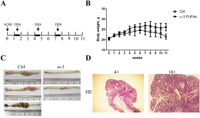
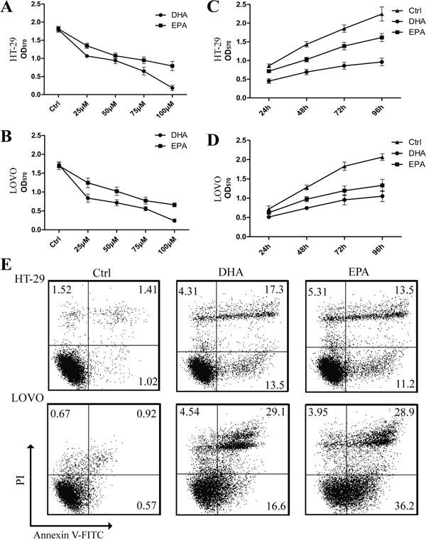
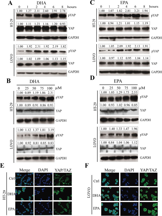
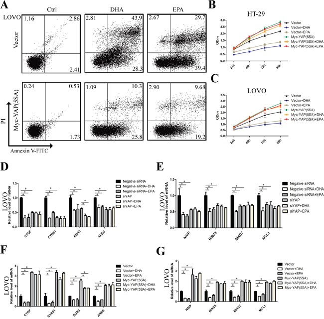
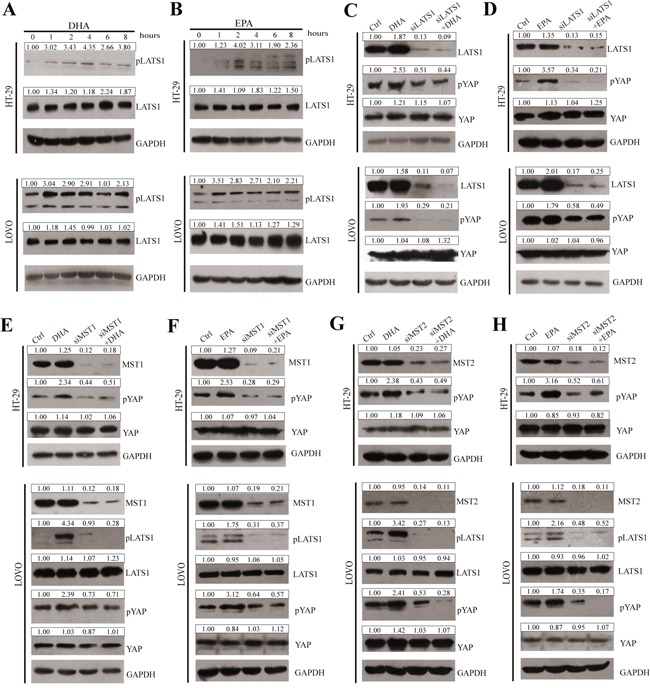
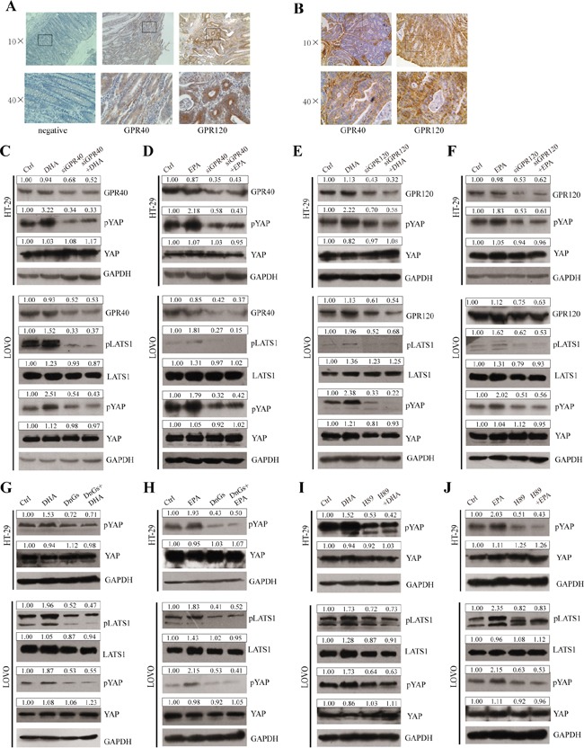
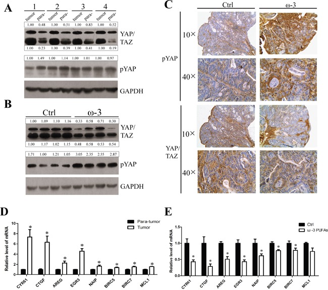
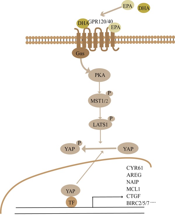
Colorectal cancer (CRC) is one of the most common cancers leading to high mortality. However, long-term administration of anti-tumor therapy for CRC is not feasible due to the side effects. Omega-3 polyunsaturated fatty acids (ω-3 PUFAs), particularly DHA and EPA, exert protection against CRC, but the mechanisms are unclear. Here, we show that ω-3 PUFAs inhibit proliferation and induce apoptosis of CRC cells in vitro and alleviate AOM/DSS-induced mice colorectal cancer in vivo. Moreover, ω-3 PUFAs promote phosphorylation and cytoplasmic retention of YAP and this effect was mediated by MST1/2 and LATS1, suggesting that the canonical Hippo Pathway is involved in ω-3 PUFAs function. We further confirmed that increase of pYAP by ω-3 PUFAs was mediated by GPRs, including GPR40 and GPR120, which subsequently activate PKA via Gαs, thus inducing the Hippo pathway activation. These data provide a novel DHA/EPA-GPR40/120-Gαs-PKA-MST1/2-LATS1-YAP signaling pathway which is linked to ω-3 PUFAs-induced inhibition of cell proliferation and promotion of apoptosis in CRC cells, indicating a mechanism that could explain the anti-cancer action of ω-3 PUFAs.

Keywords: GPR; Hippo pathway; YAP; colorectal cancer; omega-3 PUFAs.


Conflict of interest statement

No competing interests declared.

Figures

Figure 1
Figure 1. ω-3 PUFAs intake prevents AOM/DSS-induced colorectal cancer
A. Tumor induction protocol. Mice were injected with AOM on day 1 followed by three rounds of DSS treatment. After 1 week, 2.5% DSS was administered in the drinking water for 5 days, followed by 16 days of tap water in sterile. This cycle was repeated twice (5 days of 2.5% DSS followed by a 16 days recovery period and 5 days of 2.5% DSS). Mice were terminated at week 11 after injection of AOM. B. Body weight changes in mice fed with different diets. Values are means ± SEM. C. Photographs showing colons with tumors from mice fed with the control diet (left) and mice fed with the diet supplemented with ω-3 PUFAs (right). D. Representative hematoxylin and eosin stain of colorectal tumor.
Figure 2
Figure 2. ω-3 PUFAs suppress proliferation and induce apoptosis of CRC cells
A-D. HT-29 (A) and LOVO (B) cells were assessed by an MTT assay for viability following exposure for 72 h to media containing 10% FBS and varying concentrations of DHA or EPA. HT-29 (C) and LOVO (D) cells were assessed by an MTT assay for viability following exposure for 24h, 48h, 72h and 96h, respectively, to media containing 10% FBS and 75μM DHA or EPA. E. HT-29 and LOVO cells were treated with 75 μM DHA or EPA for 48 h, cell apoptosis was determined by FACS analysis. The data are expressed as the mean ± SEM for triplicate experiments. *P<0.05.
Figure 3
Figure 3. ω-3 PUFAs induce phosphorylation and cytoplasmic translocation of YAP in CRC cells
A-D. pYAP and YAP signals were examined and quantified by western blot after HT-29 and LOVO cells were treated with 75 μM DHA (A and B) or EPA (C and D) for indicated times (A and C) or different concentrations at 4h (B and D). GAPDH served as the loading control. Bands were semiquantified by image intensity area under the curve. Intensity of specific band is normalized in relation to loading control protein intensity. E and F. HT-29 (E) and LOVO (F) cells were treated with 75 μM DHA or EPA for 6 h. Expression and translocation of YAP/TAZ were determined by confocal microscopy; DAPI stained nuclei blue.
Figure 4
Figure 4. ω-3 PUFAs inhibit proliferation and induce apoptosis of CRC cells via YAP
A. LOVO cells were infected with empty vector or pQCXIH Myc-YAP (5SA) retroviral for 72 h, after infection, cells were treated with 75 μM DHA or EPA for additional 48 h, cell apoptosis was determined by FACS analysis. B and C. CRC cells were infected with empty vector or pQCXIH Myc-YAP (5SA) retroviral for 72 h, after infection, cells were treated with 75 μM DHA or EPA for 24h, 48h, 72h, or 96h respectively, cell viability was determined by MTT assay. D and E. LOVO cells were transfected with YAP siRNA for 48 h. After transfection, cells were treated with 75μM DHA or EPA for additional 24h. Total RNA was extracted and used for qRT-PCR analysis of the representative panel of pro-proliferative genes and anti-apoptosis genes. F and G. LOVO cells were infected with empty vector or pQCXIH Myc-YAP (5SA) retroviral for 72 h. After infection, cells were treated with 75 μM DHA or EPA for additional 24 h. Total RNA was extracted and used for qRT-PCR analysis of the representative panel of pro-proliferative genes and anti-apoptosis genes. The data are expressed as the mean ± SEM for triplicate experiments. *P<0.05.
Figure 5
Figure 5. ω-3 PUFAs-induced YAP phosphorylation and cytoplasm translocation is predominantly through the canonical Hippo Pathway
A and B. pLATS1 and LATS1 expressions were examined and quantified by western blot after HT-29 and LOVO cells were treated with 75μM DHA (A) or EPA (B) for indicated times. C and D. CRC cells were transfected with LATS1 siRNA for 48h, after transfection, cells were treated with 75μM DHA (C) or EPA (D) for additional 4h, pYAP, YAP and LATS1 expressions were examined and quantified by western blot in HT-29 cells and LOVO cells. E and F. CRC cells were transfected with MST1 siRNA for 48h, after transfection, cells were treated with 75μM DHA (E) or EPA (F) for additional 4h. pYAP, YAP, pLATS1, LATS1 and MST1 expressions were examined and quantified by western blot in HT-29 cells and LOVO cells. G and H. CRC cells were transfected with MST2 siRNA for 48 h. After transfection, cells were treated with 75 μM DHA (G) or EPA (H) for additional 4 h. pYAP, YAP, pLATS1, LATS1 and MST2 expressions were examined and quantified by western blot in HT-29 cells and LOVO cells. GAPDH served as the loading control. Bands were semiquantified by image intensity area under the curve. Intensity of specific band is normalized in relation to loading control protein intensity.
Figure 6
Figure 6. GPR120, GPR40, Gαs and PKA are involved in mediating ω-3 PUFAs-induced YAP phosphorylation
A and B. The expressions of GPR40 and GPR120 in paraffin-embedded CRC tissues of human CRC patients (A) and AOM/DSS-induced mouse modles (B) were examined using IHC staining, 10× and 40×magnification. C and D. GPR40 was knocked down by siRNA and further treated with 75μM DHA (C) or EPA (D) for additional 4h. The expression of pLATS1, LATS1, pYAP, YAP and GPR40 were examined by western blot and quantified in HT-29 and LOVO cells. E and F. GPR120 was knocked down by siRNA and further treated with 75μM DHA (E) or EPA (F) for additional 4h. The expression of pLATS1, LATS1, pYAP, YAP and GPR120 were examined by western blot and quantified in HT-29 and LOVO cells. G and H. Gαs function was blocked by transfected with DnGs and further treated with 75μM DHA (G) or EPA (H) for additional 4h. The expression of pLATS1, LATS1, pYAP and YAP were examined by western blot and quantified in HT-29 and LOVO cells. I and J. PKA was inhibited by the inhibitor H-89 and further treated with 75μM DHA (I) or EPA (J) for additional 4h. The expression of pLATS1, LATS1, pYAP and YAP were examined by western blot and quantified in HT-29 and LOVO cells. GAPDH served as the loading control. Bands were semiquantified by image intensity area under the curve. Intensity of specific band is normalized in relation to loading control protein intensity.
Figure 7
Figure 7. ω-3 PUFAs supplementation prevents AOM/DSS-induced colorectal cancer through activating the Hippo pathway
A and B. Total protein of the colorectal tumor tissues and para-tumor normal tissues from the control group mice (A) or total protein of colorectal tumor tissues from the control group mice and ω-3 group mice (B) were prepared and subjected to western blot analysis to detect YAP/TAZ and pYAP. GAPDH served as the loading control. Bands were semiquantified by image intensity area under the curve. Intensity of specific band is normalized in relation to loading control protein intensity. C. IHC stainings of YAP/TAZ and pYAP in colorectal tumors from the control group mice (left panel) and the ω-3 group mice (right panel), 10× and 40×magnification. D and E. Total RNA of the colorectal tumor tissues and para-tumor normal tissues from the control group mice (D), or total RNA of colorectal tumor tissues from the control group and ω-3 group mice (E) was prepared and used for qRT-PCR analysis of pro-proliferative genes and anti-apoptosis genes. The data are expressed as the mean ± SEM for triplicate experiments. *P<0.05.
Figure 8
Figure 8. Summary the ω-3 PUFAs-YAP signaling pathway revealed in this study
See Discussion for details.

References

    1. Siegel R, Ma J, Zou Z, Jemal A. Cancer statistics, 2014. CA Cancer J Clin. 2014;64:9–29. - PubMed
    1. DeSantis CE, Lin CC, Mariotto AB, Siegel RL, Stein KD, Kramer JL, Alteri R, Robbins AS, Jemal A. Cancer treatment and survivorship statistics, 2014. CA Cancer J Clin. 2014;64:252–271. - PubMed
    1. Berquin IM, Edwards IJ, Chen YQ. Multi-targeted therapy of cancer by omega-3 fatty acids. Cancer letters. 2008;269:363–377. - PMC - PubMed
    1. Gerber M. Omega-3 fatty acids, cancers: a systematic update review of epidemiological studies. The British journal of nutrition. 2012;107:S228–239. - PubMed
    1. Vaughan VC, Hassing MR, Lewandowski PA. Marine polyunsaturated fatty acids and cancer therapy. British journal of cancer. 2013;108:486–492. - PMC - PubMed

MeSH terms