. 2016 Aug 9;14(1):211.
doi: 10.1186/s12957-016-0966-6.

Surgical spacer placement prior carbon ion radiotherapy (CIRT): an effective feasible strategy to improve the treatment for sacral chordoma

Affiliations

Surgical spacer placement prior carbon ion radiotherapy (CIRT): an effective feasible strategy to improve the treatment for sacral chordoma

Cobianchi Lorenzo et al. World J Surg Oncol. .

Erratum in

Abstract

Background: Sacral chordoma (SC) is a neoplasm arising from residual notochordal cells degeneration. SC is difficult to manage mainly because of anatomic location and tendency to extensive spread. Carbon ion radiotherapy (CIRT) is highly precise to selectively deliver high biological effective dose to the tumor target sparing the anatomical structure on its path even if when SC is contiguous to the intestine, and a surgical spacer might be an advantageous tool to create a distance around the target volume allowing radical curative dose delivery with a safe dose gradient to the surrounding organs. This paper describes a double approach-open and hand-assisted laparoscopic-for a silicon spacer placement in patients affected by sacral chordoma undergoing carbon ion radiotherapy.

Methods: Six consecutive patients have been enrolled for surgical spacer placement-open (three) or hand-assisted (three)-prior carbon ion radiotherapy treatment in order to increase efficacy of carbon ion radiotherapy minimizing its side effects.

Results: Results showed that silicon spacer placement for SC treatment is feasible both via laparoscopic and laparotomic approach.

Conclusions: Its use might improve CIRT safety and thus efficacy for SC treatment.

Keywords: Carbon ion radiotherapy; Chordoma; Spacer placement.


Figures

Fig. 1
Fig. 1
Shaping and preparation of patient-tailored silicone spacer. Starting from a single, 1-mm width and 10 × 10 cm silicone sheet (a), the first plate has been created and shaped directly from tumor features (b) with surgical blade. Subsequently, the optimal width for the final spacer has been chosen (usually around 5 mm) and then singles pre-modeled silicone sheets (c) are stacked up and fixed by 1-0 Prolene suture (d–f)
Fig. 2
Fig. 2
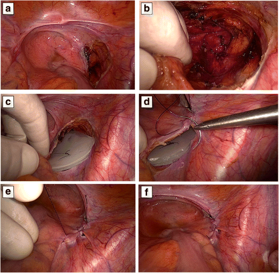
Laparoscopic hand-assisted silicone spacer placement. The peritoneum has been dissected directly above of the tumor area intended to dress through the spacer (a, b). The molded spacer has been located inside the created “peritoneal pouch” and then fastened by continue 3-0 Prolene suture (ce). The conclusion of peritoneal closure ensures the optimal fixation and stability of the silicone spacer (f)
Fig. 3
Fig. 3
Plan comparison study on different CT from the same patient selected for spacer positioning at CNAO. a Different CIRT plans depicted on axial images at the same anatomical level for the same chordoma patient. Red lines for GTV and green lines for CTV are included within the prescription dose isodose in both CIRT plans of 70.4 Gy (RBE). In the upper panel, the digestive area (rectal wall in green and intestine in blue) is contiguous to the prescribed dose isodose. In the lower panel, the digestive tract is far away from irradiated area due to the spacer (pointed by a red arrow). b Comparison of DVH for GTV (red), PTV (pink), and digestive tract (rectum green, intestine blue) dose coverage from the two CIRT plans compared in a. Coverage of the tumor is higher with the spacer CIRT plan (dotted lines) compared to the plan without it (continuous lines). With the CIRT spacer, plan dose sparing to the digestive tract is achieved

References

    1. Chambers KJ, Lin DT, Meier J, Remenschneider A, Herr M, et al. Incidence and survival patterns of cranial chordoma in the United States. Laryngoscope. 2014;124(5):1097–1102. doi: 10.1002/lary.24420. - DOI - PubMed
    1. McMaster ML, Goldstein AM, Bromley CM, Ishibe N, et al. Chordoma: incidence and survival patterns in the United States, 1973-1995. Cancer Causes Control. 2001;12:1–11. doi: 10.1023/A:1008947301735. - DOI - PubMed
    1. Walcott BP, Nahed BV, Mohyeldin A, Coumans JV, et al. Chordoma: current concepts, management, and future directions. Lancet Oncol. 2012;13:e69–e76. doi: 10.1016/S1470-2045(11)70337-0. - DOI - PubMed
    1. Hanna SA, Aston WJ, Briggs TW, Cannon SR, et al. Sacral chordoma: can local recurrence after sacrectomy be predicted? Clin Orthop Relat Res. 2008;466:2217–2223. doi: 10.1007/s11999-008-0356-7. - DOI - PMC - PubMed
    1. Chen KW, Yang HL, Lu J, Liu Y, et al. Prognostic factors of sacral chordoma after surgical therapy: a study of 36 patients. Spinal Cord. 2010;48:166–171. doi: 10.1038/sc.2009.95. - DOI - PubMed

Publication types

Substances

LinkOut - more resources