. 2016 Aug 10:6:31167.
doi: 10.1038/srep31167.

"A novel highly stable and injectable hydrogel based on a conformationally restricted ultrashort peptide"

Affiliations

"A novel highly stable and injectable hydrogel based on a conformationally restricted ultrashort peptide"

Chaitanya Kumar Thota et al. Sci Rep. .

Abstract

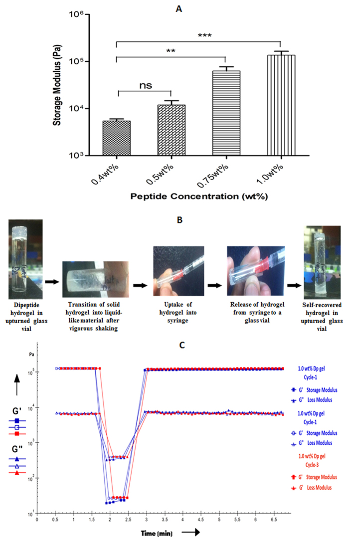
Nanostructures including hydrogels based on peptides containing non protein amino acids are being considered as platform for drug delivery because of their inherent biocompatibility and additional proteolytic stability. Here we describe instantaneous self-assembly of a conformationally restricted dipeptide, LeuΔPhe, containing an α,β-dehydrophenylalanine residue into a highly stable and mechanically strong hydrogel, under mild physiological aqueous conditions. The gel successfully entrapped several hydrophobic and hydrophilic drug molecules and released them in a controlled manner. LeuΔPhe was highly biocompatible and easily injectable. Administration of an antineoplastic drug entrapped in the gel in tumor bearing mice significantly controlled growth of tumors. These characteristics make LeuΔPhe an attractive candidate for further development as a delivery platform for various biomedical applications.


Figures

Figure 1
Figure 1. Instant self-assembly of LeuΔPhe into a highly stable hydrogel at room temperature and its possible applications in biomedical field.
Figure 2
Figure 2. Electron micrographs of Dp gel at different concentrations.
(A) TEM image of 0.3wt% Dp gel showing small fibrils of >100 nm length. (B) TEM image of 0.4wt% LeuΔPhe showing relatively longer and branched fibrils of length in micrometers. (C) TEM image of 0.5wt% Dp gel indicating presence of dense network of fibrils. (D) SEM image of 0.5wt% LeuΔPhe showing dense fibrilar network of the gel. (E–H) ESEM images showing porous fibrilar mesh of 0.5wt% Dp gel.
Figure 3
Figure 3
Mechanical strength and self-recovery of Dp gel (A) Storage modulus (G’ values) of Dp gel at different peptide concentrations (0.4wt%, 0.5 wt%, 0.75wt% and 1.0wt%), showing increase in gel strength with increasing peptide concentration Graphs represent mean ± standard deviation (n = 3). Significance values determined by t-test are marked with asterisks (ns for P-value  >  0.05, * for P-value  <  0.05, ** for P-value  <  0.01, *** for P-value  <  0.001, **** for P-value  <  0.0001). (B) Syringeability results showing recovery of gel (1.0wt%) after disruption of its structure. (C) Thixotropic time dependent step-strain data of Dp gel (1.0wt%) showing self-healing property of Dp gel. Higher G’ values and G” values (G’ > G”) initially at lower strain (0.1%) indicate solid like structure of the gel while on increasing strain to 50%, G’ and G” values were declined (G’ < G”), indicating transition of gel into liquid-like material. Finally on removal of high strain, the gel restored its original strength (G’ > G”). Three cycles were performed to verify reproducibility.
Figure 4
Figure 4
CD spectra showing effect of different parameters on formation of Dp gel and its thermal stability (A) CD spectrum showing conformational changes in the structure of LeuΔPhe on self-assembly in different salt (acetate) conditions. (B) CD spectrum showing effect of changes in pH of the buffer (sodium acetate buffer) condition on LeuΔPhe self-assembly into hydrogel. (C) CD temperature scan of Dp gel (0.5wt%) showing its stability at physiological temperatures.
Figure 5
Figure 5. Percentage release of drugs from Dp gel, indicating their continuous & slow release.
Figure 6
Figure 6
(A) Cumulative percentage release of curcumin from Dp gels (at 0.5wt% and 1wt%) showing decrease in curcumin release on increase in peptide concentration, indicating its peptide concentration based tunability. (B) Percentage cytotoxicity of released curcumin on HeLa cells from Dp gels (0.5wt%), entrapped with 0 mM, 2 mM and 4 mM curcumin, assessed by LDH assay. Graphs represent mean ± standard deviation (n = 3). Significance values determined by t-test are marked with asterisks (ns for P-value  >  0.05, * for P-value  <  0.05, ** for P-value  <  0.01, *** for P-value  <  0.001, **** for P-value  <  0.0001).
Figure 7
Figure 7
Cytotoxicity of Dp gel (A) Percentage cell viability of HEK293T cells treated with Dp gels of different peptide concentration, indicating its biocompatibility. (B) Live/Dead assay results showing no change in percentage population of live cells in the presence of Dp gel compared to control, indicating its high biocompatibility. Graph represents mean ± standard deviation (n = 3).
Figure 8
Figure 8. Tumor regression study in mice xenograft model showing changes in relative tumor volume of different groups of mice treated with different formulations with time.
Graph represents mean ± standard deviation (n = 4).

References

    1. Zhang S. Fabrication of novel biomaterials through molecular self-assembly. Nature biotechnology 21, 1171–1178 (2003). - PubMed
    1. Kobsa S. & Saltzman W. M. Bioengineering approaches to controlled protein delivery. Pediatric research 63, 513–519 (2008). - PubMed
    1. Zhang R., Tang M., Bowyer A., Eisenthal R. & Hubble J. A novel pH-and ionic-strength-sensitive carboxy methyl dextran hydrogel. Biomaterials 26, 4677–4683, (2005). - PubMed
    1. Xing Q., Yates K., Vogt C., Qian Z., Frost M. C. & Zhao F. Increasing mechanical strength of gelatin hydrogels by divalent metal ion removal. Scientific Reports 4, 1–10, (2014). - PMC - PubMed
    1. Black K. A. et al.. Biocompatibility and characterization of a Peptide amphiphile hydrogel for applications in peripheral nerve regeneration. Tissue Engineering Part A 21, 1333–1342 (2015). - PMC - PubMed

Publication types