. 2016 Nov;139(3):419-431.
doi: 10.1111/jnc.13764. Epub 2016 Oct 3.

Mechanistic characterization of nitrite-mediated neuroprotection after experimental cardiac arrest

Affiliations

Mechanistic characterization of nitrite-mediated neuroprotection after experimental cardiac arrest

Cameron Dezfulian et al. J Neurochem. 2016 Nov.

Abstract

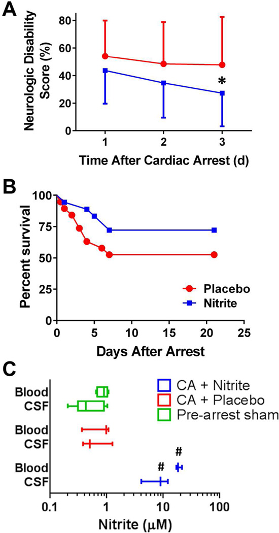
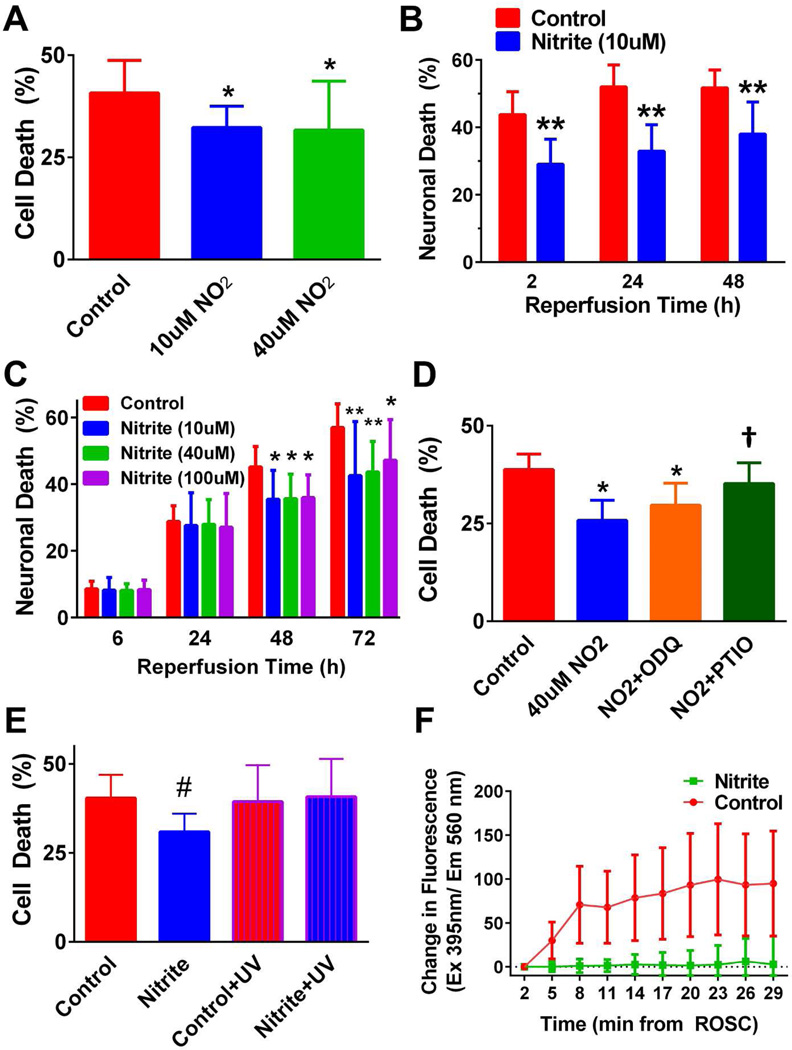
Nitrite acts as an ischemic reservoir of nitric oxide (NO) and a potent S-nitrosating agent which reduced histologic brain injury after rat asphyxial cardiac arrest (ACA). The mechanism(s) of nitrite-mediated neuroprotection remain to be defined. We hypothesized that nitrite-mediated brain mitochondrial S-nitrosation accounts for neuroprotection by reducing reperfusion reactive oxygen species (ROS) generation. Nitrite (4 μmol) or placebo was infused IV after normothermic (37°C) ACA in randomized, blinded fashion with evaluation of neurologic function, survival, brain mitochondrial function, and ROS. Blood and CSF nitrite were quantified using reductive chemiluminescence and S-nitrosation by biotin switch. Direct neuroprotection was verified in vitro after 1 and 4 h neuronal oxygen glucose deprivation measuring neuronal death with inhibition studies to examine mechanism. Mitochondrial ROS generation was quantified by live neuronal imaging using mitoSOX. Nitrite significantly reduced neurologic disability after ACA. ROS generation was reduced in brain mitochondria from nitrite- versus placebo-treated rats after ACA with congruent preservation of brain ascorbate and reduction of ROS in brain sections using immuno-spin trapping. ATP generation was maintained with nitrite up to 24 h after ACA. Nitrite rapidly entered CSF and increased brain mitochondrial S-nitrosation. Nitrite reduced in vitro mitochondrial superoxide generation and improved survival of neurons after oxygen glucose deprivation. Protection was maintained with inhibition of soluble guanylate cyclase but lost with NO scavenging and ultraviolet irradiation. Nitrite therapy results in direct neuroprotection from ACA mediated by reductions in brain mitochondrial ROS in association with protein S-nitrosation. Neuroprotection is dependent on NO and S-nitrosothiol generation, not soluble guanylate cyclase.

Keywords: cardiac arrest; cerebral ischemia; mitochondria; nitric oxide; reactive oxygen species; reperfusion injury.


Conflict of interest statement

Disclosures/Conflicts of Interest Dr. Dezfulian served on an ad hoc advisory board to Mallinckrodt which markets iNO.

Figures

Figure 1
Figure 1. Outcomes from nitrite vs. placebo randomized controlled trial
Rats were subjected to 7 min asphyxial cardiac arrest (ACA) and randomized to nitrite or placebo IV 5 min after return of spontaneous circulation (ROSC). A) Neurologic disability was lower at day 1 in nitrite rats and decreased progressively with significant difference noted by day 3. In this scale, 0% refers to no disability and 100% is death. B) Survival plots of the two groups over 3 weeks reveal most deaths occurred during the first (days 2–7) due to persistent coma or severe weight loss necessitating euthanasia. C) Blood and CSF obtained after sham or 15 min after ROSC from 8 min ACA treated with nitrite or placebo demonstrate significantly higher median blood and CSF nitrite levels with nitrite therapy compared to either group. Comparisons performed using 2-way repeated measures ANOVA with Sidak’s post-hoc adjustment (A) and 1-way ANOVA with Holm-Sidak’s post-hoc adjustment. (survival experiments n=18 nitrite, n=19 placebo; blood/CSF levels n=3 nitrite and placebo, n=5 sham; *, p<0.05 and #, p<0.01 nitrite vs placebo)
Figure 2
Figure 2. Brain mitochondrial ROS and ATP generation after ACA
Function was assessed in isolated brain mitochondria at designated times after CA and compared to non-arrested shams. A) Sample output of Amplex Red fluorescence over time when isolated mitochondria were respiring with the complex I substrates pyruvate, malate and ADP. B) Mitochondria isolated 15 min after CA from the brains of nitrite-treated rats generated similar levels of peroxide as those obtained from sham brains which was significantly less than peroxide generation from placebo-treated animals. ATP generation was significantly increased in nitrite compared to placebo treated rats at C) 15 min and D) 24 h after CA. Rates of ROS and ATP generation are normalized to complex I state 3 oxygen consumption (see Suppl Fig 2, 3). Insets show sample luminescence data in C, D. Comparisons made using Kruskal-Wallis test with Dunn’s post-hoc adjustment. (n=6 sham; n=7 in 15 min experiments; n=10 in 24 h experiments; *, p<0.05; **, p<0.01 for nitrite vs. placebo)
Figure 3
Figure 3. Free radical injury is reduced by nitrite therapy
The generation of free radicals in brain after 30 min of reperfusion from 8 min no flow ACA with placebo or nitrite therapy is compared to sham surgery using immuno-spin trapping with DMPO. A) Representative sections of hippocampus demonstrate staining corresponding to nuclei (DAPI; white), biomolecular free radical-DMPO adducts (DMPO; blue), glia (GFAP; green) and neurons (NeuN; red). DMPO staining was diffuse particularly within placebo brains and B) quantification of this signal for the entire section was normalized to nuclei count (DAPI; n=4). C) Consistent with increase oxidative stress/free radical injury, brain ascorbate levels declined substantially from sham (0 min; n=4) levels at 15 min post-CA (n=6) and was significantly lower at 60 min in placebo (n=8) compared to nitrite (n=9) treated rats. All comparisons performed using ANOVA with Holm-Sidak’s post-hoc comparison. Measurement bar indicates 500 µm (A). (*, p<0.05; **, p<0.01; ***, p<0.001)
Figure 4
Figure 4. Nitrite increases brain mitochondrial S-nitrosation
Biotin switch of brain mitochondria obtained 15 min after 8 min ACA treated with placebo or nitrite or sham. A) sample blot probed with anti-biotin (upper panel) and anti-cytochrome oxidase 4 (lower panel) antibodies. The approximate molecular weight markers are shown to the left. A negative control (−) omitted copper/ascorbate during the biotin switch which was present in the other lanes. B) Summary data quantifying the density of bands around the ~75 kDa region blotted with anti-biotin antibody normalized to COX4 (~17 kDa) demonstrate a significant increase in mitochondrial S-nitrosation in nitrite vs. placebo treated rats using ANOVA with post-hoc Sidak’s adjustment (n=4–5/group; *, p<0.05).
Figure 5
Figure 5. Nitrite protects against early and delayed neuronal death after oxygen glucose deprivation (OGD) and reduces mitochondrial superoxide generation
Cultured day in vitro 10–12 neurons underwent 1 or4h OGD and reperfusion with NBM alone (control) or supplemented with nitrite (NO2) at the concentration specified. A) NO2 significantly reduced cell death at 10 and 40 µM levels 18h after reperfusion from 4h OGD. B) 4h OGD resulted in substantial early neuronal death (after 2h reperfusion) with little subsequent increase and this early death was protected by NO2. C) When OGD was shortened to 1h, there was little early death (after 6h reperfusion) but substantial delayed neuronal death approaching levels similar to that of 4h OGD after 72h of reperfusion. Again NO2 was protective at 10 and 40 µM with slightly less protection at 100 µM. D) Mechanistic studies examining protection by 40 µM NO2 from 4h OGD demonstrated a slight, non-significant increase in cell death with 10 mM ODQ and loss of protection with 100 µM carboxy-PTIO. E) Irradiation of cells for 4 min with 6W UV light (365 nm) immediately after reperfusion from 4h OGD resulted in loss of protection provided by 40 µM NO2 after 24h. F) Neurons on coverslips were loaded with mitoSOX 15 min before 1h OGD. Superoxide generation was assessed immediately after reperfusion (time 0) through excitation at 396 nm. Values are in fluorescence units with the value at the time of reperfusion subtracted from all subsequent points (ie time 0 has value 0). Each point represents summary data from 20 fields obtained in 2 separate experiments (different cultures). with comparison’s made using 2-way repeated measures ANOVA with post-hoc Tukey’s adjustment. All comparisons performed using 1-way ANOVA (A, D, E) or 2-way repeated measures ANOVA (B, C, F) with Sidak’s (A–E) or Tukey’s post-hoc adjustment. (n=12 for all experiments excpet E where n=16; *, p<0.05; #, p<0.01; **, p<0.001 vs. control [same reperfusion time if applicable]; †, p<0.001 vs NO2; In F, all values after reperfusion differ significantly at p<0.001)

References

    1. Aufderheide TP, Nichol G, Rea TD, et al. A Trial of an Impedance Threshold Device in Out-of-Hospital Cardiac Arrest. New England Journal of Medicine. 2011;365:798–806. - PMC - PubMed
    1. Barrett RD, Bennet L, Blood AB, Wassink G, Gunn AJ. Asphyxia and Therapeutic Hypothermia Modulate Plasma Nitrite Concentrations and Carotid Vascular Resistance in Preterm Fetal Sheep. Reproductive Sciences. 2014;21:1483–1491. - PMC - PubMed
    1. Basu S, Liu X, Nozari A, Rubertsson S, Miclescu A, Wiklund L. Evidence for Time-dependent Maximum Increase of Free Radical Damage and Eicosanoid Formation in the Brain as Related to Duration of Cardiac Arrest and Cardio-pulmonary Resuscitation. Free Radical Research. 2003;37:251–256. - PubMed
    1. Belikova NA, Glumac AL, Kapralova V, Cheikhi A, Tyurina YY, Vagni VA, Kochanek PM, Kagan VE, Bayir H. A high-throughput screening assay of ascorbate in brain samples. J Neurosci Methods. 2011;201:185–190. - PMC - PubMed
    1. Bernard SA, Gray TW, Buist MD, Jones BM, Silvester W, Gutteridge G, Smith K. Treatment of Comatose Survivors of Out-of-Hospital Cardiac Arrest with Induced Hypothermia. New Engl J Med. 2002;346:557–563. - PubMed

Publication types

MeSH terms