Review
. 2017 Jan:33:3-17.
doi: 10.1016/j.arr.2016.08.002. Epub 2016 Aug 6.

Cockayne syndrome: Clinical features, model systems and pathways

Affiliations
Review

Cockayne syndrome: Clinical features, model systems and pathways

Ajoy C Karikkineth et al. Ageing Res Rev. 2017 Jan.

Abstract

Cockayne syndrome (CS) is a disorder characterized by a variety of clinical features including cachectic dwarfism, severe neurological manifestations including microcephaly and cognitive deficits, pigmentary retinopathy, cataracts, sensorineural deafness, and ambulatory and feeding difficulties, leading to death by 12 years of age on average. It is an autosomal recessive disorder, with a prevalence of approximately 2.5 per million. There are several phenotypes (1-3) and two complementation groups (CSA and CSB), and CS overlaps with xeroderma pigmentosum (XP). It has been considered a progeria, and many of the clinical features resemble accelerated aging. As such, the study of CS affords an opportunity to better understand the underlying mechanisms of aging. The molecular basis of CS has traditionally been ascribed to defects in transcription and transcription-coupled nucleotide excision repair (TC-NER). However, recent work suggests that defects in base excision DNA repair and mitochondrial functions may also play key roles. This opens up the possibility for molecular interventions in CS, and by extrapolation, possibly in aging.

Keywords: Cockayne syndrome; Mitochondria; Neurodegeneration; Parylation; Progeria; Transcription.


Figures

Figure 1
Figure 1
Shows the progressive loss of facial subcutaneous fat over time, leading to sunken eyes and the ‘wizened’ facies typical of CS patients (reproduced with permission from (Wilson et al., 2015, fig. 2a, 2b).
Figure 2
Figure 2
Fundoscopic examination of the retina shows the typical retinal pigmentary “leak” in a salt and pepper pattern. The yellow arrows indicate the areas of involvement, while the blue arrows indicate areas of relatively preserved retina (reproduced with permission from Orphanet Journal of Rare Diseases) (Hamel, 2006, fig. 1).
Figure 3
Figure 3
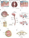
Some of the most important clinical manifestations of CS are shown. Figure 3A: Cerebral cortex - may be slightly thinned, with relative preservation of cortical neurons. Figure 3B: White matter changes - There is “tigroid leukodystrophy”, with areas that have relatively less leukodystrophy interspersed with more affected areas in a pattern resembling the stripes on a tiger. Figure 3C: Cerebellum - There is loss of Purkinje cells. Figure 3D: Eye - Cataracts and corneal opacification are seen in CS patients. Figure 3E: Neurons – There are fewer oligodendroglia, and less myelin is produced, leading to a demyelinating neuropathy. Increased numbers of astrocytes are seen in areas of myelin and oligodendroglia loss. It has not been clearly established whether the astrocytes are the cause of or (more possibly) reactive to the changes in myelin and oligodendroglia. Figure 3F: Retina – shows loss of rods and cones, the photoreceptor cells, and also of ganglion and outer nuclear cell layers. Figure 3G: Cochlea – shows loss of hair cells. This is most pronounced generally in the basal turn of the cochlea. Figure 3H: Spinal cord – may show loss of anterior horn cells. It is not clear whether this is the primary event or secondary (and retrograde) to neuronal loss.
Figure 4
Figure 4
The brain calcifications in CS are a pathognomonic feature. They are mainly in the deep white matter, especially the basal ganglia, and progress with age. Some calcification can also occur in the depths of the sulci (white arrow in figure f). Also note the progressive brain atrophy (reproduced with permission from Kubota et al) (Kubota et al., 2015, fig. 4).
Figure 5
Figure 5
Molecule to medicine. In silico analyses show association of Cockayne syndrome with mitochondrial diseases using methods such as hierarchical clustering and a support vector machine.

References

    1. Aamann MD, Muftuoglu M, Bohr VA, Stevnsner T. Multiple interaction partners for Cockayne syndrome proteins: implications for genome and transcriptome maintenance. Mech Ageing Dev. 2013;134:212–224. doi: 10.1016/j.mad.2013.03.009. - DOI - PMC - PubMed
    1. Aamann MD, Sorensen MM, Hvitby C, Berquist BR, Muftuoglu M, Tian J, de Souza-Pinto NC, Scheibye-Knudsen M, Wilson DM, Stevnsner T, Bohr VA. Cockayne syndrome group B protein promotes mitochondrial DNA stability by supporting the DNA repair association with the mitochondrial membrane. FASEB J. 2010;24:2334–2346. doi: 10.1096/fj.09-147991. - DOI - PMC - PubMed
    1. Babu V, Hofmann K, Schumacher B. A C. elegans homolog of the Cockayne syndrome complementation group A gene. DNA Repair. 2014;24:57–62. doi: 10.1016/j.dnarep.2014.09.011. - DOI - PMC - PubMed
    1. Bartenjev I, Butina MR, Potocnik M. Rare case of Cockayne syndrome with xeroderma pigmentosum. Acta Derm Venereol. 2000;80:213–214. - PubMed
    1. Batenburg NL, Thompson EL, Hendrickson EA, Zhu XD. Cockayne syndrome group B protein regulates DNA double-strand break repair and checkpoint activation. EMBO J. 2015;34:1399–1416. doi: 10.15252/embj.201490041. - DOI - PMC - PubMed