. 2016 Oct;56(10):2571-2583.
doi: 10.1111/trf.13745. Epub 2016 Aug 9.

Testosterone-dependent sex differences in red blood cell hemolysis in storage, stress, and disease

Affiliations

Testosterone-dependent sex differences in red blood cell hemolysis in storage, stress, and disease

Tamir Kanias et al. Transfusion. 2016 Oct.

Abstract

Background: Red blood cell (RBC) hemolysis represents an intrinsic mechanism for human vascular disease. Intravascular hemolysis releases hemoglobin and other metabolites that inhibit nitric oxide signaling and drive oxidative and inflammatory stress. Although these pathways are important in disease pathogenesis, genetic and population modifiers of hemolysis, including sex, have not been established.

Study design and methods: We studied sex differences in storage or stress-induced hemolysis in RBC units from the United States and Canada in 22 inbred mouse strains and in patients with sickle cell disease (SCD) using measures of hemolysis in 315 patients who had homozygous SS hemoglobin from the Walk-PHASST cohort. A mouse model also was used to evaluate posttransfusion recovery of stored RBCs, and gonadectomy was used to determine the mechanisms related to sex hormones.

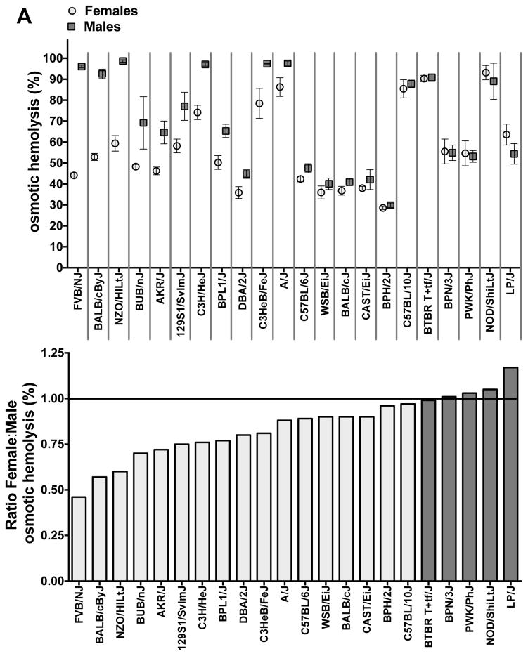
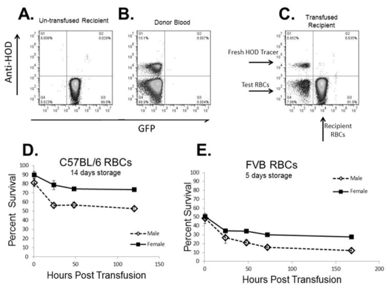
Results: An analysis of predisposition to hemolysis based on sex revealed that male RBCs consistently exhibit increased susceptibility to hemolysis compared with females in response to routine cold storage, under osmotic or oxidative stress, after transfusion in mice, and in patients with SCD. The sex difference is intrinsic to the RBC and is not mediated by plasmatic factors or female sex hormones. Importantly, orchiectomy in mice improves RBC storage stability and posttransfusion recovery, whereas testosterone repletion therapy exacerbates hemolytic response to osmotic or oxidative stress.

Conclusion: Our findings suggest that testosterone increases susceptibility to hemolysis across human diseases, suggesting that male sex may modulate clinical outcomes in blood storage and SCD and establishing a role for donor genetic variables in the viability of stored RBCs and in human hemolytic diseases.


Conflict of interest statement

Dr. James C. Zimring has a sponsored research agreement with Immucor, and serves on the scientific advisory board for Rubious Therapeutics. The rest of the authors declare no competing financial interests.

Figures

FIGURE 1
FIGURE 1. Male sex is associated with increased susceptibility to storage and stress-induced hemolysis
RBCs from male or female donors were collected from expired RBC units (ITxM, 47±5 days old) and tested for A. percent storage hemolysis (n=77 females and 70 men). B. Osmotic fragility (n=77 females and 70 men). C. Mechanical fragility (n=45 females and 50 men). A–C: Mean±SD, * p<0.05 by unpaired t-test.
FIGURE 2
FIGURE 2. The effect of sex and age on end-of-storage hemolysis in SAGM units
Data derive from leukocyte-reduced SAGM RBC units tested within 24 h of product expiry (42 days of storage) by Canadian Blood Services. A. Kernel density estimate of percent storage hemolysis in male (solid blue line) versus female (dashed red line) blood donors. n= 28,543, 16,700 males and 11,843 females B. Distribution of storage hemolysis by sex and age (5-year intervals). Mean hemolysis values for male versus female donors are based on donor age at time of donation. Errors represent 95 % CI. n= 28,267, 16,636 males and 11,631 females. * Represents significant differences (p<0.0001 by one-way ANOVA) in hemolysis between male and female donors within age range. † Represents age range at which hemolysis is significantly (p<0.0001 by one-way ANOVA and Games-Howell test analysis) different from donors of the same sex aged 20–24.
FIGURE 3
FIGURE 3. Sex differences in hemolytic propensity in 22 mouse strains
RBCs from age-matched male and female mice from 22 selected in-bred and wild-derived strains were subjected to A. Osmotic hemolysis (Pink test 24 h). B. AAPH-induced oxidative hemolysis (50 mmol/L, 3 h, 37° C). A–B. Top panels: Hemolytic scores are represented as Mean±SEM (n=3–5) in males versus females of each tested strain. Lower panels: Sex differences (data derived from top panels) are represented as the ratio of the females’ score (mean percent hemolysis) divided by the males’ score. A ratio score of “1” means no sex differences; X<1 means females hemolyzed less than males. Dark grey bars indicate strains in which no sex differences were observed or males were more resistant to hemolysis than females.
FIGURE 4
FIGURE 4. Sex differences in post-transfusion recovery of stored mouse RBCs
Mouse RBCs collected from male or female C57BL/6J or FVB/NJ strain were leukocyte reduced and stored (4° C) in CPDA-1 for 14 days or 5 days, respectively. Stored RBCs were transfused into C57BL/6J (UbiC-GFP) x FVB/NJ F1 female recipients, and post-transfusion recovery was assessed as described under Materials and Methods. A–C. Representative flow cytometric dot plots from A. Expression (Q3, 99.9 %) of green fluorescent protein (GFP) in recipient mice (C57BL/6J (UbiC-GFP) x FVB/NJ F1). B. Donor blood was gated as non-GFP/non-HOD positive RBCs (Q4, 89.9 %) whereas anti HOD-positive mouse RBCs, which were spiked into donors samples prior to transfusion to serve as a fresh tracer population, were gated in Q1. C. After transfusion, Test (donor) RBCs are visualized as HOD-negative GFP-negative RBCs, and tracer HOD RBCs are visualized as HOD-positive GFP-negative RBCs. Recipient RBCs are gated out as the only GFP-positive RBCs. D–E. Post-transfusion recovery of male versus female stored C57BL/6J (D) or FVB/NJ (E) RBCs. Each curve represents the average of 3 experiments.
FIGURE 5
FIGURE 5. Effect of orchiectomy or testosterone repletion on predisposition to hemolysis and post-transfusion recovery in FVB/NJ RBCs
A–C. FVB/NJ pups underwent gonadectomy at 4 weeks and housed with age-matched intact males or females (n=10–15 per group) for 14–16 weeks, after which all animals were sacrificed. RBCs from each group were subjected to A. osmotic stress (24 h pink test), B. AAPH-induced oxidative hemolysis (50 mM, 3 h 37° C), C. RBCs from females (n=5), males (n=10), and orchiectomized males (n=10) were subjected to diamide-induced hemolysis (0.5 mM, 75 min). A–C: Mean±SEM. * designates statistically significant (p<0.05) differences obtained by one-way ANOVA and Holm-Sidak’s multiple comparison test using males as a reference group. A. p=0.0149. B, p=0.0201. C, p=0.0008. D–F. Orchiectomy FVB/NJ mice (15–16 week old) were treated with testosterone (1 mg/Kg body weight/day; Orch+T) or with propylene glycol (Orch Sham, drug vehicle) for 32 days as described under Material and Methods. Age-matched intact FVB/NJ males treated with propylene glycol were used as a reference for the hemolytic assays. After treatments, RBCs were subjected to osmotic (D) and oxidative stress (E) assays. F. Representative image of AAPH-induced oxidative hemolysis (50 mmol/L, 2 h, 37° C) in RBCs from each mouse group. Mean±SEM; * designates statistical differences (p<0.05 unpaired t-test) between Orch+T and Orch Sham. D. p=0.0132; E. p=0.034. G–H. Percent post-transfusion recovery (Mean±SD) of leukocyte-reduced stored RBCs from orchiectomy or intact FVB/NJ males after one (G) or six (H) days of storage. **** p<0.0001 by repeated measures two-way ANOVA and Sidak’s multiple comparisons test.
FIGURE 6
FIGURE 6. Sex differences in predisposition to hemolysis in sickle cell disease
Sex differences in BERK hemizygous (HbAS) sickle cell mice (Mean±SEM; n=4 per sex). A. Mean osmotic hemolysis (%) in response to 24 h Pink test. B. Mean oxidative hemolysis (%) in response to AAPH treatment (50 mmol/L, 3 h, 37° C). C+D. Mean percent hemolysis (C) and a corresponding image (D) in response to incubation with diamide (0.5 μmol/L, 90 min, 37° C). * designates significant differences (p<0.05, unpaired t-test) in hemolysis between males and females. A. p= 0.0182. B. p<0.0001, C. p= 0.004.

Similar articles

Cited by

References

    1. Donadee C, Raat NJ, Kanias T, Tejero J, Lee JS, Kelley EE, Zhao X, Liu C, Reynolds H, Azarov I, Frizzell S, Meyer EM, Donnenberg AD, Qu L, Triulzi D, Kim-Shapiro DB, Gladwin MT. Nitric oxide scavenging by red blood cell microparticles and cell-free hemoglobin as a mechanism for the red cell storage lesion. Circulation. 2011;124:465–76. - PMC - PubMed
    1. Alexander JT, El-Ali AM, Newman JL, Karatela S, Predmore BL, Lefer DJ, Sutliff RL, Roback JD. Red blood cells stored for increasing periods produce progressive impairments in nitric oxide-mediated vasodilation. Transfusion. 2013;53:2619–28. - PMC - PubMed
    1. Roback JD, Neuman RB, Quyyumi A, Sutliff R. Insufficient nitric oxide bioavailability: a hypothesis to explain adverse effects of red blood cell transfusion. Transfusion. 2011;51:859–66. - PMC - PubMed
    1. Wither M, Dzieciatkowska M, Nemkov T, Strop P, D’Alessandro A, Hansen KC. Hemoglobin oxidation at functional amino acid residues during routine storage of red blood cells. Transfusion. 2016;56:421–6. - PubMed
    1. Gladwin MT, Kanias T, Kim-Shapiro DB. Hemolysis and cell-free hemoglobin drive an intrinsic mechanism for human disease. J Clin Invest. 2012;122:1205–8. - PMC - PubMed