. 2016 Jul 26:7:1158.
doi: 10.3389/fmicb.2016.01158. eCollection 2016.

Engineering of Helicobacter pylori Dimeric Oxidoreductase DsbK (HP0231)

Affiliations

Engineering of Helicobacter pylori Dimeric Oxidoreductase DsbK (HP0231)

Katarzyna M Bocian-Ostrzycka et al. Front Microbiol. .

Abstract

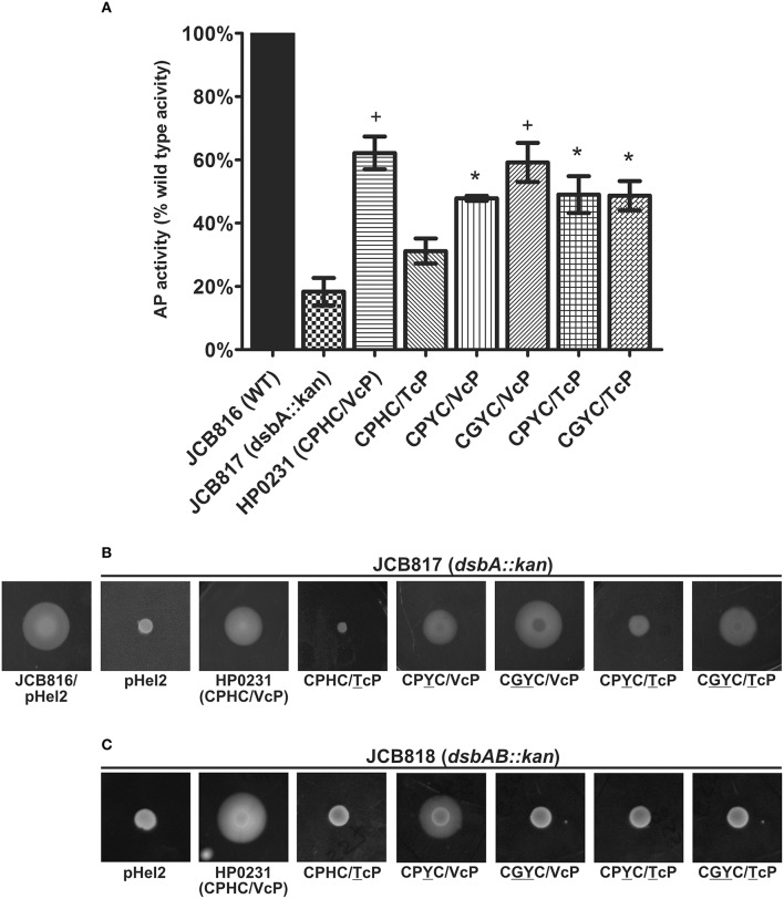
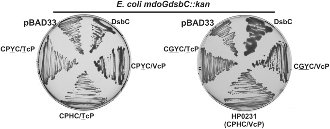
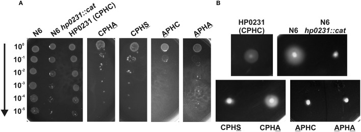
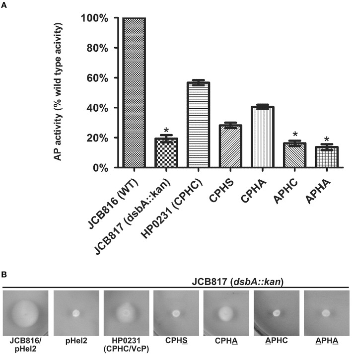
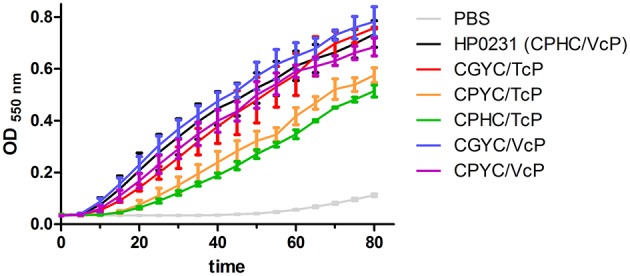
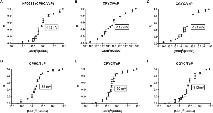
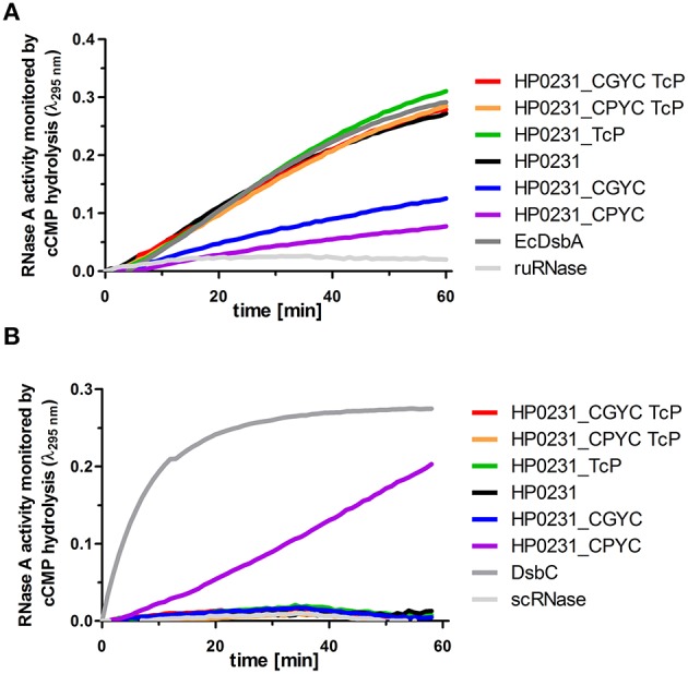
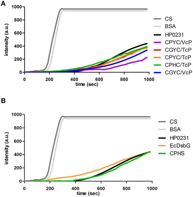
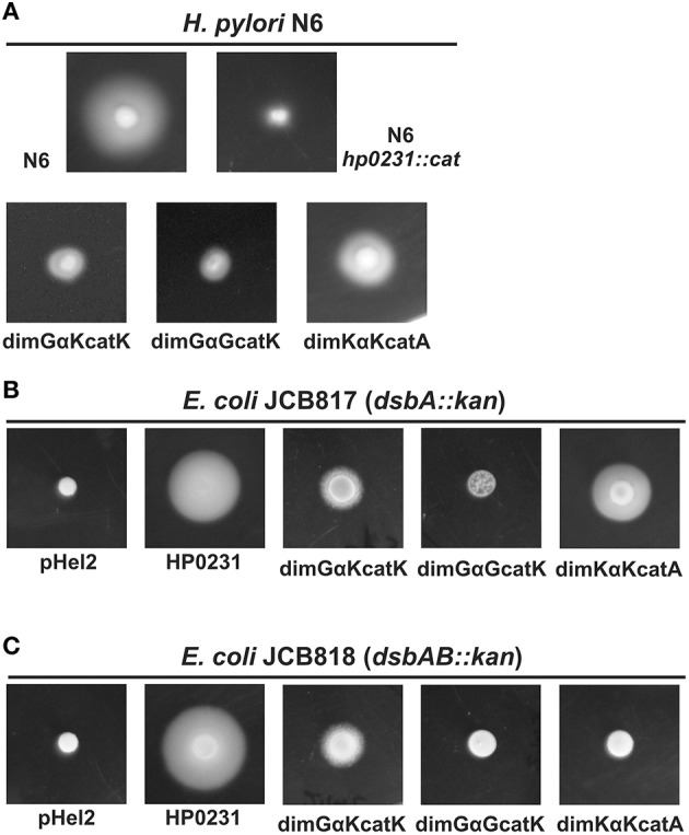
The formation of disulfide bonds that are catalyzed by proteins of the Dsb (disulfide bond) family is crucial for the correct folding of many extracytoplasmic proteins. Thus, this formation plays an essential, pivotal role in the assembly of many virulence factors. The Helicobacter pylori disulfide bond-forming system is uncomplicated compared to the best-characterized Escherichia coli Dsb pathways. It possesses only two extracytoplasmic Dsb proteins named HP0377 and HP0231. As previously shown, HP0377 is a reductase involved in the process of cytochrome c maturation. Additionally, it also possesses disulfide isomerase activity. HP0231 was the first periplasmic dimeric oxidoreductase involved in disulfide generation to be described. Although HP0231 function is critical for oxidative protein folding, its structure resembles that of dimeric EcDsbG, which does not confer this activity. However, the HP0231 catalytic motifs (CXXC and the so-called cis-Pro loop) are identical to that of monomeric EcDsbA. To understand the functioning of HP0231, we decided to study the relations between its sequence, structure and activity through an extensive analysis of various HP0231 point mutants, using in vivo and in vitro strategies. Our work shows the crucial role of the cis-Pro loop, as changing valine to threonine in this motif completely abolishes the protein function in vivo. Functioning of HP0231 is conditioned by the combination of CXXC and the cis-Pro loop, as replacing the HP0231 CXXC motif by the motif from EcDsbG or EcDsbC results in bifunctional protein, at least in E. coli. We also showed that the dimerization domain of HP0231 ensures contact with its substrates. Moreover, the activity of this oxidase is independent on the structure of the catalytic domain. Finally, we showed that HP0231 chaperone activity is independent of its redox function.

Keywords: Dsb proteins; Helicobacter pylori; chaperone activity; disulfide bonds; oxidoreductases; protein engineering; site-directed mutagenesis.


Figures

Figure 1
Figure 1
HP0231 with CPHC/TcP is not active in H. pylori hp0231::cat cells. As a positive control, H. pylori N6 hp0231::cat was transformed with pHel2 carrying the native hp0231 gene with CPHC/VcP motifs. (A) Cadmium sensitivity assay. Exponentially growing H. pylori (wt, the hp0231::cat mutant and hp0231::cat complemented in trans with hp0231 or its mutated forms). Cultures were 10-fold serially diluted, spotted on BA plates with 8 μM CdCl2, and incubated at 37°C. The mutant shows reduced growth after 3 days of incubation on plates containing cadmium chloride. All but one (HP0231 with CPHC/TcP) of the mutated forms, partially restored the cadmium resistance of the H. pylori hp0231::cat. (B) Motility assay. Bacterial motility was monitored after 4 days of incubation on 0.35% MH-agar plates containing 10% FCS. The hp0231::cat mutated strain and the same strain complemented in trans with TcP mutated form are non-motile.
Figure 2
Figure 2
Only HP0231 with the CPHC/TcP motifs did not restore the E. coli dsbA::kan wild type phenotype in two functional assays. As negative controls, E. coli dsbA::kan was transformed with an empty pHel2 vector. (A) Alkaline phosphatase (AP) assay. The bars represent average activity of three independent experiments (n = 3) with the wild type set to 100% activity. There are significant differences (p < 0.001) in relative alkaline phosphatase activity between the E. coli wt cells and the E. coli dsbA::kan mutant strain, and also the strains complemented in trans by hp0231 and hp0231-mutated forms. Error bars marked with asterisk (*) indicate no significant difference between dsbA::kan complemented with CPYC/VcP, CPYC/TcP and CGYC/TcP—these strains are slightly less active than strains with bars marked with plus sign (+); these indicate no significant difference between dsbA::kan complemented with native form of HP0231 and CGYC/VcP mutant (ANOVA followed by post-hoc Tukey's test). Alkaline phosphatase activity of wild type and dsb mutants and complemented strains was performed in M63 minimal medium. (B) Motility of the E. coli dsbA::kan complemented in trans by hp0231-mutated forms. Bacterial motility was monitored after 24 h of incubation on 0.35% LB-agar plates. The E. coli dsbA::kan/HP0231(CPHC/TcP) is non-motile, while E. coli dsbA::kan/CPYC/TcP is less motile than other strains. The figure presents a representative result. (C) Motility of the E. coli dsbAB::kan complemented in trans by hp0231-mutated forms. Only the E. coli dsbA::kan/CPYC/VcP can restore motility. The figure presents a representative result.
Figure 3
Figure 3
Only two mutated forms of HP0231 (CPYC/VcP and CGYC/VcP) function as an isomerase in an mdoGdsbC::kan strain. The E. coli mdoGdsbC::kan strain harboring various recombinant plasmids (pBAD33, pBAD33/DsbC+, pHel2/HP0231+, pHel2/HP0231-mutated forms) were grown on M63 minimal medium for 2 days at room temperature. The mucoid phenotype of the mdoGdsbC/DsbC+ strain was evaluated. OnlyHP0231 with CPYC/VcP or CGYC/VcP motifs complement the dsbC mutation.
Figure 4
Figure 4
HP0231 with CPHA/VcP motifs is active in H. pylori hp0231::cat cells. As a positive control, H. pylori N6 hp0231::cat was transformed with pHel2 carrying the native hp0231 gene. (A) Cadmium sensitivity assay. Exponentially growing H. pylori (wt, the hp0231::cat mutant and hp0231::cat complemented in trans with hp0231 or its mutated forms). Cultures were 10-fold serially diluted, spotted on BA plates with 8 μM CdCl2, and incubated at 37°C. The mutant shows reduced growth after 3 days of incubation on plates containing cadmium chloride. Mutated forms having the cysteines of the CXXC motif changed to alanine or serine are inactive in this assay. (B) Motility assay. Bacterial motility was monitored after 4 days of incubation on 0.35% MH-agar plates containing 10% FCS. Only the hp0231::cat complemented in trans with HP0231 with CPHA/VcP is motile.
Figure 5
Figure 5
HP0231 with the CPHA/VcP motifs is active in E. coli dsbA::kan cells. (A) Alkaline phosphatase (AP) assay. The bars represent average activity of three independent experiments (n = 3) with the wild type set to 100% activity. There are significant differences (p < 0.001) in relative alkaline phosphatase activity between the E. coli wt cells and the E. coli dsbA::kan mutant strain, and also the strains complemented in trans by hp0231 and hp0231-mutated forms. Error bars marked with an asterisk (*) indicate no significant difference between dsbA::kan complemented with APHC and APHA (ANOVA followed by post-hoc Tukey's test). Alkaline phosphatase activity of wild type and dsb mutants and complemented strains was performed in M63 minimal medium. (B) Motility of the E. coli dsbA::kan complemented in trans by hp0231-mutated forms. Bacterial motility was monitored after 24 h of incubation on 0.35% LB-agar plates. Only the E. coli dsbA::kan complemented with CPHA/VcP is motile. The figure presents a representative result.
Figure 6
Figure 6
Two HP0231-mutated versions (CPHC/TcP and CPYC/TcP) are less active in the insulin reduction assay. The reaction contained 131 μM insulin in potassium phosphate buffer, pH 7.0 and 2 mM EDTA. The reaction was performed in the absence or presence of 10 μM EcDsbA and 10 μM HP0231-mutated forms. Reactions were started by adding DTT to a final concentration of 1 mM. Changes in the absorbance at 650 nm as a function of time were measured. The figure presents the average of three independent experiments (n = 3). Purified EcDsbA or HP0231 were used as a control.
Figure 7
Figure 7
(A96F) The redox equilibrium of H. pylori HP0231-mutated forms with glutathione corresponds with their ability to reduce insulin in the insulin reduction assay. The fraction of reduced (R) HP0231-mutated forms was determined using the specific HP0231 fluorescence at 330 nm. The bars represent the average of three independent experiments.
Figure 8
Figure 8
RNase activity assays performed on purified HP0231 and HP0231-mutated forms. Purified EcDsbA, EcDsbC or HP0231 were used as controls. (A) Two HP0231-mutated variants CPYC/VcP and CGYC/VcP are less active in an oxidase activity assay (reduced unfolded—ruRNase activity assay) compared to HP0231 wt and its other mutated forms. Reactions were carried out in 200 μl of PBS buffer containing 100 mM Tris acetate pH 8.0, 2 mM EDTA, 0.2 mM GSSG, 1 mM GSH, 4.5 mM cCMP, ruRNaseA (10 μM) and the analyzed enzyme (20 μM). The reaction was performed in the absence or presence of 20 μM EcDsbA, 20 μM HP0231, or 20 μM HP0231-mutated forms. Changes in absorbance at 296 nm as a function of time were measured. Three independent experiments were performed. The figure presents a representative result. (B) Only one HP0231-mutated variant (CPYC/VcP) functions as an isomerase in the scrambled RNase (scRNase) activity assay. Reactions were carried out in 200 μl of PBS buffer containing 100 mM Tris acetate pH 8.0, 2 mM EDTA, 10 μM DTT, 4.5 mM cCMP, scRNaseA (40 μM) and the analyzed enzyme (20 μM). Reactions were performed in the absence or presence of 20 μM EcDsbC, 20 μM HP0231 or 20 μM HP0231-mutated forms. Changes in absorbance at 296 nm as a function of time were measured. Three independent experiments were performed. The figure presents a representative result.
Figure 9
Figure 9
(A,B) All of the HP0231-mutated forms suppress the thermal aggregation of citrate synthase (CS) at 43°C at a similar level. 30 μM CS was diluted 200-fold into prewarmed 40 mM HEPES-KOH, pH 7.5, at 43°C in the absence or presence of 0.15 μM HP0231-mutated forms. Protein aggregation was monitored with light scattering measurements using a Varian spectrofluorometer. The excitation and emission wavelengths were set to 350 nm. The excitation and emission slit widths were set to 2.5 nm. To exclude non-specific protein effects, control experiments in the presence of 1.5 μM bovine serum albumin were conducted. (A) Chaperone activity of CXXC/XcP HP0231-mutated forms. (B) Chaperone activity of CPHS/VcP mutant. Three independent experiments were performed. The figure presents a representative result. Purified EcDsbG or HP0231 were used as a controls.
Figure 10
Figure 10
In H. pylori N6 hp0231::cat in vivo complementation experiments only dimKαKcatA hybrid protein is active, but in EcDsbB-dependent manner. (A) Motility assay of hybrid proteins. H. pylori motility was monitored after 4 days of incubation on 0.35% MH-agar plates containing 10% FCS. Only the hp0231::cat/dimKαKcatA strain is relatively motile. (B,C) E. coli motility was monitored after 24 h of incubation on 0.35% LB-agar plates. The E. coli dsbA::kan/dimKαKcatA is motile, in an EcDsbB-dependent manner, while two hybrids with DsbG dimerization domains are essentially non-motile.

Similar articles

Cited by

References

    1. Atkinson H. J., Babbitt P. C. (2009). An atlas of the thioredoxin fold class reveals the complexity of function-enabling adaptations. PLoS Comput. Biol. 5:e1000541. 10.1371/journal.pcbi.1000541 - DOI - PMC - PubMed
    1. Bardwell J. C., McGovern K., Beckwith J. (1991). Identification of a protein required for disulfide bond formation in vivo. Cell 67, 581–589. 10.1016/0092-8674(91)90532-4 - DOI - PubMed
    1. Behrens W., Bönig T., Suerbaum S., Josenhans C. (2012). Genome sequence of Helicobacter pylori hpEurope strain N6. J. Bacteriol. 194, 3725–3726. 10.1128/JB.00386-12 - DOI - PMC - PubMed
    1. Berkmen M. (2012). Production of disulfide-bonded proteins in Escherichia coli. Protein Expr. Purif. 82, 240–251. 10.1016/j.pep.2011.10.009 - DOI - PubMed
    1. Bocian-Ostrzycka K. M., Grzeszczuk M. J., Dziewit L., Jagusztyn-Krynicka E. K. (2015a). Diversity of the Epsilonproteobacteria Dsb (disulfide bond) systems. Front. Microbiol. 6:570. 10.3389/fmicb.2015.00570 - DOI - PMC - PubMed